




No CrossRef data available.
Article contents
- Abstract
- Overview
- Cognitive Impairments in Visuospatial, Numerical, and Attentional Function in DS22q11.2
- Cueing
- Enumeration
- Distance Effect
- Conflict and Inhibitory Function
- Cognitive Function and Psychopathology
- Neural Substrate Changes in DS22q11.2
- Structure–Function Relationships and Cognitive Dysfunction in DS22q11.2
- Genetic Factors in Cognitive Function and Neural Structure
- References
A multilevel analysis of cognitive dysfunction and psychopathology associated with chromosome 22q11.2 deletion syndrome in children
Published online by Cambridge University Press: 01 November 2005
Article contents
- Abstract
- Overview
- Cognitive Impairments in Visuospatial, Numerical, and Attentional Function in DS22q11.2
- Cueing
- Enumeration
- Distance Effect
- Conflict and Inhibitory Function
- Cognitive Function and Psychopathology
- Neural Substrate Changes in DS22q11.2
- Structure–Function Relationships and Cognitive Dysfunction in DS22q11.2
- Genetic Factors in Cognitive Function and Neural Structure
- References
Abstract
We present a multilevel approach to developing potential explanations of cognitive impairments and psychopathologies common to individuals with chromosome 22q11.2 deletion syndrome. Results presented support our hypothesis of posterior parietal dysfunction as a central determinant of characteristic visuospatial and numerical cognitive impairments. Converging data suggest that brain development anomalies, primarily tissue reductions in the posterior brain and changes to the corpus callosum, may affect parietal connectivity. Further findings indicate that dysfunction in “frontal” attention systems may explain some executive cognition impairments observed in affected children, and that there may be links between these domains of cognitive function and some of the serious psychiatric conditions, such as attention-deficit/hyperactivity disorder, autism, and schizophrenia, that have elevated incidence rates in the syndrome. Linking the neural structure and the cognitive processing levels in this way enabled us to develop an elaborate structure/function mapping hypothesis for the impairments that are observed. We show also, that in the case of the catechol-O-methyltransferase gene, a fairly direct relationship between gene expression, cognitive function, and psychopathology exists in the affected population. Beyond that, we introduce the idea that variation in other genes may further explain the phenotypic variation in cognitive function and possibly the anomalies in brain development.
We thank the children and families that participated in our studies and the staff of the 22q and You Center at the Children's Hospital of Philadelphia. This work was supported by grants from the NIH (R01HD42974 and R01HD46159) and the Philadelphia Foundation to T.J.S., Grant PO1DC02027 to B.S.E., and Grant M01-RR00240 to the Children's Hospital of Philadelphia.
Information
- Type
- Research Article
- Information
- Copyright
- © 2005 Cambridge University Press
In recent years, a great deal has been learned about a disorder that is one of the most common genetic causes of developmental disability, mental retardation, and psychopathology. Resulting from a 1.5- to 3-Mb microdeletion on the long (q) arm of chromosome 22 (Driscoll, Budarf, & Emanuel, 1992; Driscoll et al., 1992), this disorder is most accurately characterized as the “chromosome 22q11.2 deletion syndrome” (DS22q11.2). Thus defined, the disorder encompasses previously described phenotypes including DiGeorge (1965), velocardiofacial (Shprintzen, Goldberg, Lewin, Sidoti, Berkman, Argamaso, & Young, 1978), and conotruncal anomaly face (Burn, Takao, Wilson, Cross, Momma, Wadey, Scambler, & Goodship, 1993) syndromes, and some cases of Cayler cardiofacial syndrome (Giannotti, Digilio, Marino, Mingarelli, & Dallapiccola, 1994) and Opitz G/BBB syndrome (McDonald–McGinn, Driscoll, Bason, Christensen, Lynch, Sullivan, Canning, Zavod, Quinn, & Rome, 1995). A molecular fluorescence in situ hybridization probe for the deletion set the prevalence at 1 in 4,000 live births (Burn & Goodship, 1996), an estimate currently thought to be quite conservative (e.g., Shashi, Muddasani, Santos, Berry, Kwapil, Lewandowski, & Keshavan, 2004). Furthermore, several factors point to a significant growth in the identified population of individuals with DS22q11.2 in the near future. One is that the major cause for mortality in the syndrome, congenital heart defects, is now routinely resolved surgically. Another is that, because this is a contiguous gene deletion syndrome with no effect on reproductive fitness, adults with this deletion will have a 50% chance of having an affected child. A third is that growing knowledge of the disorder is increasing rates of early identification and diagnosis. Given that DS22q11.2 typically produces developmental disability and frequently results in serious psychopathology it is essential to develop a deep understanding of the cognitive and behavioral phenotype. That should improve clinical management of the disorder and may provide important clues into gene–brain–behavior relationships. Investigation of this syndrome from a cognitive neuroscience perspective can provide valuable information for basic research scientists and for clinicians. For example, studies of DS22q11.2 are likely to help explicate the neural bases of basic cognitive processes that are disturbed in other developmental disorders that produce characteristic impairments in visuospatial and numerical cognition (e.g., fragile X, Turner, and Williams–Beuren syndromes). Such studies involving children with DS22q11.2 are also likely to shed light on developing brain structure/function relationships and to what extent these can account for the observable impairments. Furthermore, investigations of disorders like DS22q11.2 can complement research into the role of genetic variation in individual differences in cognitive function (for review, see Parasuraman & Greenwood, 2004), as well as the genetic etiology of complex neuropsychiatric disorders. In contrast to traditional psychiatric genetics studies, in which one begins with a complex cluster of symptoms (i.e., psychiatric diagnosis) and attempts to map these traits to specific genetic loci, here we are investigating the phenotypic manifestations of a disorder with a known, homogeneous genetic etiology. The effects of a known alteration in a system with an identified function (e.g., the ability to produce the catechol-O-methyltransferase [COMT] enzyme, which affects regulation of synaptic dopamine particularly in the prefrontal cortex), can thus be studied in terms of its effect on a theoretically motivated research question (i.e., the genetic mediation of dysfunction in executive cognition). As we shall make clear below, a multiple levels approach to such investigations is likely to prove fruitful as a way to integrate the findings of investigations of the neurocognitive basis of developmental disability and psychopathology.
Most of the early progress in understanding the effects of the deletion was related to its physical manifestations (see Figure 1 for an image of a child with the deletion). These typically involve cleft palate and/or velopharyngeal insufficiency, immune deficiencies, neonatal hypocalcemia, the aforementioned congenital heart defects, and facial dysmorphisms (Emanuel, McDonald–McGinn, Saitta, & Zackai, 2001; McDonald–McGinn et al., 1999). Although many of these symptoms are at present well understood and can be effectively treated and managed, far less is known about the apparent changes in brain structure and function, and their relation to the range of cognitive impairments and psychiatric disorders that have been reported. The first reports of these behavioral aspects of the phenotype came from standardized tests of intellectual function, academic achievement, and behavioral parent-report measures. For example, a study of 33 children and adults with DS22q11.2 reported a mean full-scale IQ in the borderline range of intellectual functioning (71.2 ± 12.8), with a mean Verbal IQ of 77.5 (±14.9), and a mean performance IQ of 69.1 (±12.0; Moss, Batshaw, Solot, Gerdes, McDonald–McGinn, Driscoll, Emmanuel, Zackai, & Wang, 1999). Similar results were reported in a Belgian sample that extended the age range from infancy to older adults (Swillen, Devriendt, Legius, Prinzie, Vogels, Ghesquiere, & Fryns, 1999). With regard to academic achievement, Moss et al. also reported that math composite scores (from the Wechsler Achievement Scales) were significantly lower than spelling and reading composite scores (80.1 ± 15.2 vs. 88.3 ± 16.4 and 86.7 ± 18.2, respectively). Analyses on enlarged samples of this same study population generally replicated and extended this pattern (e.g., Woodin, Wang, Aleman, McDonald–McGinn, Zackai, & Moss, 2001). In addition, within-subject comparisons of memory function indicated that rote verbal memory scores were significantly higher than those for visuospatial memory (Bearden, Woodin, Wang, Moss, McDonald–McGinn, Zackai, Emannuel, & Cannon, 2001). This was consistent with the findings of Wang, Woodin, Kreps–Falk, and Moss (2000) who, in a study of the basis of arithmetical cognitive impairments in this syndrome, found poorer performance on a test of visuospatial short-term memory than a test of auditory number recall.

A 4-year-old girl with DS22q11.2.
With regard to psychosocial and behavioral functioning, Swillen, Devriendt, Legius, Eyskens, Dumoulin, Gewillig, and Fryns (1997) reported results from the Child Behavior Checklist, indicating significantly elevated scores for the “social problems,” “attention problems,” and “withdrawn” subscales. A study focusing on younger children (13 to 63 months) with DS22q11.2 (Gerdes et al., 1999) not only reported the typical findings of delayed language, speech, and motor development, but also found that 75% of the toddlers studied exhibited behaviors during testing judged to be “highly active, emotional and disorganized,” as measured by the Bayley Scale of Infant Development (Bayley, 1969), whereas one-quarter of the toddlers in the sample were judged to be “behaviorally inhibited.” It is possible that early childhood behavioral problems in DS22q11.2 do develop into more chronic, severe mental illnesses in adolescence and early adulthood. Indeed, this syndrome is one of the highest known risk factors to date for schizophrenia, with prevalence rates on the order of 25–30% in adult patients (Bassett & Chow, 1999; Bassett, Hodgkinson, Chow, Correia, Scutt, & Weksberg, 1998; Murphy, Jones, & Owen, 1999).
However, it is not known at present whether these cases are related to the childhood behavioral disorders that have been observed in DS22q11.2. In addition, elevated rates of bipolar disorder (Papolos, Faedda, Veit, Goldberg, Morrow, Kucherlapati, & Shprintzen, 1996), anxiety disorders and attention-deficit/hyperactivity disorder (ADHD; Niklasson, Rasmussen, Oskarsdottir, & Gillberg, 2001; Swillen, Devriendt, Legius, Prinzie, Vogels, Ghesquiere, & Fryns, 1999) are frequently reported. ADHD (particularly the inattentive or combined type, rather than the hyperactive type) appears to be the most common psychiatric morbidity in children and adolescents with DS22q11.2, found in 35–55% of patients. There are estimates that the prevalence of autistic spectrum disorders is between 14 and 31% (Fine, Weissman, Gerdes, Pinto–Martin, Zackai, McDonald–McGinn, & Emannuel, in press; Niklasson, Rasmussen, Oskarsdottir, & Gillberg, 2001). Oppositional defiant disorder is reported in 8–43% of children with the syndrome (Arnold, Siegel–Bartelt, Cytrynbaum, Teshima, & Schachar, 2001; Feinstein, Eliez, Blasey, & Reiss, 2002; Niklasson, Rasmussen, Oskarsdottir, & Gillberg, 2001, 2002; Papolos et al.,1996), and one study found that about one-third meet criteria for obsessive–compulsive disorder (OCD; Gothelf et al., 2004). The reason for these elevated rates of psychopathology is not known at present.
Overview
The characterizations of DS22q11.2 based on standardized tests were of enormous importance because of the insights they provided for clinical care and the development of recommendations for education and behavioral management. However, as is always the case in the earliest stages of research, this initial picture of the DS22q11.2 phenotype was primarily descriptive, and provided little in the way of explanation of the observable problems. In particular, such descriptions provide little clarity in terms of processing accounts of how the observed behavior is generated. They offer, at best, only a very loose mapping to the underlying neural or other biological substrates on which those processes might depend. Nevertheless, such descriptions are extremely valuable for the constraints that they provide for potential explanations. Indeed, they create a clear starting point for our investigations into the mechanism by which the chromosome 22q11.2 deletion may lead to learning difficulties and pathological behavioral patterns. Of course, the goal of any such investigation is to work toward the development of behavioral and pharmacological interventions that may reduce, or even completely ameliorate, such cognitive and behavioral difficulties for individuals affected by the deletion. To that end, we are developing a neurocognitive specification of children with DS22q11.2 that characterizes what may be thought of as an intermediate state between the genetic basis of the disorder and the cognitive and behavioral impairments that are observed. Although our analysis does not concern or imply heredity in any way, the resulting model will be similar to the idea of an endophenotype. This concept is being used to simplify investigations of the biological basis of psychiatric disorders by decomposing the entirety of their behavioral manifestations into discrete components more amenable to analysis. Gottesman and Gould (2003, p. 636) have defined endophenotypes as “measurable components unseen by the unaided eye along the pathway between disease and distal genotype … [that] may be neurophysiological, biochemical, endocrinological, neuroanatomical, cognitive, or neuropsychological … in nature.” It is on these latter components that we shall focus as we attempt to investigate whether a small set of neurocognitive anomalies might cascade into a wide range of cognitive and behavioral problems. Should this prove to be the case, these specific processing characteristics might act to identify early risk for later impairment.
Thus, our aim in this paper is to show how the initial accounts of apparently separate domains of intellectual and behavioral dysfunction can be understood in terms of a more unified neurocognitive model of the disorder by examining multiple levels of analysis. (For further discussion of this approach see an earlier Special Issue of this journal and its editorial by Cicchetti & Dawson, 2002.) Multiple level accounts afford integration of two sorts in terms of explanations of dysfunction. “Horizontal integration” unifies superficially different domains of function (such as visuospatial attention and numerical processing) by specifying the underlying systems required for their implementation in terms of processes and/or biological substrates. Such accounts can also be used to directly relate so-called “typical function” and psychopathology by characterizing the variations and commonalities in those functional and biological specifications of required processes and substrates. An example of this is prefrontal cortex dependent inhibitory function, which varies in healthy subjects and which appears to be implicated in the pathophysiology of conditions such as ADHD and schizophrenia, where it operates suboptimally (e.g., Durston, 2003; Vaidya, Austin, Kirkorian, Ridlehuber, Desmond, Glover, & Gabrieli, 1998; van Veen & Carter, 2002). “Vertical integration” unifies accounts of a given functional domain in terms of clear specifications at the cognitive processing, neural, and genetic levels of analysis. In the paper that follows, we use horizontal integration throughout to generate explanations of cognitive dysfunction. The major sections of the paper, however, are arranged in “top-down” fashion as we describe each level of our vertically integrated account.
Our first level concerns the cognitive processes that underlie some of the key behaviors of interest. We focus on attentional, visuospatial, and numerical task processing because these are among the most common cognitive impairments seen in DS22q11.2. Earlier work (e.g., Simon, 1997) suggested that there may be a common, early developing, basis to these abilities. Simon suggested that a small set of object cognition and attentional competencies form the building blocks out of which those domain-specific functions are constructed. In addition, impairments in those functions unify much of the symptomatology of what is referred to as a “nonverbal learning disorder,” a label that has often been used to describe the cognitive profile of the DS22q11.2 population. The diagnostic criteria and typical characteristics presented by Rourke (e.g., Rourke, Ahmad, Collins, Hayman–Abello, Hayman–Abello, & Warriner, 2002) for nonverbal learning diorder include impairments in problem solving and concept formation, visual, and tactile perception and attention, social perception and interaction, reading comprehension, and arithmetic, along with relative strengths in single word reading and spelling. We report on studies involving low-level components of the orienting attention system to higher level ones concerned with the processing of spatially coded representations of relative magnitude. We also comment on the relationship of some of those impairments to similar ones reported in other conditions, including autistic spectrum disorders, which appear to have increased prevalence within the DS22q11.2 population (Fine et al., in press; Niklasson et al., 2001). We further review other aspects of attentional processing in DS22q11.2, especially as they relate to inhibition and control. These findings are considered in relation to similar problems observed in ADHD, schizophrenia, OCD, and other psychiatric disorders detected at elevated rates in the DS22q11.2 population.
The second level specifies the neural substrate involved in implementing these cognitive functions. We focus on measures of brain tissue volume and neural connectivity to generate potential explanations for the functional impairments, particularly in the areas of attentional orienting, selection, inhibition, and visuospatial cognition that are observed in the affected population. Therefore, we report findings from our lab and others that show changes in the structure of the brain in individuals with DS22q11.2. These changes are notable in brain regions implicated (although mostly only in adults so far) in the cognitive processes in which we have detected impairment. Again, structural and functional anomalies in similar brain areas have also been reported in individuals who do not have chromosome 22q11.2 deletions but who exhibit psychiatric disorders common in DS22q11.2 (e.g., ADHD, autism, schizophrenia, OCD).
Finally, the third level focuses on an emerging account of genetic influences on brain structure and function in DS22q11.2. In particular, we examine genes contributing to the expression of neurotransmitters that may mediate some of the key cognitive processes affected by the syndrome, and genes that may potentially regulate the development of neuroanatomical structures that are altered in DS22q11.2.
Cognitive Impairments in Visuospatial, Numerical, and Attentional Function in DS22q11.2
Our initial investigations have focused primarily on impairments in visuospatial and numerical cognition manifested by children with DS22q11.2. Our primary hypothesis is that dysfunction in the posterior parietal lobule (PPL) is critical to the disturbance of key functions required by tasks in these domains. We set out to evaluate this hypothesis by adapting a set of cognitive performance tasks and complementing those investigations with magnetic resonance imaging (MRI) studies of the brain. Details of the methods of these cognitive tasks have been previously published elsewhere (Simon, Bearden, McDonald–McGinn, & Zackai, 2005). The data presented here are from a larger sample of children, including those whose data were previously published. It includes 38 children ages 7–14 years with chromosome 22q11.2 deletions who were recruited by the 22q and You Center at the Children's Hospital of Philadelphia, and 35 typically developing controls ages 7–15 years (see Table 1 for further demographic details). Genetic diagnosis was confirmed by fluorescence in situ hybridization testing with the N25(D2S75) molecular probe. The average age of the two groups was not significantly different. As results accrued and new hypotheses emerged over the first few years of our research project we have transitioned to different versions of the tasks described below. Thus, not all children in the sample completed all of the tasks.
Demographic information about study participants

Because we were interested in examining overall performance differences in children with DS22q11.2, and because systematically elevated error rates are expected in groups with developmental difficulties, we evaluated performance on the behavioral tasks with a combination of response time (RT) and error rates. We combined the RT and error rates using the formula RT/(1−% error). This adjustment has been recommended for its maintenance of the influence of RT changes, while controlling for any speed/accuracy tradeoffs (Townsend & Ashby, 1983), and has been used to examine spatial cueing and executive control in children in previous studies (e.g., Akhtar & Enns, 1989). This approach seemed most appropriate in our studies because excluding all trials with errors would likely skew the resulting data profile of children with DS22q11.2 away from a representative characterization of their performance. Using this adjustment, the RT remains unchanged with 100% accuracy and is increased in proportion to the number of errors. For all analyses, subjects performing at a worse than chance rate on at least half of the conditions of the task, or having a condition mean RT 2.5 standard deviations above their group mean, were removed as outliers. All reported statistical results were significant at the p = .05 alpha level unless otherwise noted.
Cueing
The cueing task is an adaptation of the standard spatial cueing paradigm (e.g., Posner, 1980). Briefly, children saw a central fixation stimulus (a solid black diamond 5.6° high and wide when viewed at a distance of 36 cm) on either side of which was a square box (7.6° in diameter, formed from solid lines 0.8° thick and beginning 5° above and below the center of the diamond). Targets were 2 × 2 black and white diamond checkerboards appearing inside the boxes. Valid and invalid cues were solid white triangles (2.8° wide × 1.4° high) appearing within the black diamond and pointing left or right and neutral cues were diamonds (2° high × wide) that almost filled the whole central stimulus and provided no location information about the upcoming target. The child's task was to press one key (the leftmost on a button box) for a target on the left and another (the rightmost) for a target on the right (for full task details see Simon et al., 2005). The cue could have no predictive information (neutral cue, 12.5% of trials), accurate predictive information (valid cue, 65% of trials), or misleading information (invalid cue, 12.5% of trials). The remaining 10% trials were catch trials, to ensure vigilance, in which cues but no targets were presented. Analyses of data from 14 children with DS22q11.2 and 22 typically developing children indicated that, as predicted, the main effect of cue type was significant both for children with DS22q11.2 and controls, with valid cues producing more efficient processing than neutral cues, followed by the least efficient processing of the invalid cues. Inefficient processing is defined as that reflected by longer reaction times, which can be further inflated by the presence of errors. The RTs for the children with DS22q11.2 were significantly slower overall when compared to controls, and there was a significant interaction between group and cue type (see Figure 2). This pattern indicated that children with DS22q11.2 had increased difficulty with invalid spatial cues relative to neutral or valid cues, but that this was not the case for typically developing children. Additionally, in children with DS22q11.2, the increased difficulty for invalid cues was exaggerated for targets in the left visual field, although this difference was not statistically significant. Thus, these results suggest that children with DS22q11.2. are significantly impaired at navigating the visuospatial environment in the absence of specific indications of where to direct their attention. They are particularly handicapped under conditions where previously allocated attentional resources need to be disengaged and reallocated to other locations in a self-directed fashion.

The performance of children with DS22q11.2 and controls on the cueing task.
Cueing tasks depend on a number of cognitive processes. Target detection simply involves determining whether a predetermined target is present in the visual field. If no discrimination between target types is required, this is primarily a sensory-driven process. If identification of a specific target type or a target/distracter discrimination needs to be made, higher level target identification processes will be required, as will a decision process. When a cue appears in a location other than that of the targets, usually centrally located between alternative target locations, and indicates the direction of the subsequent target location (accurately or otherwise), target interpretation processes must be deployed to identify the direction in which the cue is pointing. This is known as an endogenous cueing design, and that is the case in our experiment described above. However, when a cue appears peripherally, typically in the location where a target will be detected, little or no interpretation is required. This kind of design, used in the Attentional Networks Task (ANT) we describe below, is known as exogenous cueing.
For an endogenous cueing experiment to be effective, some percentage of trials, typically 15–30%, must have nonpredictive cues; that is, they indicate a location other than the one in which the target will subsequently appear. This adds the requirement for attention allocation and reallocation processes. Cues are either invalid (i.e., indicating the wrong location) or neutral, in which they either indicate a nontarget (typically central) location or they cue multiple locations. In the neutral condition of experiments like our cueing task described above, the participant must decide upon a strategy to allocate attention (e.g., randomly pick one location, have attention remain centrally located until a cue appears, etc.). Invalidly cued trials require the participant to disengage his or her attention from the cued location and reallocate it elsewhere. This is the critical condition in such an experiment because of the extra attentional resources required.
Performance on cueing tasks has been shown, primarily in adults, to depend on a well-established frontoparietal brain network that includes areas of the intraparietal and superior parietal cortex and the frontal eye fields for so-called top-down or volitionally controlled attention (e.g., Corbetta & Shulman, 2002). As will be reported later, several of these regions are volumetrically reduced in the brains of children with DS22q11.2, as are some other areas that have been implicated in visuospatial processing. These include the superior parietal lobule and medial precuneus (e.g., Culham, Cavanaugh, & Kanwisher, 2001), as well as right inferior parietal and superior temporal regions (e.g., Corbetta, 1998), and the anterior and posterior cingulate (e.g., Mesulam, Nobre, Kim, Parrish, & Gitelman, 2001). Thalamic structures, specifically the pulvinar, have also been shown to be critical in adults for visuospatial processing (Petersen, Robinson, & Morris, 1987a; Ward, Danziger, Owen, & Rafal, 2002) as have other subcortical regions, especially lobules VI and VII of the cerebellar vermis, part of the neocerebellum (Akshoomoff & Courchesne, 1994).
Enumeration
In the enumeration task, the study participants were required to respond by speaking into a microphone, as quickly as possible, the number of objects (1–8) that were presented on a computer display. Target stimuli were small bright green bars presented on a red background square. The entire display subtended only 2° of the visual angle so that saccades could not be engaged during visual search for items to be counted. For each numerosity there were 20 different patterns of randomly placed targets (for more details, see Simon et al., 2005). The current dataset, collected from 29 children with DS22q11.2 and 30 control children, revealed a pattern of results consistent with predictions arising from our PPL dysfunction hypothesis. Specifically, based on findings from adult studies, we predicted that subitizing performance, or the fast and accurate enumeration of very small sets of items, would not differ between the groups because it does not depend on PPL function whereas the slower and less accurate counting, which does depend on the PPL, would be expected to produce poorer performance in children with DS22q11.2 than in controls.
The predicted minimal slope for RT for numbers in the subitizing range (1–3 or 4) along with an increased slope for RTs for larger numbers of objects in the counting range (5–8) was found in both children with DS22q11.2 and controls. The results also revealed a significant Group × Quantity interaction, with longer RTs for the DS22q11.2 group only within the counting range (5–8), for both average RT in the counting range (5–8) and counting slope between 3 and 4 items, 4 and 5 items, and 5 and 6 items. Children with DS22q11.2 tended to have a smaller subitizing span than the control children (three and four items, respectively). However, this difference was not significant (p = .084) in the current data set but it was in the previously published work (Simon et al., 2005). Moreover, as in the previously published results, no group differences were discovered for average RT in the subitizing range. In addition, in concurrence with our previous results, the subitizing range slopes for each group were not statistically significant (DS22q11.2, 1–3 items = 305.08 ms/item; control, 1–4 items = 196.99 ms/item). However, whereas the slopes between 2 and 3 objects (DS22q11.2 = 242.95 ms/item, control = 167.09 ms/item) did not differ significantly, the slopes between 3 and 4 objects (DS22q11.2 = 784.65 ms, control = 343.02 ms) did. It is worth mentioning here that the values of the slopes just reported are higher than in our previous report because they were carried out on adjusted reaction times for the current analyses. These findings indicate a reduced subitizing span in children with DS22q11.2 (see Figure 3). Thus, we again found that children with DS22q11.2 showed significant impairment when they were required to engage the likely parietally dependent orienting attention system to count three or more targets using the visual modality. This was not the case when they were required to enumerate small sets of items without the need for shifts of visuospatial attention. These results again illustrate that self-directed navigation of the visuospatial environment, this time in the service of obtaining a count of the number of items presented, is a particularly difficult and error prone task for children with DS22q11.2. Their error rate in the counting range was 7.21%, more than twice that of the 3.53% error rate seen in the controls.

The performance of children with DS22q11.2 and controls on the enumeration task.
Relative to the cueing task, some overlapping and some unique cognitive processes are required for performance on this task. Regardless of the number of target items presented for enumeration, each must be detected and individuated, designated as a target for enumeration, and subjected to further processing. When the number of items is very small (e.g., <4) there appears to be an ability in most primates and especially humans to carry out that individuation process rather effortlessly and, if not in parallel, with very little extra effort required for each additional item (e.g., Trick & Pylyshyn, 1993, 1994). Typically, for children and adults who have learned to count, an incrementing process is not required for each item in this range, and the final stage of reporting the total is done without further serial effort.
For larger sets it appears that most, if not all, of the targets are processed in a serial fashion. Initially, a particular item is chosen as the current target by individuating it from others, attention is temporarily allocated to that target, some record is made of having processed that item, the next item on a list of quantities is retrieved, and the total of items processed so far is incremented. The process is repeated until the participant believes that he or she has processed all of the items in the display. Thus, functions required to detect targets and search a 2- or 3-dimensional visuospatial environment by deciding how to allocate attention to items within that display are shared with the cueing task. Counting, like some aspects of visual search, has been shown, again primarily in adults, to depend on PPL function while subitizing does not. For example, using positron emission tomography imaging Sathian, Simon, Peterson, Patel, Hoffman, and Grafton (1999) demonstrated bilateral intraparietal activations for counting, but only extrastriate occipital activations for subitizing. Piazza, Mechelli, Butterworth, and Price (2002) reported similar occipital activations in both the subitizing and counting ranges but much stronger occipital and intraparietal activations in the counting range alone. A follow-up study (Piazza, Giacomini, Le Bihan, & Dehaene, 2003) detected no specific regions more activated for subitizing than for a color naming task, but found a large network of intraparietal, precentral, occipital, prefrontal, insular, and subcortical areas activated for counting. The intraparietal sulcus, superior frontal sulcus, part of the precentral sulcus, and inferior parietal lobule components of the frontoparietal network have also been shown to respond specifically to tasks where attentional load is increased by virtue of the number of objects the subject is required to track (Culham et al., 2001).
Distance Effect
In the distance effect task, participants are required to make a magnitude judgment for dots or digits relative to a reference number. Specifically, the task involves determining whether the value of a stimulus is greater than or less than 5. The stimuli consisted of the values 1, 4, 6, and 9, and each was presented within a 5-cm square at a viewing distance of 60 cm, thus subtending a visual angle of 4.75°. Each of the four stimuli was presented 5 times, in both Arabic numeral and dot pattern form for a total 20 instances of each, or 80 trials presented in two blocks of 40 each. If the number of dots or the value of the Arabic numeral presented was larger than 5, participants responded with a right button press. If the number of dots or Arabic numeral was smaller than 5, participants responded with a left button press (for more details, see Simon et al., 2005). The current data set for this task included 26 children with DS22q11.2 and 23 control children. As reported previously, children with DS22q11.2 demonstrated increased difficulty with the purely spatial dot notation compared to the Arabic notation. In addition, evaluation of the distance effect (i.e., greater difficulty indicated by increased RT when comparing the relative magnitude of quantities near the reference number [4, 6] versus quantities far from the reference number [1, 9]) revealed that control children demonstrated the typical distance effect for both notations (dots and Arabic numbers) and for both small and large numbers, whereas children with DS22q11.2 demonstrated a significant distance effect for small Arabic numerals only and trended toward a typical distance effect for large numbers of dots (see Figure 4). Again, our results indicated that children with DS22q11.2 do not perform in the same way as control children when making relative magnitude judgments. This may be due to difficulty in navigating the visuospatial environment (as reported for the cueing task), and in using visuospatial searches to determine numerical quantities (as reported for the enumeration task), which may indicate an anomalous mapping of quantity and space. Those impairments could conceivably create atypical spatial representations of relative magnitude or “symbolic distance.” Further investigations of this and related tasks will enable us to determine the precise neurocognitive basis for this deficit.

The performance of children with DS22q11.2 and controls on the distance effect task.
The distance effect task requires different but related processes to those involved in enumeration and cueing. The first step requires identifying the stimulus, and then either retrieving its semantic numerical value (in the case of the Arabic numbers) or computing its value by subitizing or counting in the dot pattern condition. The next step involves retrieving the value of the standard against which the other values were to be compared. Given that, in this case it was the value 5 in every trial, task performance did not require a working memory load. Both values are then placed on a mental “number line” or similar representational structure based on spatial relationships (Dehaene & Cohen, 1995). Finally, a comparison is made, with the symbolic distance between the two values on the scale determining the difficulty of the comparison. Cognitive processing in such a task has been shown in adults to be primarily dependent on posterior parietal regions, particularly the intraparietal sulcus, precuneus, and angular gyrus, as well as posterior cingulate regions (e.g., Dehaene, Spelke, Pinel, Stanescu, & Tsivkin, 1999; Göbel, Walsh, & Rushworth, 2001; Pinel, Le Clec'H, van de Moortele, Naccache, Le Bihan, & Dehaene, 1999). A study using event related potentials with children similar in age to those that we tested reported neural activity consistent with the pattern detected in adults (Temple & Posner, 1998).
Conflict and Inhibitory Function
Before we go on to examine brain anomalies in children with DS22q11.2 that might further explain the observed impairments in the cognitive functions described above, we shall examine another area of disability in this population. Although less fully investigated than the visuospatial and numerical difficulties, executive and inhibitory dysfunction are commonly observed in children with the deletion. Performance is typically weak on neuropsychological tests of cognitive flexibility and problem solving, particularly those involving a set-shifting component (e.g., Tower of Hanoi, Trails B; Woodin et al., 2001). Children with the deletion are often observed to be somewhat perseverative in their responding and are frequently described as disinhibited and distractible (Swillen, Vogels, Devriendt, & Fryns, 2000). These patterns are also characteristic of a variety of psychiatric conditions, such as ADHD, schizophrenia and OCD, which are seen in elevated rates in this population. These are all disorders proposed to have dysfunctional executive control processes, which may be related to disruption of dopaminergic pathways in the brain (Nieoullon, 2002).
Therefore, we decided to specifically examine executive control function in addition to the visuospatial processes described above. To do this we used an adaptation of the ANT previously altered for use with children (Rueda, Fan, McCandliss, Halparin, Gruber, Lercari, & Posner, 2004). It is a single task that tests three somewhat independent attentional functions: alerting, spatial cueing, and executive control. The ANT is a combination of a spatial cueing RT task (Posner, 1980) and a flanker task (Eriksen & Eriksen, 1974), and requires participants to identify the orientation (left or right) of a specific target.
For our version of the ANT, we tested 18 children with DS22q11.2 and 16 control children, and we added an invalid cue condition to the original three spatial cue types (none, valid, and neutral) and three flanker types (none, congruent, and incongruent; for complete details, see Bish, Ferrante, McDonald–McGinn, Zackai, & Simon, 2005). The purpose of adding the invalid condition was to investigate the difficulty with disengaging from a spatial location showed by children with DS22q11.2 in the cueing task, discussed above. The cue type allows for processing resources to be allocated to a particular location in space that may or may not be predictive of subsequent target location. This portion of the task is similar to the cueing task previously discussed. The presence or absence of a spatial cue preceding, and therefore signaling, the onset of a target stimulus tests the alerting system. The presence of irrelevant flankers in the ANT tests the executive control network, as processing of the incongruent flankers must be inhibited to produce correct, efficient responses (see Figure 5a for example stimuli).

(a) Examples of flanker stimuli from the attentional networks test (ANT). (b) The performance of children with DS22q11.2 and controls on the executive attention component of the ANT.
The predicted executive control problems in children with DS22q11.2 were evaluated in two ways using the ANT. First, we investigated whether children with DS22q11.2 demonstrated difficulty with trials containing incongruent flankers compared to those with congruent or no flankers. Slowed RT and/or errors on incongruent trials indicate problems with processing only task-relevant information and “filtering out” irrelevant information. Second, we investigated dynamic adaptation to stimulus conflict by examining the Gratton effect (Gratton, Coles, & Donchin, 1992). The Gratton effect is exhibited by a pattern in which performance on congruent trials is more efficient (i.e., RT is reduced) when preceded by congruent trials than by incongruent trials. In addition, performance on incongruent trials is more efficient when preceded by an incongruent trial than when preceded by a congruent trial. Such a pattern of results is thought to reflect the fact that the previous trial has set up the context for the following trial, thus allowing the child to benefit from the already established allocation of attentional resources.
Evaluation of the executive system indicated that both groups had significantly more difficulty with incongruent flankers, but a significant (p < .05) group by flanker interaction revealed that children with DS22q11.2 had significantly greater difficulties with incongruent flankers compared to controls (Figure 5b). These results are published in detail elsewhere (Bish et al., 2005), and replicate other, recently published, findings of impairment on incongruent flanker conditions in affected (Sobin, Kiley–Brabeck, Daniels, Blundell, Anyane–Yeboa, & Karayiorgou, 2004). We interpret this as indicating some dysfunction of the executive control network, in that children with DS22q11.2 were less able to inhibit the processing of irrelevant information such that it negatively affected their performance. There was also an unusual (but nonsignificant) tendency for children with DS22q11.2 to benefit from the presence of congruent flankers, while controls were slowed by the presence of congruent flankers, compared to no flankers. This finding may provide evidence for the inability of children with DS22q11.2 to narrow the focus of attention to a specific spatial location or to lower the salience of stimuli within the field of view that are known to be irrelevant to the decision processes required by the task. Examination of the Gratton effect also revealed an atypical pattern of conflict monitoring in children with DS22q11.2, who appeared to be impaired in their ability to use information from preceding incongruent trials to inhibit the processing of incongruent flankers on subsequent trials. Specifically, a significant (p < .05) difference emerged when children with DS22q11.2 faced the situation where an incongruent flanker trial was followed by another trial of the same type (condition II, Figure 6). Here, instead of benefiting from inhibition generated in response to incongruent flankers on the preceding trial and therefore performing more efficiently on the following trial, children with DS22q11.2 were negatively affected by the previous trial's conflicting information, which led to poorer performance than the controls. This explanation seems more likely than assuming that no inhibition was generated in response to the flankers because the Gratton effect was evident when children with DS22q11.2 responded to the congruent flankers.

The performance of children with DS22q11.2 and controls on the Gratton effect.
This pattern of performance suggests an inability on the part of children with DS22q11.2 to dynamically respond to a flow of facilitatory or conflicting information and to adjust their attentional resources accordingly, particularly when presented with incongruent distracting stimuli. This kind of deficit in the processing of contextual information has also been proposed as a core deficit in schizophrenia (van Veen & Carter, 2002). Thus, not only have we seen that children with DS22q11.2 have impairments in the orienting attention system associated with difficulties in other cognitive domains such as those involving numerical magnitude judgments, but also results from the ANT indicate additional executive dysfunction. Taken together, these complementary attentional difficulties might further explain the problems these children have with arithmetic and mathematics, where spatial, numerical and executive cognitive components all play a role. A research question that deserves considerable attention in future is whether, in tandem with other diagnostic criteria, patterns of performance such as those demonstrated by the Gratton effect might serve as potential early markers for those children at greatest risk for developing later psychopathology.
Operation of the orienting and alerting systems was also examined within the current dataset. With respect to the former, results revealed that, in contrast to the endogenous cueing task described above, data from the exogenous cueing component in the ANT did not indicate a significantly increased performance deficit for the invalidly cued trials. However, the pattern for valid and neutral cues is similar for the two experiments. So, while performance of children with DS22q11.2 was worse overall in terms of adjusted RT than that control children on the orienting component of the ANT, there was no particular effect of the type of cue presented. This is an interesting departure from our previous results and it may indicate a particular neural basis for the previously observed cueing task results. Studies with adults have shown that endogenous cues, which do not appear in subsequent target locations, involve hemispheric integration and competition (Kingstone, Grabowecky, Mangun, Valsangkar, & Gazzaniga, 1997). In contrast, exogenous cues, like those in the ANT, appear in the actual target location. Under conditions where cue location is random, and thus nonpredictive, responding is thought to be reflexive and can apparently be processed by the two hemispheres independently (Kingstone, Friesen, & Gazzaniga, 2000). Although the cues in our version of the ANT are somewhat predictive, this performance difference still might indicate that hemispheric interaction within the parietal lobes is particularly dysfunctional in children with DS22q11.2, while possible independent parietal response to reflexive cueing is not. This finding may also lend explanation to the enumeration results; that is, no differences between groups in the subitizing range but impaired performance in DS22q11.2 patients in the counting range. In a visual search task, similar to the enumeration task described here, Luck, Hillyard, Mangun, and Gazzaniga (1989) demonstrated that visual search for a small number of items can be processed independently in the two hemispheres by adults, but when the number of items increases and search becomes strategic and volitional, hemispheric integration is necessary.
To examine the alerting effect, we compared performance on trials with no cues to performance on trials with neutral (i.e., centrally located) cues. It is assumed that the presence of a cue signals the onset of target stimulus, thereby activating the alerting network and priming the decision and response processes. The prediction is that trials with neutral cues should produce faster responses than trials with no cues. There was a trend for both groups to show the standard alerting effect, although children with DS22q11.2 exhibited an unusual pattern when we subdivided the alerting trials into long intertrial interval (ITIs) of >1 s, and short ITIs of <1 s. For the short ITI trials there was no RT difference between children with DS22q11.2 and controls, t (39) = 0.311, p = .758. However, when we looked at long ITI trials the average “neutral cue trial minus no cue trial” RTs were extremely different. In fact, children with DS22q11 responded more slowly to trials that contained a cue than those that did not, controls = −22.55 ms, DS22q11.2 = 94.91 ms, t (39) = −2.792, p = .008. One possible explanation for this unexpected reversal is that, during the long ITI, children with DS22q11.2 were unable to maintain fixation on the central fixation point. Consequently, the centrally located spatial cue might have captured their attention. It would then act like invalid spatial cue and required reorienting to the actual location of the subsequently appearing target. The possibility that fixation maintenance is impaired with increasing duration may be related to our finding of pronounced volumetric reduction in the pulvinar region in the DS22q11.2 population (Bish, Nguyen, Ding, Ferrante, & Simon, 2004). The pulvinar nucleus has been shown, in adult humans and monkeys, to be critical for the engagement of attention at a specific location (Petersen, Robinson, & Morris, 1987b; Posner & Petersen, 1990).
The required cognitive processes for the ANT are similar to those described earlier for the cueing task, with additional processes invoked by the flanker component of the task. Critical among these is a process that detects conflict between the target stimulus and those that are presented as flankers. The appearance of flankers increases the number of items to encode, and thus should increase processing time. The direction of the flankers must subsequently be interpreted and, if they conflict with that of the target stimulus, the strong informational load indicating a response opposite to that indicated by the target must be inhibited, or the representation of the flanker information must be suppressed to allow that encoded from the target to determine the eventual response. This inhibitory process becomes more important when one considers the Gratton effect and its relevance to dynamic adjustment of attention over time, where the context, or stream of trials in the experiment, influences the allocation of attentional resources. When like trials appear, one after the other, performance improves as resources are focused on the central item so as to inhibit flankers in an incongruent trial. Changes in trial type require changes in resource allocation, and it is the nature of these changes that can be used to make predictions about performance.
Cognitive Function and Psychopathology
The kind of cognitive process analysis (and inferences about the associated neural substrate) employed here allows the researcher to generate explanations of dysfunction and make predictions about which conditions in an experiment will produce particular patterns of performance. This is because each trial type has been designed to require particular cognitive functions that are hypothesized to be critical. Results like the ones presented above can then be used to evaluate those hypotheses and explain why the particular patterns of performance emerged. This is a particularly valuable component of the cognitive neuroscience approach to explaining impairments like those seen in DS22q11.2. This method also allows investigators to use cognitive processing accounts that have been well specified for healthy individuals to generate predicted patterns of performance if specific functional deficits have been introduced by injury or developmental anomalies. Perhaps even more importantly, this kind of cognitive processing account, and the complementary neural and genetic analyses discussed below, allows us to develop an integrated account of the effects of 22q11.2 deletion on brain development, cognition, and behavior.
Several examples illustrate potential links between the kinds of cognitive processing impairments just described in the DS22q11.2 population and the most common psychopathologies reported in this population. Although there is little published research regarding the nature and prevalence of autistic spectrum disorders in DS22q11.2, the two studies mentioned earlier suggest that 14–31% of individuals with the syndrome may fit autistic spectrum criteria (Fine et al., in press; Niklasson et al., 2001). Several studies have recently shown that the spatial attentional processes negatively affected in individuals with DS22q11.2 also operate suboptimally in individuals with autism. Landry and Bryson (2004) carried out a short study of 15 young children with autism whose performance was compared to 13 chronologically aged-matched children with Down syndrome and 13 mental age-matched typically developing controls on a spatial attention task similar to our cueing task, described above. Using three monitors instead of three locations on a single screen, children saw a “fixation” stimulus on the central screen followed, after a delay of 250 ms, by another stimulus on one of the peripheral screens. In the 10 “shift” trials, the central stimulus was removed before the peripheral one appeared. In the 10 “disengage” trials, both stimuli were visible. The dependent variable was the time to initiate an eye movement to the peripheral stimulus. Typically developing children and those with autism both took longer to respond to the disengage trials. However, the largest performance deficit in the study was found in autistic children on those disengage trials. The exhibited both an extreme RT cost and increased incidence of disengage failures (18% of trials for autistic, 7.7% for typical, and 0.8% for Down syndrome children). Because the disengage function is thought to be dependent on posterior parietal function (Posner, Walker, Friedrich, & Rafal, 1984), this indicates further evidence of similar processing impairments in DS22q11.2 and autism.
Belmonte and Yurgelun–Todd (2003) describe a convergence of behavioral and neurophysiological results that also indicate a fundamental disturbance in visual attentional processing in autism. This involves impairments in the rapid shifting of attention, engagement in the presence of distracters, and difficulty with dividing attention between visual attributes. They also report that, despite near perfect performance on a specially designed spatial attention task, six adults with autism showed an altered pattern of brain activation compared to six healthy controls. During attentional processing, the controls subjects activated a typical network of superior parietal, frontal, and temporal areas while the autistic subjects activated only bilateral ventral occipital cortex. Differences in activation were greatest in the intraparietal sulcus and superior parietal lobe, areas critical for normal spatial attentional processing.
Belmonte and Yurgelun–Todd (2003) interpret these findings as indicative of “overconnected neural systems” that result in hyperarousal and reduced selectivity of attentional processing. They propose that one should consider “autistic cognition in terms of a bottom-up developmental influence of low-level attentional processing on higher order cognitive processing” (Belmonte and Yurgelun–Todd, 2003, p. 661). Although we do not wish to imply a direct parallel between cognitive processing impairments in autism and DS22q11.2, this position is very similar to the one encompassed by our PPL dysfunction hypothesis of visuospatial and numerical cognitive impairments in DS22q11.2. As stated earlier, we hypothesize that a key aspect of our neurocognitive model of DS22q11.2 involves disturbances in early developing visual attention and object cognition processes that cascade into later developing impairments in specific domains such as visuospatial and numerical cognition. Courchesne and colleagues (e.g., Courchesne, Townsend, Akshoomoff, Saitoh, Yeung–Courchesne, Lincoln, James, Haas, Schreibman, & Lau, 1994; Townsend, Courchesne, Covington, Westerfield, Harris, Lyden, Lowry, & Press, 1999) have also shown significant impairments in spatial attention for individuals with developmental cerebellar abnormality and those with autism. They point out that Purkinje cell reductions in the cerebellar vermis and hemispheres have been observed in autism and may underlie impairments in attentional shifting and social development. As discussed below, reduced volume in the cerebellum is a consistent finding in DS22q11.2, as are impairments in visual attentional processing. Given that autistic spectrum disorders are also found at elevated rates in the DS22q11.2 population, it may also be that a specific pattern in parietal and cerebellar structure and function could characterize individuals with both DS22q11.2 and autistic spectrum disorders. If so, this would add diagnostic specificity and provided further insight into the relationship between these disorders.
A number of researchers have recently examined the neurocognitive basis of the complementary executive aspects of attentional selection and control. Casey, Thomas, Welsh, Badgaiyan, Eccard, Jennings, and Crone (2000), using a flanker task similar to that used in our ANT experiment, showed a dissociation in young adults between the role of different brain regions as attentional selection and conflict processing demands were varied. As conflict increased when flankers were incompatible with targets, more activity was seen in the anterior cingulate and dorsolateral prefrontal cortex. Activity in the superior frontal and superior parietal cortex and right cerebellum was more evident when incongruent trials followed previous incongruent trials, and basal ganglia and left insula activations increased in response to sudden changes in trial type that violated expectations. These two conditions correspond to the Gratton effect, described above. Finally, more commonly reported spatial attention regions in inferior parietal and superior temporal cortex showed an inverse activation to those superior frontal and parietal regions activated by repeated incongruity. Bunge, Hazeltine, Scanlon, Rosen, and Gabrieli (2002) used a variant of the flanker task that also showed different contributions to response selection from prefrontal and parietal cortices. In a cross-sectional study, they demonstrated that children only recruited a subset of the inhibitory prefrontal cortex circuits engaged by adults and that they were less able to withhold inappropriate responses (Bunge, Dudukovic, Thomason, Vaidya, & Gabrieli, 2002). Thus, inhibitory impairments in populations such as children with DS22q11.2 may result from a stable dysfunction in prefrontal and parietal neural circuitry, or they may merely be due to immature development as a consequence of such children's already well-documented developmental delay. Such dysfunction may not only explain poorer performance on tasks like the ANT, but also may serve to flag susceptibility for later psychopathology in children with the greatest inhibitory problems.
Involvement of these brain circuits in impairments associated with various psychiatric conditions is now well established. In her review of the biological basis of ADHD, Durston (2003, p. 184) states that “[t]he most convincing evidence is for poor inhibitory or cognitive control […]. Studies using Go/NoGo, Stop, and Stroop paradigms consistently show poorer performance and slower reaction time for children with ADHD.” Her review concludes that, although poor inhibitory control and associated impairments in frontostriatal circuitry do appear to be central to ADHD, anatomical studies also implicate more posterior cerebral areas in this disorder, including the cerebellum. Numerous reports also link functioning in prefrontal and subcortical brain circuits to inhibitory cognitive functions, especially with respect to ADHD (e.g., Durston, Tottenham, Thomas, Davidson, Eigsti, Yang, Ulug, & Casey, 2003; Vaidya et al., 1998). The latter study, using a functional imaging paradigm, showed that children with ADHD produced a different pattern of activation in these areas to that seen in healthy children, which is characterized by increased frontal and reduced striatal activations during inhibition processing in a Go/No-Go task.
Although OCD and schizophrenia present with quite different clinical symptomatologies, these disorders can be viewed as sharing a common core pathology of motivation and response inhibition, characterized by an inability to inhibit impulsive or compulsive response patterns on the one hand (i.e., ADHD, OCD), and inability to spontaneously generate and maintain goal-oriented behavior on the other (i.e., schizophrenia). Notably, the neurocognitive profile of OCD appears to be characterized by specific impairments on tasks of executive and visual memory function. As this pattern of performance is qualitatively similar to that of patients with frontal lobe lesions and subcortical pathology, it has been hypothesized that dysfunction of frontal–striatal systems may be central to the pathophysiology of this disorder (Purcell, Maruff, Kyrios, & Pantelis, 1998). Although recent meta-analyses have concluded that a minority of schizophrenia patients (on the order of 25%) have frontal and temporal lobe volumes outside the normal range, a larger proportion, approximately half of the patient population, differ from healthy individuals in terms of physiological activation of these regions (Davidson & Heinrichs, 2003; Zakzanis & Heinrichs, 1999). This distinction suggests that schizophrenia may result from an abnormally functioning cerebral system, as opposed to focal neuronal loss or pathology. Hypofrontality has been one of the most consistently reported findings in functional imaging studies of schizophrenia. In addition, several studies have observed reduced prefrontal blood flow in conjunction with relatively increased striatal metabolism during performance on putatively “prefrontal” cognitive tasks, suggesting dysregulated frontal–subcortical circuitry (Velakoulis & Pantelis, 1996). Although similar behavioral patterns characterized by dysfunction in motivation and response inhibition have been observed in patients with DS22q11.2 and these disorders are known to occur at elevated rates in the DS22q11.2 population, it remains to be seen whether this is related to a common pathophysiology, and whether structural and functional anatomical findings differ between DS22q11.2 patients with and without psychiatric comorbidities.
Neural Substrate Changes in DS22q11.2
Having reviewed processing accounts of cognitive impairments observed in the DS22q11.2 population, we turn now to an examination of the possible neural basis of those disabilities. Investigation of the relationship between cognitive function and neural structure is motivated by the question of whether there are patterns of unusual brain morphology in regions that lesion and functional imaging studies have shown, mostly in adults, to be critical for the processes delineated above. In other words, it may be inferred that differences such as the amount of neural tissue in a critical brain region between individuals with DS22q11.2 and typically developing controls may be related to the differential ability to carry out cognitive operations thought to depend on that region. However, in making such inferences it is important to bear in mind that some key structure/function mapping assumptions made in studies of adult populations are unlikely to be appropriate. For example, studies of the developing brain tend to focus on functional connectivity rather than strict localization, and further assume that static structure–function mappings are inappropriate for the plastic, developing brain (for an extensive review of this issue, see Johnson, Halit, Grice, & Karmiloff–Smith, 2002). In addition, they caution against unidirectional, causal “deficit” assumptions where “damage to specific neural substrates both causes and explains the behavioral deficits observed in developmental disorders” (p. 525). Although this last constraint is an important goal for a mature theory, it is valuable to use some aspects of the deficit assumption as a means of generating candidate hypotheses for the neural basis of cognitive dysfunction, especially early in a research program like ours. Thus, to extend our limited understanding of brain/behavior interactions in this population, we next review recent findings on neuroanatomy in DS22q11.2. We pay particular attention to connections between the morphological anomalies detected and the functional impairments just described, while keeping Johnson et al.'s (2002) exhortations in mind.
Children with DS22q11.2 have been previously found to have significant overall reductions in brain volume, compared to typically developing children; Eliez, Schmitt, White, and Reiss (2000) and Kates, Burnette, Jabs, Rutberg, Murphy, Grados, Geraghty, Kaufmann, and Pearlson (2001) reported whole brain volumetric reductions of 11 and 8.5%, respectively. In both studies, the reduction in white matter was slightly greater than that in gray matter. Areas of reduced volume were concentrated in the posterior and inferior regions of the brain, including the parietal, temporal, and occipital lobes, and in the cerebellum. When total brain volume was accounted for, the frontal lobes were relatively enlarged. A more diverse pattern of differences, which included posterior reductions, as well as reduced gray matter in the insula and frontal and right temporal lobes, was reported in adults with DS22q11.2 by Van Amelsvoort, Daly, Robertson, Suckling, Ng, Critchley, Owen, Henry, Murphy, and Murphy (2001). These differences may be due to the older age of the study population, or the fact that an IQ-matched control group was employed. Both Van Amelsvoort et al. (2001) and Barnea–Goraly, Menon, Krasnow, Ko, Reiss, and Eliez' (2003) diffusion tensor imaging study reported findings consistent with increased white matter volume in the area of the splenium of the corpus callosum in the DS22q11.2 population.
We acquired MRI data that would allow us to examine the structure, volume, and white matter integrity of the brains of children with DS22q11.2 and controls (for details, see Simon, Ding, Bish, McDonald–McGinn, Zackai, & Gee, 2005). Our findings, gathered from 18 children with DS22q11.2 and 18 controls, aged between 7 and 15 years, showed that proportion of gray and white matter and cerebrospinal fluid (CSF) that made up the total brain volume was similar in both groups and was within expected component ranges (e.g., Woods, 2004) for overall brain tissue (approximately 55, 27, and 18% for gray matter, white matter, and CSF, respectively). However, also consistent with previous reports, we found that the total brain volumes for children with DS22q11.2 were significantly lower than those of controls (by 8.9%). This difference is accounted for by reductions in the gray matter (9.9%) and white matter (11.07%) components, but not in CSF (1.08%). It is reasonable to assume that, as tissue decreases, CSF values increase. The lack of a difference in CSF volume between children with DS22q11.2 and controls is likely due to smaller cranial volume in children with DS22q11.2 compared to controls. Although head circumference was not measured systematically by us, we do have such measures for three children in our sample taken at the time of our studies. Head circumference ranged from 0.2 to 0.67 standard deviations below the mean. Furthermore, Gerdes et al. (1999) reported that, in a sample of 40 children with DS22q11.2 between the ages of 13 and 63 months, 20% had a head circumference below the 5th percentile, 78% had a circumference between the 5th and 50th percentile and only one child was at the 95th percentile. Thus, it is possible that the lack of significant CSF differences may be attributable to reduced cranial volume in children with DS22q11.2.
Rather than employing the region of interest approach utilized by many of the previous studies, in which volumes of entire brain regions are quantified, we used voxel-based morphometry methods (Ashburner & Friston, 2000). This involves transforming each individual brain to fit a normalized template to reveal group differences between populations in the form of clusters of specific voxels that can be localized and identified using the standard Talairach coordinates and labels. After brain tissue is segmented into gray and white matter and CSF, the analyses are carried out. Because many of the clusters that we detected were quite large they often spanned more than a single tissue type. For example, the peak of a cluster may be in gray matter with an extent large enough to include surrounding white matter. In those cases, the complementary analyses also detected those colocalized differences, and this overlap can be observed in Figures 7, 8, 9, and 10. We will report our results in terms of the major regions in which differences were found, for a range of tissue types, where appropriate.

Voxel-based morphometry results depicting clusters of greater gray matter volumes (a) in control children than in those with DS22q11.2 and (b) in children with DS22q11.2 than in controls.

Voxel-based morphometry results depicting clusters of greater cerebrospinal fluid volumes in children with DS22q11.2 than in controls.

Voxel-based morphometry results depicting clusters of greater white matter volumes in control children than in those with DS22q11.2.

Voxel-based morphometry results depicting clusters of greater fractional anisotropy values (a) in control children than in those with DS22q11.2 and (b) in children with DS22q11.2 than in controls.
The biggest differences found between the brains of the children with DS22q11.2 and those of the controls encompassed midline structures in the posterior brain, as discussed below. Thus, our findings replicated most of those previously reported, which include volume reductions in occipital, parietal, temporal and cerebellar regions (e.g., Eliez, Blasey, Menon, White, Schmitt, & Reiss, 2001; Eliez, Blasey, Schmitt, White, Hu, & Reiss, 2001; Eliez, Schmitt, White, & Reiss, 2000; Kates et al., 2001). In our sample, the gray matter analysis showed that the most extensive areas of reduced volume in the brains of children with DS22q11.2 were found in regions from the anterior aspects of the medial cerebellum through the parahippocampal, fusiform, and lingual regions, up through the cuneus, precuneus, posterior corpus callosum, posterior parietal lobes, and following the cingulum from posterior to a small medial region of its anterior aspect. In cognitive processing studies of healthy adults, many of those areas have been directly implicated in precisely the kinds of visuospatial and numerical tasks for which we have reported impairments in children with DS22q11.2 (e.g., Allen, Buxton, Wong, & Courchesne, 1997; Corbetta, 1998; Culham et al., 2001; Dehaene, Piazza, Pinel, & Cohen, 2003; Mesulam et al., 2001; Pesenti, Thioux, Seron, & De Volder, 2000; Pinel, Le Clec'H, van de Moortele, Naccache, Le Bihan, & Dehaene, 1999; Posner & Petersen, 1990). Consistent with previous reports of relatively enlarged frontal lobes, we also found two regions of increased volume in the frontal regions of the brains of children with DS22q11.2. These showed more gray matter in the right middle and superior frontal gyri, and in the right insula and superior, middle, and transverse temporal gyrus (Figure 7b).
A complementary analysis of differences in CSF volumes detected clusters of increased CSF in children with DS22q11.2, which were colocalized with many of those of the reduced gray matter, and highlighted some other specific locations. Among the most obvious of these was an increase in all ventricular areas, especially the lateral ventricles. Ventricular dilation has been shown to have a strong relationship with impairments in visuospatial cognition in children and adults (e.g., Fletcher, Bohan, Brandt, Brookshire, Beaver, Francis, Davidson, Thompson, & Miner, 1992; Mataro, Poca, Sahuquillo, Cuxart, Iborra, de la Calzada, & Junque, 2000). Regionally specific increases in CSF were also detected in the fourth ventricle area, presumably resulting from the absence of what should have been tissue in the pons, medulla, and cerebellar regions. Because of increases in the lateral ventricles, regional CSF increases in the DS22q11.2 population also encompassed some of what would typically be caudate and anterior regions, corresponding to reduced volume in these regions (Figure 8). Although overall white matter volumes were reduced to a larger degree than were gray matter volumes in children with DS22q11.2, only a single significant cluster emerged from our voxel-based whole brain measures. This is probably because white matter reductions are more diffuse and widespread than those involving gray matter. Therefore, they were less likely to coalesce into statistically significant localized clusters given the strict thresholds that we employed. Thus, the one significant cluster, in the right middle and superior frontal gyral regions, likely represents the most concentrated area of white matter reduction (Figure 9). Three other large clusters did trend towards but not reach statistical significance (for details, see Simon et al., 2005). These were in the same cerebellar and midbrain/brainstem regions reported for gray matter reductions and CSF increases, as well as in the inferior parietal and precuneus regions. As noted above, some of these areas are critical to aspects of visuospatial and numerical cognition.
As we shall see below, the results that we believe are of most interest from this study emerged from our diffusion tensor imaging analyses. This relatively new imaging methodology can be used to compute a measure called “fractional anisotropy” (FA), which characterizes the degree of coherence of orientation of water diffusion in the brain. As a result, it can be used as an indirect measure of the organization of white matter tracts, or myelination (Basser & Pierpaoli, 1996; Pierpaoli & Basser, 1996). Although some interpretive issues regarding FA measurements remain to be addressed, larger values are typically interpreted to indicate greater amounts of white matter organized in specific orientations, thus indicating the presence of neuronal fiber tracts. Our primary diffusion tensor imaging results showed that the typically developing controls had a large cluster of higher FA values, compared to children with DS22q11.2, in an area that encompassed the corpus callosum, some cingulate white matter, and thalamic white matter in the area of the pulvinar nucleus (Figure 10a). By contrast, the children with DS22q11.2 had a pattern of increased FA that differed in terms of its location and extent. The main cluster encompassed the midline of the cingulate gyrus, from anterior to posterior, and extended into a wedge shaped cluster that included the splenium of the corpus callosum, the precuneus and large portions of the inferior parietal lobe. One smaller cluster, in the right lateral inferior parietal area of the supramarginal gyrus, was also detected (Figure 10b). This latter cluster again involves brain regions critical to visuospatial and numerical cognition that we referred to earlier although, at present, the implications of this result for cognitive function are unclear. The primary significance of these different FA patterns is that the increased lateral ventricular size of children with DS22q11.2 appears to be related to a shift in location and a change in the morphology of the corpus callosum, which may negatively impact posterior parietal connectivity. The difference can be most easily appreciated in Figure 11, which depicts the major FA clusters in each group overlaid onto the clusters of increased CSF in the DS22q11.2 population. In each case, we interpret the priary clusters of increased FA to represent the corpus callosum, which carries a significant proportion of the interhemispheric connective fiber tracts. It is clearly evident from Figure 11a is the fact that the location of the corpus callosum in the control population falls entirely within the space taken up by the significantly dilated lateral ventricles in the DS22q11.2 population. In other words, the significant group difference in FA, which compares the degree of restricted orientation of diffusion in a given voxel, appears to represent the highly oriented, myelinated fiber tracts in this location in the brains of typically developing children, whereas this location in the DS22q11.2 brain appears devoid of any neural tissue and is instead filled with isotropically diffusing CSF.

Composite depictions of clusters of greater cerebrospinal fluid volumes in children with DS22q11.2 along with clusters of greater fractional anisotropy values (a) in control children and (b) in the same children.
Figure 11b shows the apparent relationship of ventricular dilation to the displacement of the DS22q11.2 corpus callosum. It is evident from this image that the interhemispheric connections of the splenium have been shifted into a far more superior and posterior location than is true for the controls (see Figure 11). The corpus callosum also appears to have been significantly altered in terms of its overall shape and size. The mechanism underlying this change is unclear, although there is no evidence that increased cranial pressure, such is the case with hydrocephalus, is responsible. We are currently working on direct morphological analyses of the callosa in these two groups. Interestingly, Shashi et al. (2004), using different methodology, also reported abnormal callosal morphology in children with DS22q11.2. The authors manually traced and measured the area of different sections of the corpus callosum from a midsagittal MRI image. They found the isthmus, or perisplenial region, or the corpus callosum to be larger in children with DS22q11.2 than age- and gender-matched controls.
In addition, there is substantial evidence that the executive control aspect of the ANT, specifically the monitoring mechanism that is dysfunctional in children with DS22q11.2, relies heavily in adults on the appropriate functioning of the anterior cingulate (Botvinick, Nystrom, Fissell, Carter, & Cohen, 1999; Bush, Luu, & Posner, 2000; Casey et al., 2000). Our results also showed that children with DS22q11.2 have increased CSF (Figure 8) and abnormal FA (Figure 10b) in an area just inferior to the area that functional neuroimaging studies with adults have implicated in conflict monitoring (Kerns, Cohen, MacDonald, Cho, Stenger, & Carter, 2004). Thus, this anatomical difference may be related to the executive impairment we reported earlier. However, just as with other structure/function mappings found in adults, we should be cautious about assuming that the same neural substrate underlies children's performance. Evidence for activation pattern differences between children and adults have been found by several studies of tasks requiring error monitoring, inhibition or response selection (e.g., Booth, Burman, Meyer, Lei, Trommer, Davenport, Li, Parrish, Gitelman, & Mesulam, 2003; Bunge, Dudukovic, et al., 2002; Durston, Thomas, Yang, Ulug, Zimmerman, & Casey, 2002).
Finally, using the region of interest method in a separate study, we have also demonstrated that the same 18 children with DS22q11.2, whose whole brain volumetric data were described above, show a significant reduction in total thalamic volume compared to the 18 controls (Bish, Nguyen, Ding, Ferrante, & Simon, 2004). The amount of the reduction of the whole thalamus was roughly equivalent to the reduction of the overall brain volume (approximately 10%). However, specific measures of the posterior thalamus, an area containing the pulvinar nucleus, revealed significantly greater volumetric reductions (22.7%) in children with DS22q11.2 relative to controls. Pulvinar reductions have previously been linked in adults to both visuospatial processing impairments (Petersen, Robinson, & Morris, 1987a) and schizophrenia (Byne, Buchsbaum, Kemether, Hazlett, Shinwari, Mitropoulou, & Siever, 2001).
Structure–Function Relationships and Cognitive Dysfunction in DS22q11.2
Taken together, these findings appear to tell a fairly consistent story about the structure/function relationships between brain anomalies in children with DS22q11.2 and their pattern of performance on a range of visuospatial, numerical and executive cognition tasks. The brains of children with DS22q11.2 evidence regions of significantly reduced gray and white matter volume, with complementary regions of increased CSF. Those changes encompassed many of the regions that have previously been shown to be critical for the cognitive processes implicated in the tasks described above. However, because those mappings have come largely from studies with adults, and because the assumptions made in adult studies may not be readily applied to developmental neuroimaging studies (see Johnson et al., 2002), caution should be used in assuming a causal relationship between these anatomical changes and cognitive processing impairments. However, the converging evidence from these studies does enable us to adopt such explanations for the observed impairments in these cognitive domains as hypotheses to be directly tested. They are couched in terms of specific processes that can be further tested and specific brain areas in which dysfunction can be investigated. For example, it will be necessary to determine whether the deficit seen in the flanker component of the ANT is attributable to poor detection of conflicting information, which might implicate the anterior cingulate region; the inability to suppress the irrelevant information, which might implicate the dorsolateral prefrontal cortex; or difficulties with response selection, which could be associated with parietal lobe function. Of course, different, immature structure–function mappings might also apply and that question will need to be investigated directly.
Some of these questions will be investigated as we accrue functional neuroimaging data using functional MRI, electrophysiological responses such as event-related potentials and other methods. Others can be addressed by adapting or developing new experiments to evaluate predictions of performance that should result when particular cognitive processes are required. The eventual goal of programs like ours is to develop an understanding of the precise nature of observable cognitive impairments so that interventions can be developed that may improve the functioning of underperforming brain regions, and/or exploit the potential of more functional regions that may compensate for the problematic functions.
Genetic Factors in Cognitive Function and Neural Structure
The final level of analysis under consideration here concerns the influence of genetics on the putative structure/function relationships described above. In this emerging subdiscipline of cognitive neuroscience, researchers are beginning to explore how different levels of gene expression arising from variations in allelic structure can influence the function of clearly identifiable cognitive systems (Fossella, Sommer, Fan, Wu, Swanson, Pfaff, & Posner, 2002; Greenwood & Parasuraman, 2003). Greenwood and Parasuraman point to the variable heritability of a number of cognitive functions. They describe the method of selecting candidate genes on the basis of functional single nucleotide polymorphisms, which affect the protein product of the gene. Allelic variation in these candidate genes may then be examined in relation to variability in performance on particular cognitive tasks with well-identified neural bases. Greenwood and Parasuraman show, in this way, the variant forms of specific genes that have been implicated in individual differences in cognitive functions such as working memory, scene recognition, attentional orienting, cued discrimination, and a range of executive functions, including rule switching. Specific gene variants, both within and outside the chromosome 22q11 region, have also been related to elevated risk of disorders such ADHD, schizophrenia, OCD, and bipolar disorder in the general population, as well as specifically in those with DS22q11.2 (e.g., Karayiorgou et al., 1995; Lachman, Kelsoe, Remick, Sadovnick, Rapaport, Lin, Pazur, Roe, Saito, & Papolos, 1997; Liu et al., 2002; Papolos et al., 1996; Swanson, Posner, Fosella, Wasdell, Sommer, & Fan, 2001).
Such accounts provide important clues toward understanding the etiology of specific cognitive impairments in populations with identifiable genetic disorders. The genetic modulation of cognitive function can be seen to provide three possible kinds of influence. One is where there is no identified disturbance of a given structure/function relationship but where specific genes known to influence a relevant neurotransmitter system have been affected, for example, by a hemizygous deletion. We examine this possibility with respect to the role of the COMT gene in dopaminergic regulation of executive function in DS22q11.2. The other possibilities remain more speculative. One is where a particular relationship between brain anatomy and cognitive function appears to be disturbed and where the normal variation of genes unaffected by the deletion, but which modulate the cognitive functions affected by DS22q11.2, serve to further influence the range of cognitive function manifested. We discuss this possibility with respect to visuospatial cognition in DS22q11.2. The final case is where changes in both a brain structure/function relationship and reduced dosage of genes in the deleted region has been implicated in development of the relevant brain regions. Under such circumstances there might be substantial effects on the capabilities of the affected cognitive system. In each case the investigation of these relationships provides a mechanism whereby functional changes can be explained, allowing potential interventions to be developed. Such explanations would not only account for the basic mechanisms that produce adverse behavioral consequences, but they would also help to better explain variation in the observable phenotype.
The gene coding for the enzyme COMT is of particular interest with respect to cognition and behavior to DS22q11.2, given its location in the deleted region of the chromosome in patients with the disorder (Grossman, Emanuel, & Budarf, 1992), and its known role in metabolic degradation of synaptic dopamine and norepinephrine (Lachman, Papolos, Saito, Yu, Szumlanski, & Weinshilboum, 1996), key neurotransmitters relevant to human cognition and behavior (Egan, Goldberg, Kolachana, Callicott, Mazzanti, Straub, Goldman, & Weinberger, 2001). The COMT gene contains a functional polymorphism (Val158Met) that determines high and low activity of this enzyme (Lachman et al., 1996). Homozygosity for the low-activity (methionine [Met]) allele is associated with a three- to fourfold reduction of COMT enzyme activity compared with homozygotes for the high-activity (valine [Val]) variant, resulting in reduced degradation of synaptic catecholamines in individuals with the Met allele.
Recent evidence suggests that the Val variant of COMT may be associated in adults with both risk for schizophrenia (Glatt, Faraone, & Tsuang, 2003) and poorer performance on cognitive tasks known to depend on the prefrontal cortex, such as attention, working memory, and executive functions (Bilder et al., 2002; Egan, Goldberg, Kolachana, Callicott, Mazzanti, Straub, Goldman, & Weinberger, 2001; Malhotra, Kestler, Mazzanti, Bates, Goldberg, & Goldman, 2002). It has been hypothesized that the characteristic behavioral profile and high incidence of psychotic disorder in DS22q11.2 may be related to COMT haploinsufficiency (i.e., the absence of one copy of the COMT gene), which in turn leads to pronounced dopamine dysregulation in individuals with the syndrome (Graf, Unis, Yates, Sulzbacher, Dinulos, Jack, Dugaw, Paddock, & Parson, 2001; Henry, van Amelsvoort, Morris, Owen, Murphy, & Murphy, 2002). Moreover, it is unknown whether COMT genotype in the intact chromosome in DS22q11.2 patients has a similar influence on executive cognition to that observed in other populations.
In a sample of 44 children with confirmed 22q11.2 deletions (16 with the methionine allele, 28 with the valine variant), we investigated the relationship between the COMT genotype and executive cognition. Findings indicated that methionine-hemizygous individuals performed significantly better on a composite measure of executive function (comprising set shifting, verbal fluency, attention, and working memory) compared to valine hemizygotes (Bearden, Jawad, Lynch, Sokol, et al., 2004). In a smaller subset of these children (N = 38; 16 Met/-, 22 Val/-), we examined the COMT genotype in relation to behavioral symptomatology, as measured by the Child Behavior Checklist. Results indicated that the Val genotype was associated with significantly greater internalizing and externalizing behavioral symptomatology in children with 22q11.2 deletions. The valine allele status was associated with a more than fourfold increase in risk for clinically significant behavior problems in children with this syndrome (Bearden, Jawad, Lynch, et al., in press). These data are consistent with previous findings in typically developing individuals, and suggest that a functional genetic polymorphism in the chromosome 22q11 region may influence behavior and cognition in individuals with COMT haploinsufficiency. Thus, COMT is a promising candidate gene that may modulate the executive function abilities in individuals with DS22q11.2.
There has been some investigation of other genes located within the deleted region on chromosome 22q, although the relationship of these to brain structure and function remains unclear. The most comprehensive survey of the pattern of expression of genes deleted in the critical DS22q11.2. region was performed by Maynard, Haskell, Peters, Sikich, Lieberman, and LaMantia (2003). They examined CNS expression of 32 mouse orthologs of human chromosome 22q11.2 genes, especially those located in the 1.5-Mb proximal region consistently deleted in individuals with DS22q11.2, and find that none of these genes is uniquely expressed in the developing or adult mouse brain. This suggests that a large number of chromosome 22q11 genes may be relevant to anomalous brain development. Expression of 27 of the relevant genes was found to be localized in the embryonic forebrain in addition to other structures affected in the disorder, including the aortic arches, branchial arches, and limb buds. Most of these genes continue to be expressed at relatively constant levels in the fetal, postnatal, and adult brain. Notable exceptions are Tbx1, ProDH2, and T10, which increase their expression in adolescence and decline in maturity. Notably, several chromosome 22q11.2 proteins are seen in subsets of neurons, including some in regions of the forebrain that are potentially altered in schizophrenia. In this survey, the expression of the Goosecoide-like (GSCL) gene was not discussed. With the use of bacterial artificial chromosomes transgenic reporters, the GSCL gene has recently been shown to be expressed in a very restricted set of neurons that arise in the ventricular zone on day 10.5 in mice, suggesting a possible role in the behavioral phenotypes associated with DS22q11.2 (Gong et al., 2003). Thus, although the story is far from complete, there is substantial evidence that the chromosome 22q11.2 deletion disrupts the expression of multiple genes throughout the development and maturation of neurons and circuits. These disruptions could potentially result in the compromise of the cognitive and behavioral functions associated with DS22q11.2.
Finally, as mentioned above, a potentially fruitful avenue for further investigation of the role of genetic factors in neurocognitive impairments in DS22q11.2 concerns one or two candidate genes in other chromosomal regions that may modulate the potentially significant structure/function relationship between posterior parietal volume reductions and visuospatial cognitive impairments. Greenwood and Parasuraman (2003) have demonstrated that variation in dosage of the cholinergic gene CHRNA4, which is widely expressed in parietal cortex, was related to performance on a cueing task, much like the one for which we described impairments in children with DS22q11.2. It will be important to investigate whether CHRNA4 genotype modulates posterior parietal structure and function to further impact the degree of difficulty experienced by individuals with DS22q11.2. In a related study, Greenwood, Sunderland, Friz, and Parasuraman (2000) also found that aspects of visual search performance, including the cue validity effect, were affected by the allelic variation of the apolipoprotein E (APOE) gene in healthy adults. APOE is known to affect, among other things, myelin integrity, and thus could be related to white matter microstructure, which we have shown to be significantly altered in the DS22q11.2 population. Thus, the interaction of CHRNA4 and APOE genotypes may further account for individual differences in the visuospatial cognitive impairments encountered in the DS22q11.2 phenotype.
Despite the progress reported here, numerous questions remain to be answered before our neurocognitive model of DS22q11.2 will be adequately specified. However, considerable clarity already appears to be emerging from results at the cognitive processing, neural structure and connectivity, and genetic levels. Much work, in the form of replication, extension, and testing of the hypotheses and results that we have presented, is required. However, we believe that the progress that we and others have made by utilizing cognitive neuroscience techniques to better understand, and eventually remediate, some of the key developmental disabilities and behavioral problems faced by individuals with chromosome 22q11.2 deletions is an early example of a translational research program that will ultimately advance our understanding of development and psychopathology.
References
REFERENCES
Akhtar, N., &
Enns, J. T.
(1989).
Relations between covert orienting and filtering in the development
of visual attention.
Journal of Experimental Child Psychology
48,
315–334.Google Scholar
Akshoomoff, N. A., &
Courchesne, E.
(1994).
ERP evidence for a shifting attention deficit in patients with
damage to the cerebellum.
Journal of Cognitive Neuroscience
6,
388–399.Google Scholar
Allen, G.,
Buxton, R. B.,
Wong, E. C., &
Courchesne, E.
(1997).
Attentional activation of the cerebellum independent of motor
involvement.
Science
275,
1940–1943.Google Scholar
Arnold, P. D.,
Siegel–Bartelt, J.,
Cytrynbaum, C.,
Teshima, I., &
Schachar, R.
(2001).
Velo-cardio-facial syndrome: Implications of microdeletion 22q11 for
schizophrenia and mood disorders.
American Journal of Medical Genetics
105,
354–362.Google Scholar
Ashburner, J., &
Friston, K. J.
(2000).
Voxel-based morphometry—The methods.
NeuroImage
11,
805–821.Google Scholar
Barnea–Goraly, N.,
Menon, V.,
Krasnow, B.,
Ko, A.,
Reiss, A. L., &
Eliez, S.
(2003).
Investigation of white matter structure in velocardiofacial
syndrome: A diffusion tensor imaging study.
American Journal of Psychiatry
160,
1863–1869.Google Scholar
Basser, P. J., &
Pierpaoli, C.
(1996).
Microstructural and physiological features of tissues elucidated by
quantitative-diffusion-tensor MRI.
Journal of Magnetic Resonance, Series B
111,
209–219.Google Scholar
Bassett, A. S., &
Chow, E. W.
(1999).
22q11 deletion syndrome: A genetic subtype of schizophrenia.
Biological Psychiatry
46,
882–891.Google Scholar
Bassett, A. S.,
Hodgkinson, K.,
Chow, E. W.,
Correia, S.,
Scutt, L. E., &
Weksberg, R.
(1998).
22q11 deletion syndrome in adults with schizophrenia.
American Journal of Medical Genetics
81,
328–337.Google Scholar
Bayley, N.
(1969).
Bayley Infant Scales of Infant Development.
New York:
Psychological Corporation.
Bearden, C. E.,
Jawad, A. F.,
Lynch, D. R.,
Monterosso, J. R.,
Sokol, S. M.,
McDonald–McGinn, D.,
Saitta, S. C.,
Harris, S. E.,
Moss, E. M.,
Wang, P. P.,
Zackai, E.,
Emannuel, B. S., &
Simon, T. J.
(in press).
Effects of COMT genotype on behavioral symptomatology in
the 22q11.2 Deletion Syndrome.
Child Neuropsychology.
Bearden, C. E.,
Jawad, A. F.,
Lynch, D. R.,
Sokol, S. M.,
Kanes, S. J.,
McDonald–McGinn, D.,
Saitta, S. C.,
Harris, S. E.,
Moss, E. M.,
Wang, P. P.,
Zackai, E.,
Emannuel, B. S., &
Simon, T. J.
(2004).
Effects of a functional COMT polymorphism on prefrontal cognitive
function in the 22q11.2 deletion syndrome.
American Journal of Psychiatry
161,
1700–1702.Google Scholar
Bearden, C. E.,
Woodin, M. F.,
Wang, P. P.,
Moss, E.,
McDonald–McGinn, D.,
Zackai, E.,
Emannuel, B., &
Cannon, T. D.
(2001).
The neurocognitive phenotype of the 22q11.2 deletion syndrome:
Selective deficit in visual–spatial memory.
Journal of Clinical and Experimental Neuropsychology
23,
447–464.Google Scholar
Belmonte, M. K., &
Yurgelun–Todd, D. A.
(2003).
Functional anatomy of impaired selective attention and compensatory
processing in autism.
Cognitive Brain Research
17,
651–664.Google Scholar
Bilder, R. M.,
Volavka, J.,
Czobor, P.,
Malhotra, A. K.,
Kennedy, J. L.,
Ni, X.,
Goldman, R. S.,
Hoptman, M. J.,
Sheitman, B.,
Lindenmayer, J. P.,
Citrome, L.,
McEvoy, J. P.,
Kunz, M.,
Chakos, M.,
Cooper, T. B., &
Lieberman, J. A.
(2002).
Neurocognitive correlates of the COMT Val(158)Met polymorphism in
chronic schizophrenia.
Biological Psychiatry
52,
701–707.Google Scholar
Bish, J. P.,
Ferrante, S.,
McDonald–McGinn, D.,
Zackai, E., &
Simon, T. J.
(2005).
Maladaptive conflict monitoring as evidence for executive
dysfunction in children with chromosome 22q11.2 deletion syndrome.
Developmental Science
8,
36–43.Google Scholar
Bish, J. P.,
Nguyen, V.,
Ding, L.,
Ferrante, S., &
Simon, T. J.
(2004).
Thalamic reductions in children with chromosome 22q11.2 deletion
syndrome.
NeuroReport
15,
1413–1415.Google Scholar
Booth, J. R.,
Burman, D. D.,
Meyer, J. R.,
Lei, Z.,
Trommer, B. L.,
Davenport, N. D.,
Li, W.,
Parrish, T. B.,
Gitelman, D. R., &
Mesulam, M. M.
(2003).
Neural development of selective attention and response
inhibition.
NeuroImage
20,
737–751.Google Scholar
Botvinick, M.,
Nystrom, L. E.,
Fissell, K.,
Carter, C. S., &
Cohen, J. D.
(1999).
Conflict monitoring versus selection-for-action in anterior
cingulate cortex.
Nature
402(6758),
179–181.Google Scholar
Bunge, S. A.,
Dudukovic, N. M.,
Thomason, M. E.,
Vaidya, C. J., &
Gabrieli, J. D.
(2002).
Immature frontal lobe contributions to cognitive control in
children: Evidence from fMRI.
Neuron
33,
301–311.Google Scholar
Bunge, S. A.,
Hazeltine, E.,
Scanlon, M. D.,
Rosen, A. C., &
Gabrieli, J. D.
(2002).
Dissociable contributions of prefrontal and parietal cortices to
response selection.
NeuroImage
17,
1562–1571.Google Scholar
Burn, J., &
Goodship, J.
(1996).
Developmental genetics of the heart.
Current Opinion in Genetics and Development
6,
322–325.Google Scholar
Burn, J.,
Takao, A.,
Wilson, D.,
Cross, I.,
Momma, K.,
Wadey, R.,
Scambler, P., &
Goodship, J.
(1993).
Conotruncal anomaly face syndrome is associated with a deletion
within chromosome 22.
Journal of Medical Genetics
30,
822–824.Google Scholar
Bush, G.,
Luu, P., &
Posner, M. I.
(2000).
Cognitive and emotional influences in anterior cingulate
cortex.
Trends in Cognitive Sciences
4,
215–222.Google Scholar
Byne, W.,
Buchsbaum, M. S.,
Kemether, E.,
Hazlett, E. A.,
Shinwari, A.,
Mitropoulou, V., &
Siever, L. J.
(2001).
Magnetic resonance imaging of the thalamic mediodorsal nucleus and
pulvinar in schizophrenia and schizotypal personality disorder.
Archives of General Psychiatry
58,
133–140.Google Scholar
Casey, B. J.,
Thomas, K. M.,
Welsh, T. F.,
Badgaiyan, R.,
Eccard, C. H.,
Jennings, J. R., &
Crone, E. A.
(2000).
Dissociation of response conflict, attentional control, and
expectancy with functional magnetic resonance imaging (fMRI).
Proceedings of the National Academy of Sciences of the United States
of America
97,
8728–8733.Google Scholar
Cicchetti, D., &
Dawson, G.
(2002).
Multiple levels of analysis.
Development and Psychopathology
14,
417–420.Google Scholar
Corbetta, M.
(1998).
Frontoparietal cortical networks for directing attention and the eye
to visual locations: Identical, independent or overlapping neural
systems?
Proceedings of the National Academy of Sciences of the United States
of America
95,
831–838.Google Scholar
Corbetta, M., &
Shulman, G. L.
(2002).
Control of goal-directed and stimulus-driven attention in the
brain.
Nature Reviews: Neuroscience
3,
201–215.Google Scholar
Courchesne, E.,
Townsend, J.,
Akshoomoff, N. A.,
Saitoh, O.,
Yeung–Courchesne, R.,
Lincoln, A. J.,
James, H. E.,
Haas, R. H.,
Schreibman, L., &
Lau, L.
(1994).
Impairment in shifting attention in autistic and cerebellar
patients.
Behavioral Neuroscience
108,
848–865.Google Scholar
Culham, J. C.,
Cavanaugh, P., &
Kanwisher, N. G.
(2001).
Attention response functions: Characterizing brain areas using fMRI
activation during parametric variations of attentional load.
Neuron
32,
737–745.Google Scholar
Davidson, L. L., &
Heinrichs, R. W.
(2003).
Quantification of frontal and temporal lobe brain-imaging findings
in schizophrenia: A meta-analysis.
Psychiatry Research
122,
69–87.Google Scholar
Dehaene, S., &
Cohen, L.
(1995).
Towards an anatomical and functional model of number
processing.
Mathematical Cognition
1,
83–120.Google Scholar
Dehaene, S.,
Piazza, M.,
Pinel, P., &
Cohen, L.
(2003).
Three parietal circuits for number processing.
Cognitive Neuropsychology
20,
487–506.Google Scholar
Dehaene, S.,
Spelke, E. S.,
Pinel, P.,
Stanescu, R., &
Tsivkin, S.
(1999).
Sources of mathematical thinking: Behavioral and brain imaging
evidence.
Science
284,
970–974.Google Scholar
DiGeorge, A.
(1965).
A new concept of the cellular basis of immunity.
Journal of Pediatrics,
67,
907.Google Scholar
Driscoll, D. A.,
Budarf, M. L., &
Emanuel, B. S.
(1992).
Consistent deletions and microdeletions of 22q11.
American Journal of Human Genetics
50,
924–933.Google Scholar
Driscoll, D. A.,
Spinner, N. B.,
Budarf, M. L.,
McDonald–McGinn, D. M.,
Zackai, E. H.,
Goldberg, R. B.,
Shprintzen, R. J.,
Saal, H. M.,
Zonana, J.,
Jones, M. C.,
Mascarello, J. T., &
Emanuel, B. S.
(1992).
Deletions and microdeletions of 22q11.2 in velo-cardio-facial
syndrome.
American Journal of Human Genetics
44,
261–268.Google Scholar
Durston, S.
(2003).
A review of the biological bases of ADHD: What have we learned from
imaging studies?
Mental Retardation and Developmental Disabilities Research
Reviews
9,
184–195.Google Scholar
Durston, S.,
Thomas, K. M.,
Yang, Y.,
Ulug, A. M.,
Zimmerman, R. D., &
Casey, B. J.
(2002).
A neural basis for the development of inhibitory control.
Developmental Science,
F9–F16.Google Scholar
Durston, S.,
Tottenham, N. T.,
Thomas, K. M.,
Davidson, M. C.,
Eigsti, I. M.,
Yang, Y.,
Ulug, A. M., &
Casey, B. J.
(2003).
Differential patterns of striatal activation in young children with
and without ADHD.
Biological Psychiatry
53,
871–878.Google Scholar
Egan, M. F.,
Goldberg, T. E.,
Kolachana, B. S.,
Callicott, J. H.,
Mazzanti, C. H.,
Straub, R. E.,
Goldman, D., &
Weinberger, D. R.
(2001).
Effect of COMT Val108/158 Met genotype on frontal lobe function
and risk for schizophrenia.
Proceedings of the National Academy of Sciences of the United States
of America
98,
6917–6922.Google Scholar
Eliez, S.,
Blasey, C. M.,
Menon, V.,
White, C. D.,
Schmitt, J. E., &
Reiss, A. L.
(2001).
Functional brain imaging study of mathematical reasoning abilities
in velocardiofacial syndrome (del22q11.2).
Genetics in Medicine
3,
49–55.Google Scholar
Eliez, S.,
Blasey, C. M.,
Schmitt, E. J.,
White, C. D.,
Hu, D., &
Reiss, A. L.
(2001).
Velocardiofacial syndrome: Are structural changes in the temporal
and mesial temporal regions related to schizophrenia.
American Journal of Psychiatry
158,
447–453.Google Scholar
Eliez, S.,
Schmitt, J. E.,
White, C. D., &
Reiss, A. L.
(2000).
Children and adolescents with velocardiofacial syndrome: A
volumetric study.
American Journal of Psychiatry
157,
409–415.Google Scholar
Emanuel, B. S.,
McDonald–McGinn, D.,
Saitta, S. C., &
Zackai, E. H.
(2001).
The 22q11.2 deletion syndrome.
Advances in Pediatrics
48,
39–73.Google Scholar
Eriksen, B. A., &
Eriksen, C. W.
(1974).
Effects of noise letters upon the identification of a target letter
in a nonsearch task.
Perception & Psychophysics
16,
143–149.Google Scholar
Feinstein, C.,
Eliez, S.,
Blasey, C., &
Reiss, A. L.
(2002).
Psychiatric disorders and behavioral problems in children with
velocardiofacial syndrome: Usefulness as phenotypic indicators of
schizophrenia risk.
Biological Psychiatry
51,
312–318.Google Scholar
Fine, S.,
Weissman, A.,
Gerdes, M.,
Pinto–Martin, J.,
Zackai, E.,
McDonald–McGinn, D., &
Emanuel, B. S.
(2005).
Autism spectrum disorders and symptoms in children with moloecularly
confirmed 22q11.2 deletion syndrome.
Journal of Autism and Developmental Disorders
35,
461–470.Google Scholar
Fletcher, J. M.,
Bohan, T. P.,
Brandt, M. E.,
Brookshire, B. L.,
Beaver, S. R.,
Francis, D. J.,
Davidson, K. C.,
Thompson, N. M., &
Miner, M. E.
(1992).
Cerebral white matter and cognition in hydrocephalic
children.
Archives of Neurology
49,
818–824.Google Scholar
Fossella, J.,
Sommer, T.,
Fan, J.,
Wu, Y.,
Swanson, J. M.,
Pfaff, D. W., &
Posner, M. I.
(2002).
Assessing the molecular genetics of attention networks.
BMC Neuroscience,
3,
14.Google Scholar
Gerdes, M.,
Solot, C. B.,
Wang, P. P.,
Moss, E. M.,
LaRossa, D.,
Randall, P.,
Goldmuntz, E.,
Clark, B. J.,
Driscoll, D. A.,
Jawad, A.,
Emmanuel, B. S.,
McDonald–McGinn, D. M.,
Batshaw, M. L., &
Zackai, E. H.
(1999).
Cognitive and behavior profile of preschool children with chromosome
22q11.2 deletion.
American Journal of Medical Genetics
85,
127–133.Google Scholar
Giannotti, A.,
Digilio, M. C.,
Marino, B.,
Mingarelli, R., &
Dallapiccola, B.
(1994).
Cayler cardiofacial syndrome and del 22q11: Part of the CATCH22
phenotype.
American Journal of Medical Genetics
53,
303–304.Google Scholar
Glatt, S. J.,
Faraone, S. V., &
Tsuang, M. T.
(2003).
Association between a functional
catechol-O-methyltransferase gene polymorphism and schizophrenia:
Meta-analysis of case–control and family-based studies.
American Journal of Psychiatry
160,
469–476.Google Scholar
Göbel, S.,
Walsh, V., &
Rushworth, M. F. S.
(2001).
The mental number line and the human angular gyrus.
NeuroImage
14,
1278–1289.Google Scholar
Gong, S.,
Zheng, C.,
Doughty, M. L.,
Losos, K.,
Didkovsky, N.,
Schambra, U. B.,
Nowak, N. J.,
Joyner, A.,
Leblanc, G.,
Hatten, M. E., &
Heintz, N.
(2003).
A gene expression atlas of the central nervous system based on
bacterial artificial chromosomes.
Nature
425(6961),
917–925.Google Scholar
Gothelf, D.,
Presburger, G.,
Zohar, A. H.,
Burg, M.,
Nahmani, A.,
Frydman, M.,
Shohat, M.,
Inbar, D.,
Aviram–Goldring, A.,
Yeshaya, J.,
Steinberg, T.,
Finkelstein, Y.,
Frisch, A.,
Weizman, A., &
Apter, A.
(2004).
Obsessive–compulsive disorder in patients with
velocardiofacial (22q11 deletion) syndrome.
American Journal of Medical Genetics
126B,
99–105.Google Scholar
Gottesman, I. I., &
Gould, T. D.
(2003).
The endophenotype concept in psychiatry: Etymology and strategic
intentions.
American Journal of Psychiatry
160,
636–645.Google Scholar
Graf, W. D.,
Unis, A. S.,
Yates, C. M.,
Sulzbacher, S.,
Dinulos, M. B.,
Jack, R. M.,
Dugaw, K. A.,
Paddock, M. N., &
Parson, W. W.
(2001).
Catecholamines in patients with 22q11.2 deletion syndrome and the
low-activity COMT polymorphism.
Neurology
57,
410–416.Google Scholar
Gratton, G.,
Coles, M. G. H., &
Donchin, E.
(1992).
Optimizing the use of information: Strategic control of activation
of responses.
Journal of Experimental Psychology: General
121,
480–506.Google Scholar
Greenwood, P. M., &
Parasuraman, R.
(2003).
Normal genetic variation, cognition and aging.
Behavioral and Cognitive Neuroscience Reviews
2,
278–306.Google Scholar
Greenwood, P. M.,
Sunderland, T.,
Friz, J. L., &
Parasuraman, R.
(2000).
Genetics and visual attention: Selective deficits in healthy adult
carriers of the epsilon 4 allele of the apolipoprotein E gene.
Proceedings of the National Academy of Sciences of the United States
of America
97,
11661–11666.Google Scholar
Grossman, M. H.,
Emanuel, B. S., &
Budarf, M. L.
(1992).
Chromosomal mapping of the human
catechol-O-methyltransferase gene to 22q11.1–q11.2.
Genomics
12,
822–825.Google Scholar
Henry, J. C.,
van Amelsvoort, T.,
Morris, R. G.,
Owen, M. J.,
Murphy, D. G., &
Murphy, K. C.
(2002).
An investigation of the neuropsychological profile in adults with
velo-cardio-facial syndrome (VCFS).
Neuropsychologia
40,
471–478.Google Scholar
Johnson, M. H.,
Halit, H.,
Grice, S. J., &
Karmiloff–Smith, A.
(2002).
Neuroimaging of typical and atypical development: A perspective from
multiple levels of analysis.
Developments in Psychopathology
14,
521–536.Google Scholar
Karayiorgou, M.,
Morris, M. A.,
Morrow, B.,
Shprintzen, R. J.,
Goldberg, R.,
Borrow, J.,
Gos, A.,
Nestadt, G.,
Wolyniec, P. S.,
Lasseter, V. K.,
Eisen, H.,
Childs, B.,
Kazazian, H. H.,
Kucherlapati, R.,
Antonarakis, S. E., &
Housman, D. E.
(1995).
Schizophrenia susceptibility associated with interstitial deletions
of chromosome 22q11.
Proceedings of the National Academy of Sciences of the United Sates
of America
92,
7612–7616.Google Scholar
Kates, W. R.,
Burnette, C. P.,
Jabs, E. W.,
Rutberg, J.,
Murphy, A. M.,
Grados, M.,
Geraghty, M.,
Kaufmann, W. E., &
Pearlson, G. D.
(2001).
Regional cortical white matter reductions in velocardiofacial
syndrome: A volumetric MRI analysis.
Biological Psychiatry
49,
677–684.Google Scholar
Kerns, J. G.,
Cohen, J. D.,
MacDonald, A. W., 3rd,
Cho, R. Y.,
Stenger, V. A., &
Carter, C. S.
(2004).
Anterior cingulate conflict monitoring and adjustments in
control.
Science
303(5660),
1023–1026.Google Scholar
Kingstone, A.,
Friesen, C. K., &
Gazzaniga, M. S.
(2000).
Reflexive joint attention depends in lateralized cortical
connections.
Psychological Science
11,
159–166.Google Scholar
Kingstone, A.,
Grabowecky, M.,
Mangun, G. R.,
Valsangkar, M. A., &
Gazzaniga, M. S.
(1997).
Paying attention to the brain. The study of selective visual
attention in cognitive neuroscience. In
J. Burak &
J. T. Enns (Eds.),
Attention, development, and psychopathology.
New York:
Guilford Press.
Lachman, H. M.,
Kelsoe, J. R.,
Remick, R. A.,
Sadovnick, A. D.,
Rapaport, M. H.,
Lin, M.,
Pazur, B. A.,
Roe, A. M.,
Saito, T., &
Papolos, D. F.
(1997).
Linkage studies suggest a possible locus for bipolar disorder near
the velo-cardio-facial syndrome region on chromosome 22.
American Journal of Medical Genetics
74,
121–128.Google Scholar
Lachman, H. M.,
Papolos, D. F.,
Saito, T.,
Yu, Y. M.,
Szumlanski, C. L., &
Weinshilboum, R. M.
(1996).
Human catechol-O-methyltransferase pharmacogenetics: Description of
a functional polymorphism and its potential application to
neuropsychiatric disorders.
Pharmacogenetics
6,
243–250.Google Scholar
Landry, R., &
Bryson, S. E.
(2004).
Impaired disengagement of attention in young children with
autism.
Journal of Child Psychology and Psychiatry
45,
1115–1122.Google Scholar
Liu, H.,
Heath, S. C.,
Sobin, C.,
Roos, J. L.,
Galke, B. L.,
Blundell, M. L.,
Lenane, M.,
Robertson, B.,
Wijsman, E. M.,
Rapoport, J. L.,
Gogos, J. A., &
Karayiorgou, M.
(2002).
Genetic variation at the 22q11 PRODH2/DGCR6 locus presents an
unusual pattern and increases susceptibility to schizophrenia.
Proceedings of the National Academy of Sciences of the United States
of America
99,
3717–3722.Google Scholar
Luck, S. J.,
Hillyard, S. A.,
Mangun, G. R., &
Gazzaniga, M. S.
(1989).
Independent hemispheric attentional systems mediate visual search in
split-brain patients.
Nature
342,
543–545.Google Scholar
Malhotra, A. K.,
Kestler, L. J.,
Mazzanti, C.,
Bates, J. A.,
Goldberg, T., &
Goldman, D.
(2002).
A functional polymorphism in the COMT gene and performance
on a test of prefrontal cognition.
American Journal of Psychiatry
159,
652–654.Google Scholar
Mataro, M.,
Poca, M. A.,
Sahuquillo, J.,
Cuxart, A.,
Iborra, J.,
de la Calzada, M. D., &
Junque, C.
(2000).
Cognitive changes after cerebrospinal fluid shunting in young adults
with spina bifida and assumed arrested hydrocephalus.
Journal of Neurology, Neurosurgery, and Psychiatry
68,
615–621.Google Scholar
Maynard, T. M.,
Haskell, G. T.,
Peters, A. Z.,
Sikich, L.,
Lieberman, J. A., &
LaMantia, A. S.
(2003).
A comprehensive analysis of 22q11 gene expression in the developing
and adult brain.
Proceedings of the National Academy of Sciences of the United States
of America
100,
14433–14438.Google Scholar
McDonald–McGinn, D. M.,
Driscoll, D. A.,
Bason, L.,
Christensen, K.,
Lynch, D.,
Sullivan, K.,
Canning, D.,
Zavod, W.,
Quinn, N., &
Rome, J.
(1995).
Autosomal dominant “Opitz” GBBB syndrome due to a
22q11.2 deletion.
American Journal of Medical Genetics
59,
103–113.Google Scholar
McDonald–McGinn, D. M.,
Kirschner, R.,
Goldmuntz, E.,
Sullivan, K.,
Eicher, P.,
Gerdes, M.,
Moss, E. M.,
Solot, C. B.,
Wang, P. P.,
Jacobs, I.,
Handler, S.,
Knightly, C.,
Heher, K.,
Wilson, M.,
Ming, J. E.,
Grace, K.,
Driscoll, D. A.,
Pasquariello, P.,
Randall, P.,
LaRossa, D.,
Emanuel, B. S., &
Zackai, E. H.
(1999).
The Philadelphia Story: The 22q11.2 deletion: Report on 250
patients.
Genetic Counselling
10,
11–24.Google Scholar
Mesulam, M. M.,
Nobre, A. C.,
Kim, Y.-H.,
Parrish, T. B., &
Gitelman, D. R.
(2001).
Heterogeneity of cingulate contributions to spatial
attention.
NeuroImage
13,
1055–1072.Google Scholar
Moss, E. M.,
Batshaw, M. L.,
Solot, C. B.,
Gerdes, M.,
McDonald–McGinn, D. M.,
Driscoll, D. A.,
Emmanuel, B. S.,
Zackai, E. H., &
Wang, P. P.
(1999).
Psychoeducational profile of the 22q11.2 microdeletion: A complex
pattern.
The Journal of Pediatrics
134,
193–198.Google Scholar
Murphy, K. C.,
Jones, L. A., &
Owen, M. J.
(1999).
High rates of schizophrenia in adults with velo-cardio-facial
syndrome.
Archives of General Psychiatry
56,
940–945.Google Scholar
Nieoullon, A.
(2002).
Dopamine and the regulation of cognition and attention.
Progress in Neurobiology
67,
53–83.Google Scholar
Niklasson, L.,
Rasmussen, P.,
Oskarsdottir, S., &
Gillberg, C.
(2001).
Neuropsychiatric disorders in the 22q11 deletion syndrome.
Genetics in Medicine
3,
79–84.Google Scholar
Niklasson, L.,
Rasmussen, P.,
Oskarsdottir, S., &
Gillberg, C.
(2002).
Chromosome 22q11 deletion syndrome (CATCH 22): Neuropsychiatric and
neuropsychological aspects.
Developmental Medicine and Child Neurology
44,
44–50.Google Scholar
Papolos, D. F.,
Faedda, G. L.,
Veit, S.,
Goldberg, R.,
Morrow, B.,
Kucherlapati, R., &
Shprintzen, R. J.
(1996).
Bipolar spectrum disorders in patients diagnosed with
velo-cardio-facial syndrome: Does a hemizygous deletion of chromosome
22q11 result in bipolar affective disorder?
American Journal of Psychiatry
153,
1541–1547.Google Scholar
Parasuraman, R., &
Greenwood, P. M.
(2004).
Molecular genetics of visuospatial attention and working
memory. In
M. I. Posner (Ed.),
Cognitive neuroscience of attention.
New York:
Guilford Press.
Pesenti, M.,
Thioux, M.,
Seron, X., &
De Volder, A.
(2000).
Neuroanatomical substrates of arabic number processing, numerical
comparison and simple addition: A PET study.
Journal of Cognitive Neuroscience
12,
461–479.Google Scholar
Petersen, S. E.,
Robinson, D. L., &
Morris, J. D.
(1987a).
Contributions of the pulvinar to visual spatial attention.
Neuropsychologia
25,
97–105.Google Scholar
Petersen, S. E.,
Robinson, D. L., &
Morris, J. D.
(1987b).
Contributions of the pulvinar to visual spatial attention.
Neuropsychologia,
25(1A),
97–105.Google Scholar
Piazza, M.,
Giacomini, E.,
Le Bihan, D., &
Dehaene, S.
(2003).
Single trial classification of parallel pre-attentive and serial
attentive processing using functional magnetic resonance imaging.
Proceedings of the Royal Society of London, Series B: Biological
Sciences,
1237–1245.Google Scholar
Piazza, M.,
Mechelli, A.,
Butterworth, B., &
Price, C. J.
(2002).
Are subitizing and counting implemented as separate or functionally
overlapping processes?
NeuroImage
15,
435–446.Google Scholar
Pierpaoli, C., &
Basser, P. J.
(1996).
Toward a quantitative assessment of diffusion anisotropy.
Magnetic Resonance in Medicine
36,
893–906.Google Scholar
Pinel, P.,
Le Clec'H, G.,
van de Moortele, P.,
Naccache, L.,
Le Bihan, D., &
Dehaene, S.
(1999).
Event-related fMRI analysis of the cerebral circuit for number
comparison.
NeuroReport
10(7),
1473–1479.Google Scholar
Posner, M. I.
(1980).
Orienting of attention.
Quarterly Journal of Experimental Psychology
32,
3–25.Google Scholar
Posner, M. I., &
Petersen, S. E.
(1990).
The attention system of the human brain.
Annual Review of Neuroscience
13,
25–42.Google Scholar
Posner, M. I.,
Walker, J. A.,
Friedrich, F. J., &
Rafal, R. D.
(1984).
Effects of parietal injury on covert orienting of attention.
The Journal of Neuroscience
4,
1863–1874.Google Scholar
Purcell, R.,
Maruff, P.,
Kyrios, M., &
Pantelis, C.
(1998).
Cognitive deficits in obsessive–compulsive disorder on tests
of frontal–striatal function.
Biological Psychiatry
43,
348–357.Google Scholar
Rourke, B. P.,
Ahmad, S. A.,
Collins, D. W.,
Hayman–Abello, B. A.,
Hayman–Abello, S. E., &
Warriner, E. M.
(2002).
Child clinical/pediatric neuropsychology: Some recent
advances.
Annual Review of Psychology
53,
309–339.Google Scholar
Rueda, M. R.,
Fan, J.,
McCandliss, B. D.,
Halparin, J. D.,
Gruber, D. B.,
Lercari, L. P., &
Posner, M. I.
(2004).
Development of attentional networks in childhood.
Neuropsychologia
42,
1029–1040.Google Scholar
Sathian, K.,
Simon, T. J.,
Peterson, S.,
Patel, G. A.,
Hoffman, J. M., &
Grafton, S. T.
(1999).
Neural evidence linking visual object enumeration and
attention.
Journal of Cognitive Neuroscience
11(1),
36–51.Google Scholar
Shashi, V.,
Muddasani, S.,
Santos, C. C.,
Berry, M. N.,
Kwapil, T. R.,
Lewandowski, E., &
Keshavan, M. S.
(2004).
Abnormalities of the corpus callosum in nonpsychotic children with
chromosome 22q11 deletion syndrome.
NeuroImage
21,
1399–1406.Google Scholar
Shprintzen, R. J.,
Goldberg, R. B.,
Lewin, M. L.,
Sidoti, E. J.,
Berkman, M. D.,
Argamaso, R. V., &
Young, D.
(1978).
A new syndrome involving cleft palate, cardiac anomalies, typical
faces, and learning disabilities: Velo-cardio-facial syndrome.
Cleft Palate Journal
15,
56–62.Google Scholar
Simon, T. J.
(1997).
Reconceptualizing the origins of number knowledge: A
“non-numerical” approach.
Cognitive Development
12,
349–372.Google Scholar
Simon, T. J.,
Bearden, C. E.,
McDonald–McGinn, D. M., &
Zackai, E. H.
(2005).
Visuospatial and numerical cognitive deficits in chromosome 22q11.2
deletion syndrome.
Cortex
41,
131–141.Google Scholar
Simon, T. J.,
Ding, L.,
Bish, J. P.,
McDonald–McGinn, D. M.,
Zackai, E. H., &
Gee, J. C.
(2005).
Volumetric, connective and morphologic changes in the brains of
children with chromosome 22q11.2 deletion syndrome: An integrative
study.
NeuroImage
25,
169–180.Google Scholar
Sobin, C.,
Kiley–Brabeck, K.,
Daniels, S.,
Blundell, M.,
Anyane–Yeboa, K., &
Karayiorgou, M.
(2004).
Networks of attention in children with the 22q11 deletion
syndrome.
Developmental Neuropsychology
26,
611–626.Google Scholar
Swanson, J.,
Posner, M.,
Fosella, J.,
Wasdell, M.,
Sommer, T., &
Fan, J.
(2001).
Genes and attention deficit hyperactivity disorder.
Current Psychiatry Reports
3,
92–100.Google Scholar
Swillen, A.,
Devriendt, K.,
Legius, E.,
Eyskens, B.,
Dumoulin, M.,
Gewillig, M., &
Fryns, J. P.
(1997).
Intelligence and psychosocial adjustment in velocardiofacial
syndrome: A study of 37 children and adolescents with VCFS.
The Journal of Medical Genetics
34,
453–458.Google Scholar
Swillen, A.,
Devriendt, K.,
Legius, E.,
Prinzie, P.,
Vogels, A.,
Ghesquiere, P., &
Fryns, J. P.
(1999).
The behavioural phenotype in velo-cardio-facial syndrome (VCFS):
From infancy to adolescence.
Genetic Counselling
10,
79–88.Google Scholar
Swillen, A.,
Vogels, A.,
Devriendt, K., &
Fryns, J. P.
(2000).
Chromosome 22q11 deletion syndrome: Update and review of the
clinical features, cognitive–behavioral spectrum, and psychiatric
complications.
American Journal of Medical Genetics
97,
128–135.Google Scholar
Temple, E., &
Posner, M. I. P.
(1998).
Brain mechanisms of quantity are similar in 5-year-old children and
adults.
Proceedings of the National Academy of Sciences of the United States
of America
95,
7836–7841.Google Scholar
Townsend, J.,
Courchesne, E.,
Covington, J.,
Westerfield, M.,
Harris, N. S.,
Lyden, P.,
Lowry, T. P., &
Press, G. A.
(1999).
Spatial attention deficits in patients with acquired or
developmental cerebellar abnormality.
Journal of Neuroscience
19(13),
5632–5643.Google Scholar
Townsend, J. T., &
Ashby, F. G.
(1983).
Stochastic modeling of elementary psychological processes.
New York:
Cambridge University Press.
Trick, L. M., &
Pylyshyn, Z. W.
(1993).
What enumeration studies can tell us about spatial attention.
Evidence for limited capacity preattentive processing.
Journal of Experimental Psychology: Human Perception and
Performance
19,
331–351.Google Scholar
Trick, L. M., &
Pylyshyn, Z. W.
(1994).
Why are small and large numbers enumerated differently? A limited
capacity preattentive stage in vision.
Psychological Review
101,
80–102.Google Scholar
Vaidya, C. J.,
Austin, G.,
Kirkorian, G.,
Ridlehuber, H. W.,
Desmond, J. E.,
Glover, G. H., &
Gabrieli, J. D.
(1998).
Selective effects of methylphenidate in attention deficit
hyperactivity disorder: A functional magnetic resonance study.
Proceedings of the National Academy of Sciences of the United States
of America
95,
4494–14499.Google Scholar
Van Amelsvoort, T.,
Daly, E.,
Robertson, D.,
Suckling, J.,
Ng, V.,
Critchley, H.,
Owen, M. J.,
Henry, J.,
Murphy, K. C., &
Murphy, D. G. M.
(2001).
Structural brain abnormalities associated with the deletion at
chromosome 22q11.
British Journal of Psychiatry
178,
412–419.Google Scholar
van Veen, V., &
Carter, C. S.
(2002).
The anterior cingulate as a conflict monitor: fMRI and ERP
studies.
Physiology & Behavior
77,
477–482.Google Scholar
Velakoulis, D., &
Pantelis, C.
(1996).
What have we learned from functional imaging studies in
schizophrenia? The role of frontal, striatal and temporal areas.
The Australian and New Zealand Journal of Psychiatry
30,
195–209.Google Scholar
Wang, P. P.,
Woodin, M. F.,
Kreps–Falk, R., &
Moss, E. M.
(2000).
Research on behavioral phenotypes: Velocardiofacial syndrome
(deletion 22q11.2).
Developmental Medicine and Child Neurology
42,
422–427.Google Scholar
Ward, R.,
Danziger, S.,
Owen, V., &
Rafal, R. D.
(2002).
Deficits in spatial coding and feature binding following damage to
the spatiotopic maps in the human pulvinar.
Nature Neuroscience
5,
99–100.Google Scholar
Woodin, M. F.,
Wang, P. P.,
Aleman, D.,
McDonald–McGinn, D. M.,
Zackai, E. H., &
Moss, E. M.
(2001).
Neuropsychological profile of children and adolescents with the
22q11.2 microdeletion.
Genetics in Medicine
3,
34–39.Google Scholar
Zakzanis, K. K., &
Heinrichs, R. W.
(1999).
Schizophrenia and the frontal brain: A quantitative review.
Journal of the International Neuropsychological Society
5,
556–566.Google Scholar
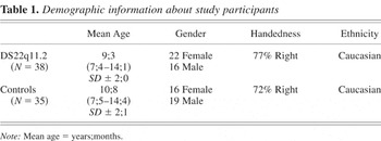
Demographic information about study participants
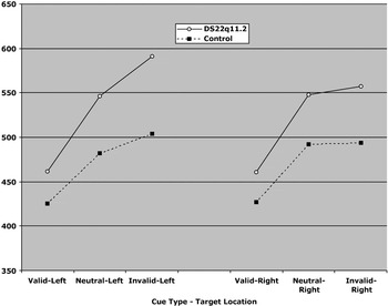
The performance of children with DS22q11.2 and controls on the cueing task.
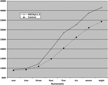
The performance of children with DS22q11.2 and controls on the enumeration task.
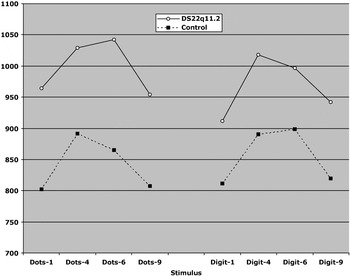
The performance of children with DS22q11.2 and controls on the distance effect task.
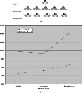
(a) Examples of flanker stimuli from the attentional networks test (ANT). (b) The performance of children with DS22q11.2 and controls on the executive attention component of the ANT.
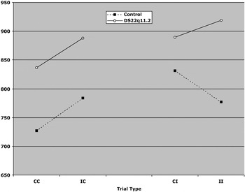
The performance of children with DS22q11.2 and controls on the Gratton effect.
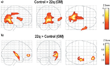
Voxel-based morphometry results depicting clusters of greater gray matter volumes (a) in control children than in those with DS22q11.2 and (b) in children with DS22q11.2 than in controls.
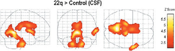
Voxel-based morphometry results depicting clusters of greater cerebrospinal fluid volumes in children with DS22q11.2 than in controls.
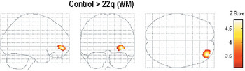
Voxel-based morphometry results depicting clusters of greater white matter volumes in control children than in those with DS22q11.2.
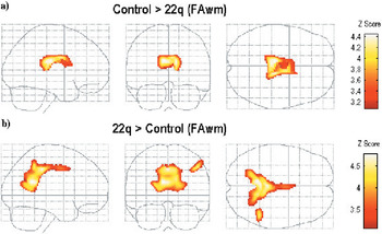
Voxel-based morphometry results depicting clusters of greater fractional anisotropy values (a) in control children than in those with DS22q11.2 and (b) in children with DS22q11.2 than in controls.
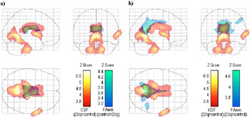
Composite depictions of clusters of greater cerebrospinal fluid volumes in children with DS22q11.2 along with clusters of greater fractional anisotropy values (a) in control children and (b) in the same children.
 You have
Access
- 66
- Cited by