Achondroplasia is the prototype of human bone growth disorders, the so-called chondrodysplasias, and the most common form of dwarfism in humans (Ref. Reference Horton, Hall and Hecht1). Depicted in artworks from cultures dating back to ancient times, it was not delineated clinically until the middle of the 20th century (Refs Reference Kozma2, Reference Rimoin3). Similarly, although suspected to reflect a disturbance of cartilage-mediated (endochondral) bone growth for a century, the nature of this disturbance has only come to light in the last 15 years. Indeed, achondroplasia is caused by mutations of the transmembrane receptor fibroblast growth factor receptor 3 (FGFR3), an important regulator of linear bone growth (Refs Reference Shiang4, Reference Rousseau5, Reference Bellus6). The last decade has witnessed a steady increase in understanding the biology of FGFR3 and how it regulates the endochondral ossification machinery that is responsible for bone growth. An important consequence of this research has been the identification of potential targets for therapeutic intervention for achondroplasia, or more specifically the development of strategies for normalising the bone growth disturbance resulting from mutant FGFR3. Although some of these strategies are still at the conceptual stage, proof of concept has been established in animals for other strategies, which are rapidly approaching clinical trials in children. This review will briefly analyse the clinical aspects of achondroplasia, examine recent advances in understanding FGFR3 biology and its role in regulating linear bone growth and explore the current status of FGFR3-targeted therapies for achondroplasia.
Achondroplasia
Clinical features
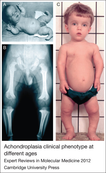
Achondroplasia is usually recognised at birth because of its distinctive clinical and radiographic features. However, prenatal diagnosis is becoming more common as the use of routine prenatal ultrasound increases in developed world (Ref. Reference Horton, Hall and Hecht1). Newborn infants with achondroplasia typically present with disproportionate shortening of the limbs, a long and narrow trunk, a large head with frontal bossing and a hypoplastic midface. The hands are short and broad, often displaying a three-pronged (trident) configuration and many joints, except for the elbow, are hyperextensible. Infants are often hypotonic and many exhibit a thoracolumbar gibbus. The typical clinical features are shown in Figure 1.
Achondroplasia clinical phenotype at different ages. (A) Infant with large head, prominent forehead, midface hypoplasia, small chest and limb shortening, especially proximally. (B) Typical radiographic findings from a child with achondroplasia including short tubular bones, metaphyseal irregularities of distal femur and proximal tibia and rounded iliac bones. (C) Three years old with typical features listed in A accompanied by tibial bowing. Reproduced from Ref. Reference Horton, Hall and Hecht1 (© 2007 Elsevier) with permission from Elsevier.

Skeletal X-rays of the newborn infant reveal characteristic abnormalities. The long bones of the limbs are short, particularly the proximal bones, with metaphyseal irregularities especially at the knees. The vertebral bodies are normal in height and width, but the pedicles are short. The pelvis is abnormal with small and square iliac wings, a horizontal acetabulum and a narrow sacrosciatic notch. The cranium is large with a prominent forehead, midface hypoplasia and contraction of the base of the skull with a small foramen magnum. Although usually not necessary, the diagnosis can be confirmed from mutational analysis of DNA. Mutational diagnosis can also be used for prenatal diagnosis particularly in couples at risk of homozygous achondroplasia.
Natural history
Children and adults with achondroplasia display a wide array of complications affecting many different organ systems. Most of these complications are consequences of abnormal linear bone growth. They tend to appear or evolve at predicted ages, thus making it possible for care givers to anticipate – and thereby minimise or even prevent – their impact on a patient's health and well-being. Indeed, anticipatory care, leading to early detection and treatment of complications, is currently the mainstay of achondroplasia management.
Most infants with achondroplasia have large heads, but the incidence of true hydrocephalus is relatively low. The most severe complication of infancy and early childhood is spinal cord compression at the cervical medullary junction. It is common and might require surgical decompression in some cases, although controversy exists over its extent and the indications for surgery. Compression usually manifests itself as long-track signs and central apnea. In older children, apnea, snoring, and other sleep disturbances might reflect airway narrowing caused by midfacial and pharyngeal hypoplasia. One consequence of this hypoplasia is a relative adenoid and tonsil hypertrophy; another is muscular upper airway obstruction caused by compression of nerves such as the hypoglossal nerve that enervate these muscles as they exit the skull.
Midfacial underdevelopment also results in short Eustachian tubes, which commonly leads to repeated and chronic otitis media during childhood often with hearing loss, which might contribute to delay in speech. Dental crowding is common owing to the relatively small jaw. Many individuals with achondroplasia develop tibial and fibula bowing during childhood. It typically progresses with age often requiring surgical correction to straighten bones and realign the knees and ankles.
Most adults with achondroplasia live happy, productive lives, although there are complications that typically arise or worsen during adulthood. Spinal stenosis with neurological signs and claudication in the legs increases from around 10% in adolescents to about 80% by the sixth decade. About a third of patients require lumbar laminectomy. Obesity is common, and does not appear to simply reflect short stature because it is more common in achondroplasia than in other conditions with comparable stature. The mechanism is unknown, although there has been speculation that it involves elusive pancreatic islet cell functions of FGFR3.
Women with achondroplasia appear to have normal fertility. However, their narrow pelvic outlet usually necessitates delivery by caesarian section. This situation is compounded because of the possibility that offspring may also have achondroplasia with a characteristically large head at birth.
Longevity studies suggest that overall and age-specific mortality rates are increased at all ages (Ref. Reference Wynn7). The increase in adults comes primarily from cardiovascular disease. The specific risk factors related to achondroplasia are not known.
Genetics of achondroplasia
Achondroplasia is inherited in an autosomal-dominant manner. It was mapped to the distal short arm of chromosome 4 in 1994 and heterozygous mutations were discovered in FGFR3 shortly afterwards (Refs Reference Shiang4, Reference Rousseau5, Reference Bellus6, Reference Velinov8). Almost all patients with the typical clinical features of achondroplasia have a glycine to arginine substitution at position 380 (G380R), which maps to the transmembrane domain of the receptor (Ref. Reference Bellus9). Additional FGFR3 mutations were subsequently detected in thanatophoric dysplasia types I and II, hypochondroplasia and in other rarer disorders including severe achondroplasia with developmental delay and acanthosis nigricans (SADDAN), Muenke craniosynostosis and Crouzon syndrome with acanthosis nigricans (Refs Reference Horton, Hall and Hecht1, Reference Bellus9, Reference Tavormina10, Reference Vajo, Francomano and Wilkin11). In contrast to the constitutional mutations of FGFR3 responsible for these conditions, somatic mutations of FGFR3 occur in cancer, especially bladder, cervical and colorectal cancer (Refs Reference Dodurga12, Reference Sonvilla13, Reference Chesi14).
Achondroplasia has an incidence of one in 10 000 to one in 30 000 live births; and it results from a spontaneous mutation to nonachondroplastic parents in an estimated 80% of cases (Refs Reference Horton, Hall and Hecht1, Reference Bellus9). This high rate of mutation combined with a recognised association with older paternal age initially suggested that FGFR3 is one of the most mutable genes in humans. However, analyses of FGFR mutations in human sperm suggest an alternative explanation (Refs Reference Goriely15, Reference Wilkie16). These studies suggest that the achondroplasia FGFR3 mutation occurs at a normal rate in relatively young men and that it gives spermatogonia bearing this mutation a competitive advantage over other spermatogonia. This advantage leads to clonal expansion of mutant cells over time and a progressive increase in the relative abundance of sperm bearing the mutation with paternal age.
FGFR3
Receptor
FGFR3 is one of four transmembrane FGF receptors (FGFR1–FGFR4) that contain an extracellular ligand-binding domain, a transmembrane domain and an intracellular split tyrosine kinase domain (Refs Reference Eswarakumar, Lax and Schlessinger17, Reference Ornitz18, Reference Chen19). The structure of FGFR3 and signalling pathways most relevant to bone growth are illustrated in Figure 2. Alternative mRNA splicing of the extracellular domain generates ‘b’ and ‘c’ isoforms with altered ligand affinity. The c isoform is primarily expressed in tissues of mesenchymal origin including skeletal tissues.
FGFR3 structure and signal transduction. Fibroblast growth factor (FGF) and heparin binding to the extracellular domain of FGF receptor 3 (FGFR3) induce dimerisation, kinase activation and transphosphorylation of tyrosine residues (in yellow) leading to activation of downstream signalling pathways such as signal transducer and activator of transcription (STAT). Membrane bound FRS2 constitutively associates with the intracellular juxtamembrane domain of FGFR3, is phosphorylated by the receptor upon activation and serves as scaffolding platform for signal transduction cascades through the MAPK–MEK–ERK–p38 pathway. C-type natiuretic peptide (CNP) binding to natiuretic peptide receptor-B (NPR-B) induces the generation of the second messenger cGMP, which activates PKG leading to attenuation of the MAPK pathway through RAF1.

The function of FGFR3 during skeletal development and postnatal growth was identified by analysing the consequences of its mutation in mice. Indeed, the global knockout of FGFR3 produced large mice with longer than normal limb bones (Refs Reference Colvin20, Reference Deng21). By contrast, targeted overexpression of FGFR3 bearing the achondroplasia mutation to cartilage of transgenic mice produced a small mouse with short bones resembling those seen in human achondroplasia (Ref. Reference Naski22). Similarly, cartilage targeted overexpression of a ligand (FGF9) that activates FGFR3 generated a dwarf mouse (Ref. Reference Garofalo23). These observations established that FGFR3 is an important negative regulator of endochondral bone growth and that the mutations that cause achondroplasia and related dwarfing conditions involve gain of this negative function.
Ligands
FGFR3 is primarily activated by paracrine FGF ligands, which include FGF1–FGF10, FGF16–FGF18, FGF20 and FGF22 (Ref. Reference Itoh and Ornitz24). The physiologic ligands for FGFR3 are not known, but FGF2, FGF4, FGF9 and especially FGF18 (see below) are the best candidates based on their distribution of expression, affinity for FGFR3 and the phenotypes of mice lacking these ligands (Refs Reference Ornitz18, Reference Ornitz25, Reference Liu26, Reference Ohbayashi27, Reference Liu28, Reference Krejci29). Heparan sulfate proteoglycans are required for FGF–FGFR3 interactions and they probably influence binding specificity and ligand localisation. Recent evidence indicates that heparan sulfate decorating chondrocyte-derived perlecan mediates binding of FGF18 to FGFR3 in the growth plate (Ref. Reference Chuang30).
The current view is that ligand binding to FGFRs induces dimerisation of receptor monomers, leading to transphosphorylation of key tyrosine residues within the receptor kinase domain, which further activates its tyrosine kinase activity (Refs Reference Eswarakumar, Lax and Schlessinger17, Reference Plotnikov31, Reference Hart, Robertson and Donoghue32, Reference Mohammadi, Olsen and Ibrahimi33, Reference Horton and Degnin34, Reference Bae and Schlessinger35). Signals are propagated downstream by the recruitment of signalling molecules to these phosphorylated residues or to closely linked docking proteins, especially fibroblast growth factor substrate 2 (FRS2), which binds to the receptor and provides additional tyrosine residues that are phosphorylated in response to receptor activation.
Mechanisms of signal transduction
Activation of several signalling pathways has been detected downstream of FGFR3, including the STAT, MAPK, PLCγ, PI3K, PKC and AKT pathways (see Box 1) (Refs Reference Eswarakumar, Lax and Schlessinger17, Reference Su36, Reference Legeai-Mallet37, Reference Sahni38, Reference Hart39, Reference Choi40). The first two have received the most attention in the context of skeletal growth plate function. STAT signals are thought to inhibit chondrocyte proliferation acting at least partially through the potent cyclin-dependent kinase inhibitor p21 (WAF1/CIP1) (Ref. Reference Su36). MAPK signals negatively influence chondrocyte proliferation, terminal differentiation and postmitotic matrix synthesis utilising both p38 and ERK arms of this pathway (Refs Reference Sahni38, Reference Murakami41, Reference Zhang42, Reference Minina43).
Box 1 Abbreviations used for signalling pathways
AKT, family of serine/threonine kinases, also known as protein kinases B (PKB)
ERK, extracellular signal-related protein kinase
MAPK, mitogen-activated protein kinase
PI3K, phosphoinositide 3-kinase
PKC, protein kinase C
PLCγ, phospholipase Cγ
STAT, signal transducer and activator of transcription
The STAT, MAPK and potentially other signalling pathways are believed to act through their influence on expression of other genes, i.e. effector genes that upregulate antiproliferative activities and downregulate growth-promoting molecules. Indeed, a large number of genes in both categories have been identified from expression screens of chondrocytic cells in which FGFR3 was stimulated by ligand or mutation (Refs Reference Dailey44, Reference Schibler45). However, the mechanistic link between the signalling pathways and the transcriptional machinery that mediates their effects on endochondral bone growth is poorly understood.
In contrast to the well-established paradigm that FGFR3 functions through signalling cascades that reside in the cytoplasm but terminate in the nucleus, there is new evidence that it might also act through direct interaction with target genes and/or transcriptional machinery within the nucleus. Degnin and colleagues have recently reported that upon activation, FGFR3 undergoes sequential cleavages to liberate an intracellular domain fragment capable of translocating to the nucleus (Ref. Reference Degnin, Laederich and Horton46). The first cleavage removes most of the extracellular domain of the receptor; the second cleavage is carried out by γ-secretase within the cell membrane. These findings suggest a plausible mechanism whereby FGFR3 can reach the nucleus, which is otherwise prevented by its membrane anchorage. Similar proteolytic processing has been reported for other receptor tyrosine kinase receptors, in some cases leading to the identification of nuclear functions that are distinct from those mediated by kinase cascades (Refs Reference Lyu, Yamamoto and Lu47, Reference Carpenter and Liao48, Reference Parkhurst, Zampieri and Chao49). Time will be needed to determine the physiological significance of this putative direct signalling pathway for FGFR3.
Modulation of downstream signals
Several factors have been postulated to modify the strength of signals downstream of FGFR3, including SOCS (Suppressors of cytokine-inducible stimulation of STAT) and SNAIL (SNAIL1) proteins (Refs Reference Ben-Zvi50, Reference de Frutos51). SOCS proteins are expressed in response to cytokine-induced stimulation of STAT phosphorylation; they act as a negative feedback mechanism to temper STAT signals. SOCS1 and SOCS3 are induced by FGF treatment of chondrocytic cells. They appear to be constitutively bound to FGFR3 and may influence trafficking of the receptor (Ref. Reference Ben-Zvi50). De Frutos and co-workers suggest that SNAIL1 is required for FGFR3 function in chondrocytes and that it lies downstream of FGFR3 but upstream of both major signalling pathways – STAT1 and MAPK – but how it functions in this role is poorly understood (Ref. Reference de Frutos51).
The best-defined pathway for modulating FGFR3 signals in the context of bone growth involves C-type natiuretic peptide (CNP, NPPC) (Fig. 2). In fact, CNP appears to modulate the MAPK signals that mediate the achondroplastic skeletal phenotype in mice and presumably in humans (Refs Reference Horton, Hall and Hecht1, Reference Potter, Abbey-Hosch and Dickey52, Reference Yasoda53).
CNP is one of three structurally related peptides that possess potent natriuretic (sodium excretion promoting), diuretic and vasodilating properties and were originally defined in the context of regulating blood pressure and volume (Refs Reference Potter, Abbey-Hosch and Dickey52, Reference Pejchalova, Krejci and Wilcox54). In contrast to atrial and brain natriuretic peptides [ANP and BNP (atrial and brain natiuretic peptide), respectively], which function in an endocrine fashion, CNP acts predominantly in a paracrine or autocrine manner in several tissues including the growth plate (Ref. Reference Sellitti, Koles and Mendonca55). Through interaction with its cognate receptor natiuretic peptide receptor-B (NPR-B), CNP induces accumulation of intracellular cGMP. Of note, homozygosity for mutations of NPR-B is responsible for acromesomelic dysplasia, type Maroteaux (OMIM 602875) (Ref. Reference Bartels56). Both CNP and NPR-B are expressed in the proliferative and prehypertrophic zones of the growth plate setting up a potential autocrine or paracrine regulatory circuit (Ref. Reference Yasoda57). Signals downstream of the two receptors intersect at the level of RAF1 such that the CNP and NPR-B signals antagonise MAPK signalling downstream of FGFR3 activation (Refs Reference Pejchalova, Krejci and Wilcox54, Reference Miyazawa58, Reference Krejci59).
Further evidence of CNP's antagonism of growth inhibitory effects of FGFR3 in the growth plate comes from mouse genetics. Mice lacking CNP or its receptor NPR-B exhibit dwarfism, whereas transgenic mice overexpressing CNP in the cartilaginous growth plate display skeletal overgrowth (Refs Reference Yasoda53, Reference Chusho60). Many tissues express another natiuretic receptor (NPR-C) that lacks a functional intracellular domain and acts as clearance receptor. In the growth plate, it appears to antagonise CNP–NPR-B signalling (Ref. Reference Potter, Abbey-Hosch and Dickey52).
Impact of the mutation
As discussed earlier, the achondroplasia mutations act by exaggerating the negative functions of FGFR3 on endochondral ossification (Refs Reference Horton, Hall and Hecht1, Reference Horton and Degnin34, Reference Horton, Unsicker and Krieglstein61, Reference Laederich and Horton62). The consequence of these mutations on receptor activation and the effects of aberrant signalling on the growth plate are explained below.
Consequences to FGFR3
The achondroplasia mutation maps to the transmembrane domain of the FGFR3 protein. It has been proposed that the amino acid substitution increases the stability of the receptor dimer, although this finding has been challenged (Refs Reference Webster and Donoghue63, Reference You, Li and Hristova64). Another mechanism involves delayed turnover of activated receptor, which can lead to an increase in overall signal output. For instance, Monsonego-Ornan and colleagues have suggested that the achondroplasia mutation slows receptor internalisation leaving it on the surface to signal (Ref. Reference Monsonego-Ornan65). Others have described a defect in c-Cbl-mediated receptor ubiquitylation that delays trafficking of mutant FGFR3 to lysosomes for degradation (Refs Reference Cho66, Reference Guo67). Similarly, Ben-Zvi et al. have suggested that SOCS3 and SOCS1 (suppressor of cytokine signalling) directly interact with FGFR3 and increase the stability and signalling of FGFR3 (Ref. Reference Ben-Zvi50).
Laederich and co-workers have recently documented that FGFR3 is stabilised by the heat shock protein 90 (Hsp90), making it an Hsp90 client protein (Ref. Reference Degnin, Laederich and Horton68). This cytoplasmic chaperone is known to stabilise ‘inherently unstable’ configurations of proteins, of which the most relevant here are kinases in their activated state. For a signalling kinase such as FGFR3, Hsp90 association can prevent kinase degradation and exaggerate signalling output. This mechanism has been implicated for a number of oncogenic kinases in several different cancers (Refs Reference Kim69, Reference Trepel70). As a result, Hsp90 has been considered a therapeutic target in many of these cancers.
Consequence to skeletal growth
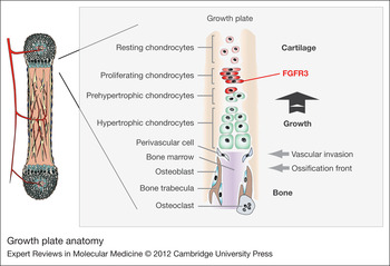
The process of endochondral ossification is responsible for both the formation and linear growth of most of the skeleton. However, the negative regulatory influence of FGFR3 is exerted mainly during the growth phase when it reduces the rate of cartilage template formation and turnover necessary for bone elongation. The anatomy of the growth plate with its landmarks and relevant cells are depicted in Figure 3. FGFR3 is expressed primarily in the late proliferating and early prehypertrophic cells. Most evidence to date suggests that FGFR3 inhibits both the proliferation and terminal differentiation of growth plate chondrocytes and synthesis of extracellular matrix by these cells (Refs Reference Ornitz18, Reference Horton71, Reference Kronenberg72). Terminal differentiation includes chondrocyte hypertrophy and is sometimes referred to as hypertrophic differentiation. This inhibitory function is consistent with its expression in cells exiting the cell cycle. However, it is also proposed that FGFR3 induces premature terminal differentiation reducing the number of cells that contribute to template synthesis (Ref. Reference Minina43, Reference Dailey73).
Growth plate anatomy. Linear bone growth results from chondrocyte proliferation and differentiation sequentially through the growth plate zones, that is resting, proliferative, prehypertrophic, hypertrophic and vascular invasion zones. Fibroblast growth factor receptor 3 (FGFR3) is primarily expressed in proliferating chondrocytes where it tightly regulates proliferation and transition to terminal (hypertrophic) differentiation. Some FGFR3 is expressed in prehypertrophic chondrocytes, where mutant FGFR3 dysregulates their differentiation and matrix secretion.

Although all the kinase cascade pathways that normally propagate FGFR3 signals presumably transmit the exaggerated output of the mutant receptors, there is evidence that the ERK and p38 arms of the MAPK pathway are especially important. Indeed, transgenic mouse strains in which expressions of constitutively active MEK1 and MEK6 were targeted to cartilage displayed ‘achondroplastic’ skeletal phenotypes; MEK1 and MEK6 lie upstream of ERK and p38 phosphorylation cascades, and would be expected to constitutively activate these downstream targets (Refs Reference Zhang42, Reference Murakami74).
Implications for treating achondroplasia
A number of nonsurgical strategies designed to reduce excessive FGFR3 output have been considered as possible treatments to normalise bone growth in achondroplasia (Fig. 4). They include strategies to interfere with FGFR3 synthesis, block its activation, inhibit its tyrosine kinase activity, promote its degradation and antagonise its downstream signals. Many have been borrowed conceptually from the cancer treatment field, which is not surprising because the biology of mutant FGFR3 is virtually identical to that of many oncogenic kinases at the molecular level.
Strategies for therapeutic intervention. (1) chemical inhibitors to reduce fibroblast growth factor receptor 3 (FGFR3) tyrosine kinase activity; (2) antibodies to block receptor activation; (3) exogenous C-type natiuretic peptide (CNP) to enhance CNP-mediated antagonism of downstream signals; (4) RNAi to reduce FGFR3 production; (5) Hsp90 inhibitors to induce degradation of activated receptor; and (6) agents to disrupt direct nuclear signalling of FGFR3. See text for discussion.

FGFR3 kinase inhibitors (Strategy 1)
The rationale for this strategy is that activation and signalling of FGFR3 depend on its kinase activity; consequently, chemical inhibition of its kinase activity should block its inhibitory output and restore bone growth. The strategy has been successful for many oncogenic kinases (Ref. Reference Hartmann75). However, because the structure of the tyrosine kinases domain is highly conserved among RTK subfamilies, kinase inhibitors frequently crossreact with other RTKs (Ref. Reference Collins and Workman76). Such crossreactivity is not problematic in cancer therapy, because cancer cells frequently depend on multiple RTKs, but it could be more of a problem in the treatment of children with achondroplasia, which involves only the single receptor.
FGFR3 selective inhibitors have been reported. Aviezer and colleagues described synthesis of an oxindole-based inhibitor selective for FGFR3 relative to FGFR1 (Ref. Reference Aviezer, Golembo and Yayon77). The compound stimulated cartilage growth in cultured limb bones from a knock-in mouse model of achondroplasia. This success has not been extended to live mice. Novartis reports on their website the generation of a highly specific orally bioavailable FGFR3 kinase inhibitor with efficacy in in vivo models of cancer in the nanomolar range (Ref. 78).
Monoclonal antibodies (Strategy 2)
This strategy is based on the successful results obtained from treating breast cancer patients with trastuzumab (trade name Herceptin) (Ref. Reference Aviezer, Golembo and Yayon77). Trastuzumab is a humanised monoclonal antibody to the extracellular domain of the HER2/neu (ErbB2), a transmembrane receptor often amplified on breast cancer cells (Ref. Reference Hall and Cameron79). HER2 transmits mitogenic signals to the cancer cells in the absence of external mitogens, and trastuzumab reverses the adverse effects of the overactive receptor. Rauchenberger et al. reported the generation of a comparable antibody to FGFR3 that effectively blocked ligand-induced FGFR3 activation (Ref. Reference Rauchenberger80); however, there have been no subsequent reports on its capacity to stimulate bone growth in vivo.
As mentioned earlier, activating FGFR3 mutations are relatively common in bladder cancer, where they are thought to promote proliferation and survival of the tumour cells (Ref. Reference Cappellen81). Blocking antibodies similar to those just described have been used successfully to reduce proliferation in cells derived from these tumours, further supporting the utility of this approach in reducing FGFR3 output (Refs Reference Martinez-Torrecuadrada82, Reference Qing83, Reference Hadari and Schlessinger84).
CNP antagonism of FGFR3 signals (Strategy 3)
The CNP-based therapeutic strategy evolved from a series of mouse genetics investigations in the Nakao laboratory in Kyoto, Japan dating back over a decade. The initial observation revealed that transgenic mice overexpressing BNP in the liver from a human serum amyloid P promoter and having very high concentrations of plasma BNP exhibited postnatal skeletal overgrowth with elongation of long bone growth plates (Ref. Reference Suda85). Tibial organ culture experiments revealed that CNP was more potent than BNP in stimulating growth, suggesting that CNP was the physiological ligand in growing bones (Ref. Reference Yasoda57). Mice lacking CNP were analysed next, revealing severe postnatal dwarfism that was rescued when the strain was crossed with mice overexpressing CNP from a transgene driven by the cartilage-specific COL2A1 promoter/enhancer (Ref. Reference Chusho60).
These findings established the ability of CNP to stimulate endochondral ossification in vivo. However, noting the phenotypic similarity of CNP-null mice to mouse models of achondroplasia, they raised the possibility that CNP could overcome the bone growth deficit caused by overactive FGFR3 in achondroplasia. To explore this possibility, mice overexpressing CNP in cartilage were crossed with mice displaying an achondroplastic phenotype resulting from overexpression of FGFR3 bearing the achondroplasia mutation in cartilage (Ref. Reference Yasoda53). The same Col2A1 promoter/enhancer was used to drive the expression of both mutant FGFR3 and CNP. The growth deficiency of the achondroplastic mice was corrected by the local overexpression of CNP and a dose effect was demonstrated in that bone growth was greater in mice homozygous compared with heterozygous for the CNP transgene insertion. The results suggested that the antagonism was primarily directed at MAPK-mediated FGFR3 signals.
Follow-up studies have shown a growth plate activity dose response to circulating CNP produced in the liver from the liver-specific transgene promoter (Ref. Reference Kake86). Comparison of two different strains harbouring the CNP transgene (one producing plasma concentrations approximately two-fold increased over normal, the other producing levels >2000-fold normal level of plasma CNP) revealed relatively greater growth response from the latter strain; however, the overgrowth led to deformities, such as severe kyphosis. Mice from this strain also exhibited reduced heart size, which is consistent with cardiovascular consequences of such a high dose of CNP, but this complication was not detected in the mildly overexpressing transgenic mouse strain.
The experimental mice with mild and chronic elevation of plasma CNP were analysed further as a preclinical treatment model for achondroplasia (Ref. Reference Kake86). The results showed that the increased bone growth was associated with qualitatively normal skeletal radiographs and growth plate structure. Growth plate differentiation markers were essentially normal, although the zones were slightly thicker than those of wild-type controls. These results are consistent with the concept that CNP ‘speeds up’ growth plate activity to generate more cartilage template over a given period of time, which translates into more bone growth. They also support the notion that CNP primarily modifies matrix production rather than chondrocyte proliferation or terminal differentiation. An additional observation was that these mice displayed a larger foramen magnum size than displayed by control mice.
Most recently, the Nakao group has explored delivery of exogenous CNP through continuous intravenous infusion (Ref. Reference Yasoda87). Recombinant CNP (CNP-22) was administered as a continuous infusion into jugular veins of wild-type and transgenic achondroplastic mice for 4 weeks starting at 3 weeks of age. Wild-type mice treated at a dose of 1 μg/kg/min had 12% greater naso-anal lengths and over 30% thicker growth plates than vehicle-only-treated controls. Significant rescue of the bone growth deficiency was also observed after 3 weeks of treatment for the achondroplastic mice even at a 10-fold lower dose of CNP.
Although encouraging, these studies with exogenous CNP point out a major obstacle in its potential use to treat human achondroplasia. Native CNP is rapidly cleared from the blood; its half-life in plasma is estimated to be 2.6 min (Ref. Reference Hunt88). To overcome this obstacle, efforts are underway to modify the structure of native CNP and thereby prolong its survival. One example of such a modification is the CNP analogue BMN 111 developed by BioMarin (Navato, California), which from preliminary reports stimulates bone growth in achondroplastic mouse models when given subcutaneously once daily (Ref. Reference Lorget89).
These investigations build a strong case for CNP as a viable therapeutic candidate for stimulating bone growth in achondroplasia. Two pharmaceutical firms, BioMarin in the USA and Chugai in Japan, are leading efforts to bring CNP-based therapeutics to human trials, which are likely to begin in the near future. In addition to a bone growth response, an important question in these trials will involve complications of long-term CNP therapy. It is likely that higher than physiological levels of plasma CNP will be required to produce sufficient antagonism of FGFR3 signals in growth plate chondrocytes to stimulate bone growth. If so, there is concern that this elevation could lead to complications resulting from excessive stimulation of its cognate receptor NPR-B in other tissues. It is reassuring that patients with acromesomelic dysplasia-type Maroteaux who display chronically elevated levels of blood CNP do not have such complications (Ref. Reference Olney90). However, they lack functional NPR-B and would be expected to be ‘protected’ from this scenario. Potentially relevant is the report of a 14-year-old girl with relatively tall stature and a Marfanoid habitus, and a de novo balanced t(2;7)(q37.1;q21.3) translocation (Ref. Reference Bocciardi91). The gene encoding CNP maps to near the translocation breakpoint on chromosome 2. The authors observed a doubling of plasma CNP levels compared with normal and substantially increased expression of this gene in cultured fibroblasts; they speculated that the elevated CNP caused the tall stature.
It is also conceivable that pharmacological levels of circulating CNP could activate NPR-A, which is normally activated by A-type or B-type natriuretic peptides. However, the risk for this complication is likely diminished by the reduced sensitivity of NPR-A to CNP compared with these other physiological ligands (Refs Reference Potter, Abbey-Hosch and Dickey52, Reference Suga92). It is also conceivable that chronically elevated concentrations of CNP could saturate NPR-C, the clearance receptor, and alter the normal turnover dynamics of any or all of the natriuretic peptides. Long-term clinical studies will be needed to resolve this issue.
Other potential strategies
There are three additional strategies that are currently under consideration. They are mentioned briefly because they offer theoretical advantages over strategies currently under investigation.
RNAi (Strategy 4)
The first approach is to silence the expression of FGFR3 using techniques that interfere with translation of its mRNA transcripts. These approaches generally fall under the rubric of RNA interference (RNAi), a natural process used by cells to protect themselves from foreign DNA, such as viruses and, more pertinent here, to silence endogenous genes (Refs Reference Davidson and McCray93, Reference Boudreau, Rodriguez-Lebron and Davidson94, Reference Lochmatter and Mullis95). In the latter context, RNAi is triggered by endogenous micro RNAs (miRNAs) that are sequentially pared down to short RNA duplexes. These duplexes are incorporated into RNA-induced silencing complexes that bind to and silence target transcripts. The silencing mechanism depends on the degree of complementation; near-perfect base pairing of the miRNA to the target transcript promotes transcript cleavage, whereas imperfect complementation leads to translational repression and mRNA decay (Ref. Reference Boudreau, Rodriguez-Lebron and Davidson94). This cellular machinery can also be engaged by artificial RNAs that mimic endogenous miRNAs, so-called small interfering-RNAs or siRNAs. Because targeting is sequence specific, any gene can theoretically be silenced by designing an appropriate siRNA. Moreover, because of the sequential nature of miRNA biogenesis, artificial RNAs can be designed to engage the RNAi machinery at different points in the pathway.
The clinical application of RNAi technology to date has focused mainly on viral diseases and cancer and to a lesser extent on genetic disease. A number of clinical trials are underway. The appeal of RNAi for achondroplasia is the possibility of selectively silencing the FGFR3 allele harbouring the achondroplasia mutation leaving the wild-type FGFR3 allele to function normally. Despite this appeal there are many issues that would need to be resolved before such therapy could be implemented. Some are common to RNAi therapies in general, such as those involving mode of delivery and stability of the siRNA delivery platform. Others are unique to achondroplasia and/or FGFR3. For instance, to what extent can the mutant allele be selectively silenced and does it matter, as long as FGFR3 function is appropriately reduced? Does FGFR3 need to be silenced to different degrees in different growth plates or at different ages and if so, how could this be achieved? And how would it be terminated at the end of puberty when bone growth ceases?
Hsp90 inhibition (Strategy 5)
Manipulation of chaperone complexes to promote the folding or unfolding of disease-causing proteins has been tested for its therapeutic potential in both in vitro and in vivo experimental models (reviewed in Ref. Reference Hartl, Bracher and Hayer-Hartl96). The chaperone Hsp90 has been the focus of many of these experiments, in part because of the readily available inhibitors of Hsp90 chaperone function. Inhibition of Hsp90 induces the degradation of its ‘client’ proteins; this has been used to destabilise oncogenes in cancer and to reduce the accumulation of aggregation-prone proteins in some neurodegenerative disease animal models. The recent identification of FGFR3 as a client of the chaperone Hsp90 indicates that its stability – and thus signalling capacity – is dependent on the function of Hsp90 (Ref. Reference Laederich97). This observation calls into question whether Hsp90 inhibitors could be used to destabilise FGFR3 and thus reduce its growth inhibitory signals in achondroplasia. Further research is warranted into the effect of Hsp90 inhibitors on the growth plate. A few publications have reported an effect of Hsp90 inhibitors on cartilage (Refs Reference Calamia98, Reference Herzog99). Ongoing clinical trials of Hsp90 inhibitors in cancer should establish the safety of Hsp90 inhibitors in humans.
Modulating FGFR3 cleavage and/or nuclear function (Strategy 6)
Recently, it was identified that FGFR3, like select other transmembrane signalling receptors, can undergo regulated intramembrane proteolysis (RIP) in response to ligand stimulation, switching the functional localisation of FGFR3 from the membrane to the nucleus (Ref. Reference Degnin, Laederich and Horton68). A key step in this process is the liberation of a cytoplasmic fragment from its membrane anchorage by the enzyme gamma secretase. The consequence of RIP on the function of FGFR3 in the growth plate remains unknown. However, observational data in the bovine growth plate indicate that cleavage events of FGFR3 occur in a spatially regulated pattern, implying an important role for FGFR3 cleavage in growth plate biology (Ref. Reference Pandit100). Altering the cleavage events of FGFR3 might permit manipulation of FGFR3 function to influence endochondral ossification. A number of drugs have been developed to alter the activity of γ-secretase because of its role in other diseases. These drugs have potential to modulate FGFR3 RIP and its contribution to achondroplasia. However, γ-secretase influences normal human physiology in many ways, so the safety of these pharmaceuticals in children is of concern (Ref. Reference Wolfe101). In addition to influencing the cleavage of FGFR3, the nuclear function and/or potential genes regulated by nuclear FGFR3 might become new drug targets for achondroplasia.
Challenges to bringing FGFR3-based therapies to the bedside
Acknowledging that drugs targeting FGFR3 are on the horizon as discussed above, there remain significant barriers to establishing the use of these drugs in achondroplasia. The greatest challenge might be the ethical considerations of conducting long-term clinical trials in children who do not have a terminal illness. Thus, any potential treatment must be thoroughly tested in preclinical models and proved safe and effective for children. Even when safety and efficacy are demonstrated, performing pharmacodynamic and pharmacokinetic studies to optimise treatment protocols in children is challenging because growth and metabolic rates vary by age and developmental stage (Ref. Reference Mulla102).
A related issue is specificity in terms of both receptors and tissues. Clearly, a drug that acts only on FGFR3 in growing bones has advantages over drugs that poorly distinguish FGFR3 signalling activity from that of other FGFRs or target FGFR3 activity in tissues unrelated to bone growth. In the first instance, drugs such as CNP that target signalling pathways downstream of FGFR3 not known to be utilised by other FGFRs may have fewer side effects related to suppression of other FGFRs than tyrosine kinase inhibitors and antisense strategies that lack complete selectivity for FGFR3. However, such drugs might be more likely to disturb functions of other receptors and signalling pathways that are modulated by CNP. At this point in time, these questions are largely theoretical, but they will need to be addressed in the future.
Although the tissue distribution of FGFR3 expression is not well defined beyond early development, tissue surveys in adults suggest that it is expressed in many nonskeletal tissues including the gastrointestinal tract, pancreatic islet of Langerhans, steroidogenic cells of the adrenal cortex, Schwann cells of sympathetic ganglia and in the central nervous system, mainly in glial cells (Ref. Reference Horton, Hall and Hecht1). Accordingly, it would seem desirable to target anti-FGFR3 therapies in achondroplasia to skeletal tissues, namely to growth plate cartilage, and away from other tissues. Tissue targeting has been successful in cancer therapy (Refs Reference Goldberg, Langer and Jia103, Reference Alexis104), but targeting avascular cartilage might be more challenging than delivering agents to cancers that tend to be highly vascularised. Targeting drugs to the growth plate remains in the conceptual stage; however, peptides have been identified that home to cartilage (Ref. Reference Rothenfluh105).
Finally, it is conceivable that drugs developed to treat other conditions but never considered for achondroplasia could be effective therapeutic agents in achondroplasia. Indeed, the number of drugs listed in the US Pharmacopeia for which safety in children has already been established is large. The biggest obstacle in identifying candidate agents for further investigation is a lack of growth plate functional assays suitable for high-throughput screening. Such assays are challenging even without the high-throughput component. Nevertheless, as they are developed in the future, this approach could be fruitful and circumvent some of the barriers to bringing new drugs into clinical practice.
Acknowledgements
The authors are supported by research grants from the Shriners Hospitals for Children (W.A.H.). M.B.L. is a recipient of a predoctoral fellowship from the PhRMA Foundation.
Conflicts of interest
Dr Horton received consultancy fees from Chugai on two occasions. He does not hold nor did he receive any stocks from Chugai.