Introduction
Head and neck squamous cell carcinoma (SCC) is the most common malignant neoplasm of the upper aerodigestive tract.Reference Don, Yoshimi, Lufkin, Fu and Calcaterra1 Lymph node metastasis plays an important role in determining patient prognosis.Reference Snow, Annyas, Van Slooten, Bartelink and Hart2 A reduction in five-year survival of approximately 50 per cent is reported in the presence of lymph node metastasis.Reference Puri, Fan and Hanna3 A further increase in loco-regional recurrence, contralateral nodal disease and a reduction in overall survival is evident when extranodal spread of lymph node metastasis is present.Reference Puri, Fan and Hanna3
In a study by Snow et al. Reference Snow, Annyas, Van Slooten, Bartelink and Hart2 of 484 cases of radical neck dissection, 75 per cent of lymph nodes greater than 3 cm and 20 per cent of lymph nodes up to 1 cm demonstrated extranodal spread on histological examination.Reference Snow, Annyas, Van Slooten, Bartelink and Hart2 Extranodal spread in the clinically negative neck has more recently been reported in as much as 20 per cent of neck dissection specimens.Reference Puri, Fan and Hanna3
Head and neck SCC metastasises primarily via lymphatics. This is initiated with a breach of the primary tumour basement membrane, allowing cells access to the peri-tumoural lymphatics. The lymphatic endothelium's absence of a basement membrane and numerous gap junctions facilitate this access.Reference Puri, Fan and Hanna3
Tumour cells then embolise to the primary echelon lymph nodes, line the subcapsular sinuses, spread along interfollicular sinuses within the cortex and finally invade the medullary sinuses. As the node increases in size, nodal architecture is replaced by tumour and direct invasion of the capsule follows. Afferent lymphatics are deflected away from the enlarging node, with further embolisation of tumour cells to subsequent lymph node levels and distant sites.Reference Puri, Fan and Hanna3 A second, less common mode of spread involves tumour cells lying in the subcapsular sinus spreading quickly and directly through the capsule, without the bulk of disease involving the node first.
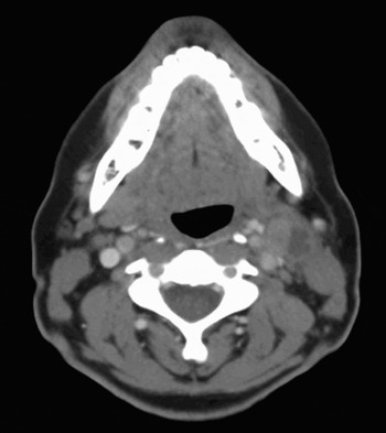
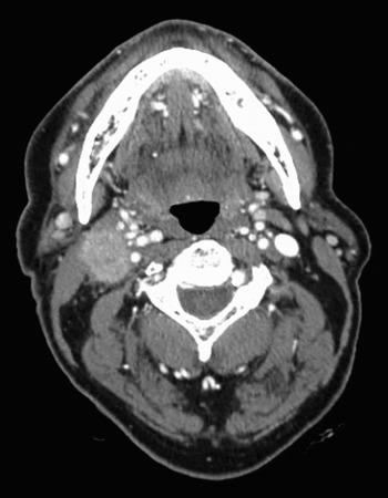
Radiologically, certain imaging features prompt suspicion of tumour spread beyond the lymph node. Indistinct nodal margins, with or without marginal enhancement, and streaky changes in adjacent fat planes are the initial features (Figure 1). Muscular infiltration, vascular invasion and bone destruction are seen in more advanced disease (Figure 2). Diagnosis of extranodal spread based on computed tomography (CT) or magnetic resonance imaging (MRI) is less reliable following surgery, irradiation or recent infection.Reference Som4
Left cervical metastatic SCC with extranodal spread.

Advanced right cervical metastatic SCC with muscular and vascular invasion.

Few published studies have addressed specifically the sensitivity and specificity of using CT imaging to detect extranodal spread of head and neck SCC. In the German literature, Steinkamp et al. compared 165 neck dissections to CT images of the same patients, looking specifically for extranodal spread.Reference Steinkamp, van der Hoeck, Bock and Felix5 They reported that CT had a sensitivity of 81 per cent, a specificity of 73 per cent and an overall accuracy of 76 per cent in detecting extranodal spread. Carvalho et al. reported a lower sensitivity (63 per cent) and specificity (60 per cent) for detection of extranodal spread by CT.Reference Carvalho, Baldwin, Carter and Parsons6
Computed tomography remains the preferred modality for assessment of cervical lymph node involvement by head and neck SCC, because of its superior spatial resolution.Reference Steinkamp, Beck, Werk and Felix7–Reference Stucksen, Kovacs, Adams and Baum10 Steinkamp's group reported a sensitivity of 74 per cent and a specificity of 72 per cent for detection of extranodal spread using MRI, in 110 patients.Reference Steinkamp, Beck, Werk and Felix7 Yousem et al. assessed radiological detection of extranodal spread, and reported a 90 per cent accuracy for CT compared with a 78 per cent accuracy for MRI.Reference Yousem, Som, Hackney, Schwaibold and Hendrix11 Standard positron emission tomography (PET) is less sensitive in the clinically negative neck due to limitations of spatial resolution, resulting in an inability to detect micrometastasis in lymph nodes.Reference Kutler, Wong, Schoder and Kraus12 Combined PET and CT offers the advantages of both imaging modalities, but is presently of limited availability in many countries, including New Zealand and parts of the United Kingdom.
Ultrasound has advantages, such as high spatial resolution, multiplanar scanning, power Doppler and the ability to perform fine needle aspiration for cytology.Reference Richards and Peacock13 There is some evidence that ultrasound scanning is superior to MRI and as accurate as CT;Reference Akoglu, Dutpek, Bekis, Degirmenci, Ada and Guneri9 however, the accuracy of this imaging modality is dependent on the expertise and experience of sonographers and radiologists.
The purpose of this study was to determine the sensitivity and specificity of CT in the detection of extranodal spread of metastatic head and neck SCC. Histology reports were used as the ‘gold standard’ for comparison. The sensitivity and specificity of CT in detecting lymph node involvement was also recorded.
Materials and methods
Patients who had undergone a cervical lymph node dissection for SCC at Christchurch Hospital otorhinolaryngological department between March 1995 and July 2005 were identified.
Patients who had undergone a staging neck CT scan prior to surgery, obtainable for reporting, were included. Patients with a histological diagnosis other than SCC were excluded.
Scans were performed on a variety of scanners. Image thickness varied between 3 and 5 mm. Intravenous contrast was given unless contraindicated by a history of allergy or renal impairment; doses ranged from 50 to 100 ml, delivered via hand injection or pump. Most patients received 50 ml of Ultravist 300 (Schering, Berlin, Germany).
Although a range of radiologists had originally assessed the scans, for the purposes of this study two experienced head and neck radiologists independently and ‘blindly’ assessed the images, without access to the original reports. They commented on the presence or absence of nodal abnormality, and whether or not they thought extranodal spread of tumour was present. Criteria used for diagnosis of abnormal nodes were multifactorial, but included size (short axis) >11 mm in level two and >10 mm elsewhere, the presence of low attenuation (not central fat), and alteration in architecture from normal ovoid morphology to a more spherical shape. Clusters of nodes were also considered significant, particularly if located close to a primary lesion or within the anatomical drainage pathway. Criteria for extranodal spread included: thick-walled, enhancing nodal margins; loss of margin definition; and alterations in adjacent fat.
Histopathological examination was the gold standard against which radiology reporting was compared. All specimens were labelled and orientated prior to formalin fixation. Specimens were reviewed at a multidisciplinary meeting, usually by a single pathologist.
An attempt was made to clarify the level of involved nodes. Node level was routinely reported, both radiologically and histopathologically, using standard anatomical boundaries. Radiological levels were recorded in 89 of the patients. Nodes which the radiologists assessed as showing features of extranodal spread were the same nodes identified histologically, in all but one neck by radiologist A and in all but two necks by radiologist B.
Results and analysis
Standard binary classification was used to estimate the sensitivity, specificity and positive predictive value of CT for the detection of nodal involvement and the presence or absence of extranodal spread. Ninety-five per cent confidence intervals were exact binomial intervals.
A total of 263 neck dissections was performed upon 238 patients at Christchurch hospital otorhinolaryngological department over the 10-year period between March 1995 and July 2005. Twenty-five patients were excluded as they had a non-SCC histological diagnosis. Eighty-nine patients were excluded because their CT images were unavailable.
A total of 149 neck dissections were statistically analysed. Radiologist A assessed 135 necks and radiologist B assessed 127 necks; 113 necks were assessed by both radiologists.
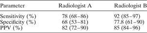
In the hands of radiologists A and B, the sensitivity of CT for the detection of nodes was respectively 78 per cent (69/88) and 92 per cent (84/91), and the specificity was respectively 68 per cent (32/47) and 77.8 per cent (28/36). The positive predictive value was respectively 82 per cent (69/84) and 85 per cent (84/92). Table I shows 95 per cent confidence intervals. Agreement between the two radiologists, for the 113 scans both evaluated, was moderate (κ = 0.55, 95 per cent confidence intervals = 0.38–0.71).
Results for CT detection of nodal involvement

95% confidence intervals are shown in parentheses. CT = computed tomography; PPV = positive predictive value
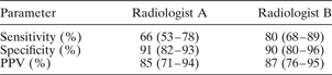
All dissections were included in the analysis of the detection of extranodal spread, not just those for which the radiologist had detected a positive node. In the hands of radiologists A and B, the sensitivity of CT for the detection of extranodal spread was respectively 66 per cent (39/59) and 80 per cent (48/60), and the specificity was respectively 91 per cent (69/76) and 90 per cent (60/67). The positive predictive value for diagnosing extranodal spread was respectively 85 per cent (39/46) and 87 per cent (48/55). Table II shows 95 per cent confidence intervals. Agreement was slightly higher for judgement of extranodal spread than for detection of nodes (κ = 0.67, 95 per cent confidence intervals = 0.53–0.81).
Results for CT detection of extranodal spread

95% confidence intervals are shown in parentheses. CT = computed tomography; PPV = positive predictive value
Discussion
This study investigated CT detection of extranodal involvement of metastatic cervical nodes, as assessed by two experienced head and neck radiologists. A previous, unpublished, local review of written reports by a variety of general radiologists, involving CT scanning of a similar group of patients, showed a much lower sensitivity (as low as 20 per cent in some cases). However, this was not a formal study. Many of the radiologists involved were from peripheral centres and were not specifically looking for extranodal spread when they assessed the CT scans. In the current study, involving experienced head and neck radiologists, the sensitivity of CT detection of extranodal spread was significantly improved. Technological advances in imaging modalities have vastly improved the diagnostic accuracy of CT scanning. In addition, clinicians' heightened expectations of imaging have further increased the demand for radiological subspecialisation, and emphasised the integral role of radiologists within the multidisciplinary team.
In the current study, many patients were excluded because their CT images were unavailable for viewing. This was partly due to the introduction of a computerised image system in the radiology department, towards the end of the study period, with phasing out of hard copy films. Many scans had been performed at peripheral hospitals, as our department was the tertiary referral centre for the majority of head and neck cancer patients in the South Island of New Zealand. Variability in image quality and slice thickness made reporting more difficult in some cases. Excluding patients presenting after July 2005, when a new, multislice, helical scanner was introduced, maintained some consistency with respect to image slices.
The patients included in this study received definitive treatment independent of any additional analysis of their CT scans or histology images; hence, this study had no impact on their treatment. New Zealand ethical guidelines for observational studies permit the secondary use of data for quality assurance, outcome analysis or resource review performed by workers employed or contracted by the service provider holding the information (see www.newhealth.govt.nz/neac).
The main therapeutic implication of our current findings involves the role of adjuvant treatment. Extranodal spread and microscopically involved resection margins are the most significant prognostic predictors of poor outcome, and also two of the strongest indications for adjuvant radiotherapy.Reference Bernier, Cooper, Pajak, van Glabbeke, Bourhis and Forastiere14 Improvement in the radiological accuracy of pre-operative detection of extracapsular spread aids planning of adjuvant radiotherapy (which may include dental extractions where appropriate to facilitate prompt commencement of treatment).
Some studies have reported improved loco-regional control with surgery plus adjuvant radiotherapy, but no significant impact on survival. This is because both surgery and radiotherapy are local treatments.Reference Puri, Fan and Hanna3
The incidence of distant metastasis is significant when extranodal spread is present. Myers et al. Reference Myers, Greenberg, Vivian and Roberts15 found that 25 per cent of patients had distant metastasis when extranodal spread was present, three to four times more than the metastasis rate with nodal involvement alone. This has lead to the issue of adjuvant chemo-radiotherapy in high-risk patients.Reference Puri, Fan and Hanna3, Reference Richards and Peacock13 Two trials on opposite sides of the Atlantic have investigated the concurrent use of cisplatin with radiotherapy, and have identified extranodal spread and positive margins as the most important prognostic indicators.Reference Richards and Peacock13 Loco-regional control rates and disease-free survival rates both improved with the addition of chemotherapy, compared with radiotherapy alone. However, the toxicity associated with this chemotherapy was severe.Reference Richards and Peacock13, Reference Cooper, Pajak, Forastiere, Jacobs, Campbell and Saxman16 Pre-operative, radiological detection of extranodal spread may identify patients suitable for the addition of adjuvant chemotherapy.
Standard PET scanning has no advantage over CT for initial, routine staging of nodal involvement in head and neck SCC. However, PET is very sensitive for detection of distant metastatic and synchronous primary lesions, both of which are more frequently seen in patients with extranodal spread. Computed tomography detection of extranodal spread may therefore be an indication for PET scanning in these patients.
• Lymph node metastasis from head and neck squamous cell carcinoma (SCC) is associated with an approximately 50 per cent reduction in five-year survival
• The presence of extranodal spread further reduces overall survival and increases loco-regional recurrence
• The presence of extranodal spread is a strong indication for adjuvant treatment
• Few published studies have assessed the accuracy of computed tomography (CT) scanning in detecting extranodal spread of head and neck SCC
• This study reports the sensitivity, specificity and positive predictive value of CT in detecting extranodal spread, when assessed by experienced radiologists
• Such pre-operative information aids patient education, planning of adjuvant treatment and allocation of resources in the management of head and neck SCC
In head and neck SCC cases, the presence of lymph node metastases and extranodal spread has a significant impact on rates of loco-regional control, distant metastasis and overall survival. Radiological detection of such extranodal spread has not been well reported in the literature. In the current study, the positive predictive value of CT for the diagnosis of extranodal spread, in the hands of experienced radiologists, was between 85 and 87 per cent. This pre-operative information is useful for surgical planning as well as for predicting the need for adjuvant treatment. Radiotherapy is standard adjuvant treatment in patients with histological evidence of extranodal spread, and the role of adjuvant chemo-radiotherapy is emerging. Technological advances in imaging continue, adding to our diagnostic precision in this challenging group of patients.