Introduction
Premature ovarian insufficiency (POI) is a reproductive disorder in which aberrant loss of primordial follicle reserve culminates in ovarian irregularity and, subsequently, subfertility and infertility earlier than normal menopausal age (Torrealday et al., Reference Torrealday, Kodaman and Pal2017; Chae-Kim and Gavrilova-Jordan, Reference Chae-Kim and Gavrilova-Jordan2019). Premature ovarian insufficiency could have diverse genetic, environmental and idiopathic etiologies, however chemotherapy and radiotherapy are considered as major culprits for POI in patients with cancer who are undergoing oncotherapy (Molina et al., Reference Molina, Barton and Loprinzi2005; Perez-Andujar et al., Reference Perez-Andujar, Newhauser, Taddei, Mahajan and Howell2013). Although various strategies such as oocyte and embryo freezing and ovarian tissue cryopreservation and autotransplantation have been applied in practice for fertility preservation in women with POI, these techniques may not be the appropriate options for patients with cancer due to urgency for commencement of oncotherapy and/or risk of malignant cell reintroduction (Jeruss and Woodruff, Reference Jeruss and Woodruff2009; Bockstaele et al., Reference Bockstaele, Tsepelidis, Dechene, Englert and Demeestere2012; Xiao et al., Reference Xiao, Zhang, Romero, Smith, Shea and Woodruff2015; Amorim and Shikanov, Reference Amorim and Shikanov2016; Fathi et al., Reference Fathi, Valojerdim, Ebrahimi, Eivazkhani, Akbarpour, Tahaei and Abtahi2017; Chae-Kim and Gavrilova-Jordan, Reference Chae-Kim and Gavrilova-Jordan2019). Alternatively, in vitro activation of primordial follicles for in vitro production of embryos could be a potential innocuous technique to preserve fertility in patients with POI (Telfer et al., Reference Telfer, McLaughlin, Ding and Thong2008; Amorim and Shikanov, Reference Amorim and Shikanov2016; McLaughlin et al., Reference McLaughlin, Albertini, Wallace, Anderson and Telfer2018). In this regard, in vitro activation of primordial follicles and their development up to metaphase II (MII) stage oocytes has been accomplished in pubertal women (Telfer et al., Reference Telfer, McLaughlin, Ding and Thong2008; McLaughlin et al., Reference McLaughlin, Albertini, Wallace, Anderson and Telfer2018), however it has not been defined in prepubertal girls (Luyckx et al., Reference Luyckx, Scalercio, Jadoul, Amorim, Soares, Donnez and Dolmans2013; Tefler and Zelinski, 2013), and therefore this aspect warrants further studies.
Dissimilar to some mammalian species, germ cell cyst breakdown leading to formation of primordial follicles begins from the late fetal period and proceeds up to the early neonatal period in mouse ovary (Pepling, Reference Pepling2006; Findlay et al., Reference Findlay, Hutt, Hickey and Anderson2015). Therefore, the 1-day-old mouse ovary only contains primordial follicles (Eppig and O’Brien, Reference Eppig and O’Brien1996; Kerr et al., Reference Kerr, Duckett, Myers, Britt, Mladenovska and Findlay2006) and, in turn, serves as an appropriate model to optimize the activation of primordial follicles under in vitro conditions because the process of primordial follicle activation would occur following the subjection of 1-day-old mouse ovaries to various experimental conditions (Eppig and O’Brien, Reference Eppig and O’Brien1996; O’Brien et al., Reference O’Brien, Pendola and Eppig2003; Atrabi et al., Reference Atrabi, Akbarinejad, Khanbabaee, Dalman, Andrade Amorim, Najar-Asl, Valojerdi and Fathi2019; Alborzi et al., Reference Alborzi, Atrabi, Akbarinejad, Khanbabaei and Fathi2020). In this context, the use of granulosa cell conditioned medium (GCCM) for in vitro culture of 1-day-old mouse ovaries for 5 days has been observed to increase the activation of primordial follicles in cultured ovaries, but the activated follicles did not progress further than the primary stage (Atrabi et al., Reference Atrabi, Akbarinejad, Khanbabaee, Dalman, Andrade Amorim, Najar-Asl, Valojerdi and Fathi2019). Here, it is noteworthy that preantral follicles are required for two-dimensional or three-dimensional culture of isolated follicles to produce MII-stage oocytes (Eppig and O’Brien, Reference Eppig and O’Brien1996; O’Brien et al., Reference O’Brien, Pendola and Eppig2003; Amorim and Shikanov, Reference Amorim and Shikanov2016). We, therefore, reasoned that a culture system for 1-day-old mouse ovaries in GCCM required some modification and hypothesized that the supplementation of GCCM with various factors would contribute to the development of ovarian follicles and that an extension of the culture period beyond 5 days might assist in the enhancement of growth of follicles up to the preantral stage.
In this context, pyruvate is a biochemical component that serves as an essential source of energy metabolism in oocytes during various stages of follicular development from the primordial to the antral stage (Biggers et al., Reference Biggers, Whittingham and Donahue1967; Harris et al., Reference Harris, Adriaens, Leese, Gosden and Picton2007; Johnson et al., Reference Johnson, Freeman, Gardner and Hunt2007; Cinco et al., Reference Cinco, Digman, Gratton and Luderer2016). In addition, ITS, which is a combination of insulin, transferrin and selenium, has been used in culture medium for the growth of various cells and tissues including isolated ovarian follicles and ovarian tissue (Eppig and O’Brien, Reference Eppig and O’Brien1996; O’Brien et al., Reference O’Brien, Pendola and Eppig2003; Fortune et al., Reference Fortune, Yang and Muruvi2011) and could enhance the activation of follicles in ovarian tissues cultured under in vitro conditions (Fortune et al., Reference Fortune, Yang and Muruvi2011). Furthermore, testosterone is a steroid hormone that has been reported to induce the activation of primordial follicles through its interaction with androgen receptors in oocytes (Magamage et al., Reference Magamage, Zengyo, Moniruzzaman and Miyano2011), alleviate follicular atresia (Qureshi et al., Reference Qureshi, Nussey, Bano, Musonda, Whitehead and Mason2008), and enhance the transition of ovarian follicles from the primary to the preantral stage in cultured ovarian grafts (Yang and Fortune, Reference Yang and Fortune2006).
Accordingly, the present study was conducted to investigate whether the incorporation of pyruvate, ITS or testosterone into GCCM could improve the development of follicles in cultured mouse ovaries. Additionally, the period of culture was extended to 11 days to evaluate the effect of time over the course of in vitro culture.
Materials and methods
Animals
NMRI mice (Royan Institute, Tehran, Iran) were used in the present study, and were maintained under stable conditions (temperature: 20–25ºC; humidity: 40–60%) under a 12-h light/dark cycle.
Preparation of GCCM
Ovaries were harvested from 30-day-old mice, which were sacrificed by decapitation. Immediately after removal, ovaries were placed in α-minimum essential medium (α-MEM; Gibco, Paisley, UK) containing 10% fetal bovine serum (FBS; Gibco, Paisley, UK), 50 μg/ml penicillin (Sigma-Aldrich, St. Louis, USA) and 50 μg/ml streptomycin (Sigma-Aldrich, St. Louis, USA) under mineral oil (Sigma-Aldrich, St. Louis, USA). Preantral follicles (100–120 µm in diameter) were isolated mechanically under a stereomicroscope from the surrounding tissues (Nikon, Tokyo, Japan). The isolated follicles were cultured in 500 μl of the same medium for 10–14 days in four-well plates. After 72 h of culturing follicles, granulosa cells were attached to the bottom of plate, whereas oocytes and other follicular remnants remained floating in the supernatant, and were further eliminated by medium replacement. These adherent granulosa cells were cultured for up to four passages and GCCM was obtained from passage numbers three and four and were used fresh for in vitro culture of 1-day-old mouse ovaries. GCCM was collected from plates with 70–80% of granulosa cell confluency and 48 h after medium replacement.
In vitro culture of 1-day-old mouse ovaries
Here, 1-day-old mouse ovaries were cultured in 96-well plates containing 200 µl of various experimental media including CON (as control group which contained merely GCCM), PYR (which contained GCCM plus 0.23 mM sodium pyruvate; Sigma-Aldrich, St. Louis, USA), ITS (which contained GCCM plus 1% ITS; Gibco, Paisley, UK), and TES (which contained GCCM plus 10–7 M testosterone; Iran Hormone Co., Tehran, Iran) for 11 days. Culture medium was changed every 2 days and approximately three-quarters of the medium was replaced by fresh medium each time. Ovaries were retrieved for histological and molecular assessments on days 5 and 11 of culture.
Histological examination
Histological examination was applied to evaluate the morphological dynamics of ovarian follicles over the course of the culture period. Ten ovaries from each experimental group at each timepoint were fixed in Bouin’s solution and formalin, dehydrated and embedded in paraffin. Then, paraffin-embedded ovaries were serially sectioned at 6-µm thickness and stained with haematoxylin and eosin (H&E) stain. Assessment of histological sections was implemented using a light microscope (Nikon, Tokyo, Japan). In this context, primordial, transitional, primary and preantral follicles were considered as follicles, in which oocytes were surrounded by a single layer of squamous granulosa cells, a single layer of both squamous and cuboidal granulosa cells, a single layer of cuboidal granulosa cells, and two or more layers of cuboidal granulosa cells, respectively. In addition, follicular activation was defined as the cumulative proportion of all types of activated follicles including transitional, primary and preantral follicles.
Real-time PCR (RT-PCR)
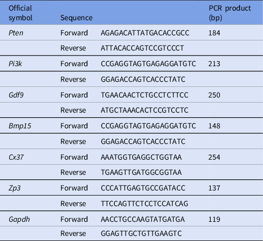
RT-PCR was applied to analyze the gene expression of factors contributing to the development of ovarian follicles, including Pten, Pi3k, Gdf9, Bmp15, Cx37 and Zp3 (Table 1). Four ovaries from each experimental group at each timepoint were used for RT-PCR. Following retrieval, ovaries were stored in RNAlater (Qiagen, Hilden, Germany) at −80°C. Total RNA extraction was performed using the RNeasy Plus Micro Kit (Qiagen, Hilden, Germany) and cDNA was synthesized according to a standard protocol suggested by the manufacturer for oligo(dT) in a reaction volume of 20 µl. In each reaction, 2 µl of synthesized cDNA was subjected to RT-PCR using the Power SYBR Green PCR Master Mix on an ABI Step One Plus thermocycler and according to the manufacturer’s instructions (Applied Biosystems, Warrington, UK). Reactions were run for 40 cycles at 94°C for 10 min, 94°C for 30 s, and 60°C for 1 min and each sample was run in duplicate. Glyceraldehyde 3-phosphate dehydrogenase (Gapdh) was used as a housekeeping gene (Table 1).
Mouse primer sequences for real-time PCR

Statistical analysis
Data were analyzed using the GLM procedure. LSMEANS statement was used for multiple comparisons. All analyses were conducted in SAS version 9.4 software (SAS Institute Inc., Cary, NC, USA). Data are presented as mean ± the standard error of the mean (SEM). Differences were considered statistically significant at P-values < 0.05.
Results
Follicular development in cultured ovaries
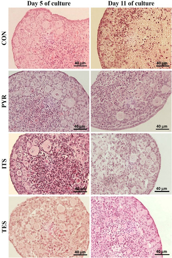
Histological examination revealed the development of various stages of ovarian follicles, including primordial, transitional, primary and preantral follicles, in 1-day-old mouse ovaries cultured for 5 and 11 days (Fig. 1).
Histological sections of ovaries cultured in the CON, PYR, ITS and TES groups on days 5 and 11.

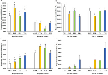
The proportion of primordial follicles decreased over time in all experimental groups (P < 0.01; Fig. 2). On day 5, the proportion of primordial follicles was higher in the PYR group compared with the CON and ITS groups (P < 0.01), but on day 11 it did not differ among groups (P > 0.05; Fig. 2A). On day 5, the proportion of transitional follicles was higher in the CON group than the PYR and TES groups (P < 0.05), and on day 11, it was higher in the CON, ITS and TES groups than in the PYR group (P < 0.05; Fig. 2B). The proportion of primary follicles increased from day 5 to day 11 in the CON, PYR and ITS groups (P < 0.05), but not in the TES group (P > 0.05; Fig. 2C). On day 5, the proportion of primary follicles was higher in the TES than in the CON group (P = 0.041); however on day 11 it was higher in the PYR group than in the CON and TES groups (P < 0.05; Fig. 2C). The proportion of preantral follicles was not influenced by the interaction effect of group by time (P > 0.05; Fig. 2D). Considering the main effect of groups, the proportion of preantral follicles was higher in the TES group compared with the CON, PYR and ITS groups (P < 0.05; Fig. 2D). Moreover, considering the main effect of time, the percentage of preantral follicles increased from day 5 to day 11 (P = 0.005; Fig. 2D).
Proportion of primordial, transitional, primary and preantral follicles in the CON, PYR, ITS and TES groups on days 5 and 11. Different letters (a and b) indicate statistically significant differences among experimental groups at the specified time point (P < 0.05). Asterisk (*) denotes statistically significant difference between days 5 and 11 in the specified experimental group (P < 0.05).

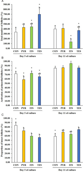
In the ITS and TES groups, the total number of follicles dwindled from day 5 to day 11 (P < 0.05), but this did not occur in the CON and PYR groups (P > 0.05; Fig. 3A). On day 5, the total number of follicles was greater in the TES than in the CON group (P = 0.002); however on day 11 it was higher in the CON and PYR groups than in the ITS group (P < 0.05; Fig. 3A).
Total number of follicles, activation of primordial follicles and proportion of morphologically intact follicles in the CON, PYR, ITS and TES groups on days 5 and 11. Different letters (a and b) indicate statistically significant differences among experimental groups at the specified time point (P < 0.05). Asterisk (*) denotes statistically significant difference between days 5 and 11 in the specified experimental group (P < 0.05).

The activation of primordial follicles increased from day 5 to day 11 in all groups (P < 0.01; Fig. 3B). The activation of primordial follicles was higher in the CON and ITS groups than in the PYR group on day 5 (P < 0.01), but this difference was not observed on day 11 (P > 0.05; Fig. 3B).
The proportion of intact follicles decreased over time in the CON group (P < 0.0001), but not in the PYR, ITS and TES groups (P > 0.05; Fig. 3C). On day 5, the proportion of intact follicles was greater in the CON group compared with the PYR, ITS and TES groups (P < 0.05); however on day 11 it was higher in the TES than in the CON group (P = 0.039; Fig. 3C).
Gene expression of factors contributing to follicular development
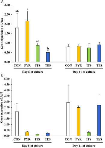
On day 5, gene expression of Pten was higher in the PYR than in the TES group (P = 0.034), but on day 11 it did not differ among experimental groups (P > 0.05; Fig. 4A). Expression of Pi3k was not influenced by the interaction of group by time (P > 0.05; Fig. 4B). However, considering the main effect of time, expression increased from day 5 to day 11 (P = 0.006), and considering the main effect of group, expression was higher in the CON than in the ITS group (P = 0.007; Fig. 4B).
Relative gene expression of Pten and Pi3k as factors regulating the activation of primordial follicles in the CON, PYR, ITS and TES groups on days 5 and 11. Different letters (a and b) indicate statistically significant differences among experimental groups at the specified time point (P < 0.05).

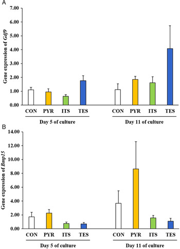
The interaction effect of group by time did not affect the expression of Gdf9 and Bmp15 (P > 0.5; Fig. 5). However, given the main effect of time, Gdf9 and Bmp15 expression increased from day 5 to day 11 (P < 0.05; Fig. 5). Moreover, considering the main effect of group, Gdf9 expression was higher in the TES group compared with the CON and ITS groups (P < 0.05; Fig. 5A), whereas Bmp15 expression was higher in the PYR group compared with the ITS and TES groups (P < 0.05; Fig. 5B).
Gene expression of Gdf9 and Bmp15, as oocyte-derived factors contributing to the development of follicles in the CON, PYR, ITS and TES groups on days 5 and 11.

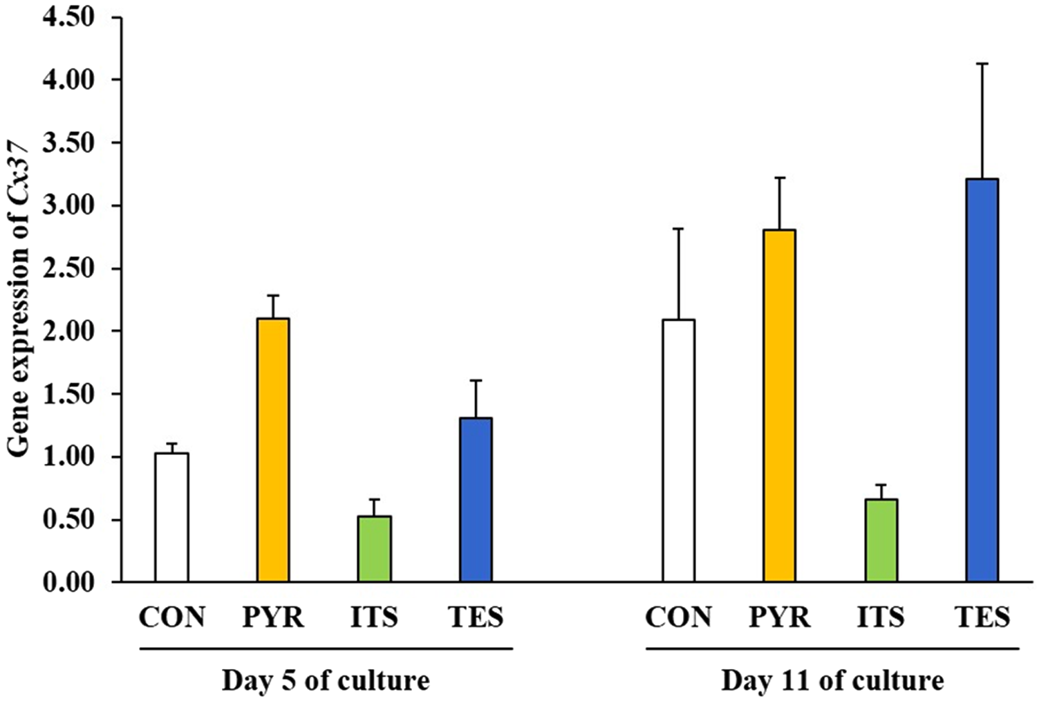
Expression of Cx37 was not influenced by the interaction effect of group by time (P > 0.05; Fig. 6). But considering the main effect of time, it increased from day 5 to day 11 (P = 0.005; Fig. 6). In addition, given the main effect of group, Cx37 expression was higher in the PYR and TES groups than in the ITS group (P < 0.01; Fig. 6).
Gene expression of Cx37 in the CON, PYR, ITS and TES groups on days 5 and 11.

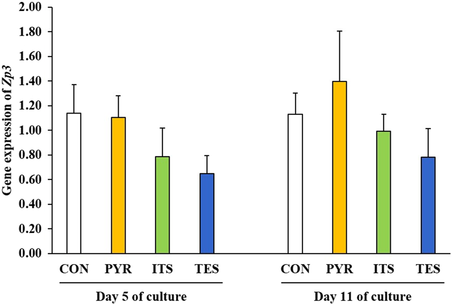
No significant effect of group, time and interaction of group by time was observed on gene expression of Zp3 (P > 0.05; Fig. 7).
Gene expression of Zp3 in the CON, PYR, ITS and TES groups on days 5 and 11.

Discussion
The present study was carried out to understand whether the addition of pyruvate, ITS or testosterone to GCCM could improve the development of follicles in 1-day-old mouse ovaries cultured under in vitro conditions. In this context, it was revealed that testosterone increased the number of follicles at the early stages of in vitro culture on day 5. Similarly, in utero exposure to androgens during the early stages of gestation led to an increment in follicular formation in ovine fetuses (Comim et al., Reference Comim, Hardy, Robinson and Franks2015). Furthermore, testosterone did not influence the activation rate of follicles in the present study, even though it suppressed the expression of Pten as the main negative regulator of follicular activation (Zhang and Liu, Reference Zhang and Liu2015; Kim and Kurita, Reference Kim and Kurita2018). Nevertheless, testosterone increased the proportion of primary follicles on day 5 and, more importantly, enhanced the development of follicles up to the preantral stage during culture. Similarly, Yang and Fortune (Reference Yang and Fortune2006) evaluated the effects of testosterone on the development of follicles in cultured bovine ovarian grafts and found that testosterone could stimulate the growth of primary follicles towards the preantral stage. Upregulation of Gdf9 could have contributed to greater follicular formation and development by testosterone as GDF9 has been indicated to play a role not only in follicle formation (Yan et al., Reference Yan, Wang, DeMayo, DeMayo, Elvin, Carino, Prasad, Skinner, Dunbar, Dube, Celeste and Matzuk2001; Bayne et al., Reference Bayne, Kinnell, Coutts, He, Childs and Anderson2015; Zhao et al., Reference Zhao, Du, Huang, Zhang, Teng, Niu, Wang and Xia2016) but also in the growth of activated follicles (Gilchrist et al., Reference Gilchrist, Lane and Thompson2008; Cook-Andersen et al., Reference Cook-Andersen, Curnow, Su, Chang and Shimasaki2016; Otsuka et al., Reference Otsuka, McTavish and Shimasaki2011). Moreover, gene expression of Cx37, the gap junction regulating reciprocal cross-talk between oocyte and surrounding granulosa or cumulus cells for normal follicular development (Teilmann, Reference Teilmann2005; Simon et al., Reference Simon, Chen and Jackson2006), was elevated in ovaries treated with testosterone. Zhang et al. (Reference Zhang, Xu, Kuai, Wang, Xue and Shang2016) has also observed a dose-dependent increase in Cx37 expression in response to testosterone exposure. Alternatively, greater expression of Cx37 in testosterone-treated ovaries might have resulted from a higher proportion of preantral follicles in the TES group, as Cx37 expression initiates from the primordial stage and continuously increases as the follicle approaches the preantral stage (Teilmann, Reference Teilmann2005; Simon et al., Reference Simon, Chen and Jackson2006).
Although the rate of follicular activation was reduced in the PYR group, pyruvate enhanced the progression of follicular development up to the primary stage by day 11 of culture. Moreover, pyruvate augmented the gene expression of Bmp15, which is derived from oocytes from follicles beyond the primordial stage (Dube et al., Reference Dube, Wang, Elvin, Lyons, Celeste and Matzuk1998). Interestingly, BMP15 is a growth factor enabling cumulus cells to shift to glycolysis, a metabolic pathway, in which ATP is generated by conversion of glucose to pyruvate and, therefore, increases the pyruvate content available for metabolism and the development of oocyte (Sugiura et al., Reference Sugiura, Pendola and Eppig2005, Reference Sugiura, Su, Diaz, Pangas, Sharma, Wigglesworth, O’Brien, Matzuk, Shimasaki and Eppig2007). Taken together, these phenomena implied that there might be a positive feedback association between BMP15 and pyruvate, which eventually could lead to increase in secretion of BMP15 and the production of pyruvate, the two elements required for normal development of ovarian follicles, particularly during the final stages (Sugiura et al., Reference Sugiura, Pendola and Eppig2005, Reference Sugiura, Su, Diaz, Pangas, Sharma, Wigglesworth, O’Brien, Matzuk, Shimasaki and Eppig2007; Gilchrist et al., Reference Gilchrist, Lane and Thompson2008; Otsuka et al., Reference Otsuka, McTavish and Shimasaki2011; Sanfins et al., Reference Sanfins, Rodrigues and Albertini2018). Additionally, similar to testosterone-treated ovaries, Cx37 expression was greater in pyruvate-treated ovaries, and could have stemmed from an upregulatory effect of pyruvate on Cx37 and/or a higher proportion of primary follicles in the PYR group (Teilmann, Reference Teilmann2005; Simon et al., Reference Simon, Chen and Jackson2006).
Unexpectedly, ITS did not affect the rate of follicular activation and even downregulated Pi3k expression, as the main positive regulator of primordial follicle activation (Zhang and Liu, Reference Zhang and Liu2015; Kim and Kurita, Reference Kim and Kurita2018), compared with the CON group, which only contained GCCM. Conversely, the insulin component of ITS has been observed to augment the activation of primordial follicles in fetal ovarian tissue in bovine (Fortune et al., Reference Fortune, Yang and Muruvi2011). In addition, the addition of ITS to GCCM has led to an inferior total number of follicles on day 11. Moreover, ovaries treated with ITS had lesser gene expression of Pi3k, Gdf9, Bmp15 and Cx37. Collectively, it appeared that when ITS was used as a supplement to GCCM, it could not positively contribute to ovarian follicle growth and instead adversely influenced the development of follicles in cultured 1-day-old mouse ovaries, which disagreed with previous studies that indicated the beneficial effects of incorporation of ITS into base medium for the culture of ovarian follicles (Eppig and O’Brien, Reference Eppig and O’Brien1996; O’Brien et al., Reference O’Brien, Pendola and Eppig2003; Fortune et al., Reference Fortune, Yang and Muruvi2011). It could be speculated that there might be component(s) in GCCM that counteracted the beneficial effects of ITS on in vitro follicular growth.
Regardless of treatments, not only did time positively affect the activation of primordial follicles, but it also enhanced the development of follicles up to the preantral stage. In this regard, no preantral follicles were observed after 5 days of culture in the CON group, similar to findings in a previous study (Atrabi et al., Reference Atrabi, Akbarinejad, Khanbabaee, Dalman, Andrade Amorim, Najar-Asl, Valojerdi and Fathi2019), however 0.08% of follicles progressed to the preantral stage by day 11 of culture in the CON group in the present study. This ascending pattern in percentage of preantral follicles over time was also found for the PYR, ITS and TES groups, despite the fact that preantral follicles were already detected on day 5 in the respective groups. Furthermore, this time-sensitive progressive changes in morphology of follicles coincided with the upregulation of Gdf9, Bmp15 and Cx37, factors contributing to the normal development of ovarian follicles (Teilmann, Reference Teilmann2005; Simon et al., Reference Simon, Chen and Jackson2006; Gilchrist et al., Reference Gilchrist, Lane and Thompson2008; Cook-Andersen et al., Reference Cook-Andersen, Curnow, Su, Chang and Shimasaki2016; Otsuka et al., Reference Otsuka, McTavish and Shimasaki2011; Sanfins et al., Reference Sanfins, Rodrigues and Albertini2018). Therefore, the extension of the culture period to 11 days assisted in the improved development of preantral follicles, which were required for the production of MII-stage oocytes under in vitro conditions for oncofertility purposes (Eppig and O’Brien, Reference Eppig and O’Brien1996; O’Brien et al., Reference O’Brien, Pendola and Eppig2003; Jeruss and Woodruff, Reference Jeruss and Woodruff2009; Amorim and Shikanov, Reference Amorim and Shikanov2016).
In conclusion, the present study has revealed the beneficial effects of pyruvate and testosterone on the development of ovarian follicles to the primary and preantral stages, respectively, in 1-day-old murine ovaries. Nonetheless, ITS failed to improve primordial follicles activation and support follicular growth. Furthermore, the extension of the culture period to 11 days provided follicles the opportunity for progressive development up to the preantral stage.
Author contributions
M.J. Atrabi: Conceptualization, methodology, investigation, data curation and writing – original draft. P. Alborzi: Investigation. V. Akbarinejad: Conceptualization, methodology, formal analysis, writing – original draft and writing – review and editing. R. Fathi: Supervision, conceptualization, methodology, writing – review and editing.
Acknowledgements
The authors would like to thank the staff at the Department of Embryology, Reproductive Biomedicine Research Center, Royan Institute for Reproductive Biomedicine, ACECR, Tehran, Iran for their kind assistance.
Financial support
This project was financially supported by the Royan Institute.
Ethical standards
All experiments and procedures were approved by the Royan Institute Ethics Committee (IR.ACECR.ROYAN.REC.1397.130).
Conflict of interest
None.