Introduction
The discovery of the genetic code about 50 years ago suggested that gene isolation and transfer into living organisms would become major tools for biologists. The first gene transfer into the mouse revealed the possibility of creating animals that could stably harbour foreign DNA and that had modified phenotypic properties (Gordon et al., Reference Gordon, Scangos, Plotkin, Barbosa and Ruddle1980; Palmiter et al., Reference Palmiter, Brinster, Hammer, Trumbauer, Rosenfeld, Birnberg and Evans1982).
Pronuclear DNA injection was also used to produce the first transgenic farm animal (Hammer et al., Reference Hammer, Pursel, Rexroad, Wall, Bolt, Ebert, Palmiter and Brinster1985). Despite the inherent inefficiency of microinjection technology, several genetically modified large animals have been generated for applications in livestock and biomedicine (Houdebine, Reference Houdebine2007).
Several alternatives to pronuclear DNA injection have been developed to improve the efficiency and to reduce the cost of generating transgenic livestock (reviewed in Kues & Niemann, Reference Kues and Niemann2011). To date, somatic cell nuclear transfer (SCNT) holds the greatest promise for significant improvements in the generation of transgenic cattle. However, this method also has proved to be too costly and inefficient for everyday use (Rideout et al., Reference Rideout, Eggan and Jaenisch2001).
For further improvement of transgenic technology in large animals, it is necessary to develop simple, inexpensive and efficient methods. Thus, the use of cell-penetrating peptides (CPPs) constitutes a promising alternative. CPPs are molecules that display the ability to internalize into eukaryotic cells through an energy-independent mechanism and to efficiently carry biologically active and therapeutically relevant molecules inside the cell (Schwarze & Dowdy, Reference Schwarze and Dowdy2000). CPPs are represented by multiple sequences of short and positively charged peptides, rich in arginine and lysine residues, that penetrate through usually impermeable cellular membranes and accumulate in the cytoplasm and/or in the nucleus of the cell (Gupta et al., Reference Gupta, Levchenko and Torchilin2005).
Kerkis et al. (Reference Kerkis, Kerkis, Rádis-Baptista, Oliveira, Vianna-Morgante, Pereira and Yamane2004) described that crotamine, a myotoxin isolated from the venom of South American rattlesnake (Crotalus durissus terrificus), is a CPP presenting both cytoplasmic and nuclear localization. It was also demonstrated that this toxin is capable of binding electrostatically to plasmid DNA to form a peptide–DNA complex and cargo delivery into cells (Nascimento et al., Reference Nascimento, Hayashi, Kerkis, Oliveira, Oliveira, Rádis-Baptista, Nader, Yamane, Tersariol and Kerkis2007). Moreover, crotamine in the micromolar range penetrates cells during the G1/S period, binding to centrosomes and chromosomes. Interestingly, in vitro, the exposure of murine stem cells and embryonic fibroblasts to crotamine concentrations between 10 and 0.01 μM exhibited no toxicity even after 72 h (Kerkis et al., Reference Kerkis, Kerkis, Rádis-Baptista, Oliveira, Vianna-Morgante, Pereira and Yamane2004). Furthermore, CHO-K1 and mice melanoma B16F10 cells exposed to crotamine showed no cytotoxic effect for concentrations up to 1 μM for 24 h (Hayashi et al., Reference Hayashi, Nascimento, Kerkis, Oliveira, Oliveira, Pereira, Rádis-Baptista, Nader, Yamane, Kerkis and Tersariol2008; Nascimento et al., Reference Nascimento, Sancey, Pereira, Rome, Oliveira, Oliveira, Nader, Yamane, Kerkis, Tersariol, Coll and Hayashi2012). Even though a few studies have assessed the effects of crotamine on some cell types, only one has described that1 μM crotamine is able to penetrate intact mouse embryos with no toxicity (Kerkis et al., Reference Kerkis, Kerkis, Rádis-Baptista, Oliveira, Vianna-Morgante, Pereira and Yamane2004). In the consulted literature, no studies have been conducted to evaluate the effects of crotamine on cattle preimplantation embryo. Therefore, the aim of this study was to investigate the effects of crotamine, using different exposure times and concentrations, on the quality of in vitro fertilized (IVF) bovine embryos to assess developmental competence and gene expression.
Materials and methods
Unless indicated otherwise, all chemicals were obtained from Sigma Chemical (St. Louis, MO, USA). This study was approved by the Ethics Committee for Animal Use of the State University of Ceará (No. 12641799-7) and is in accordance with ethical principles of animal experimentation adopted by this committee.
Obtention of crotamine and labelling with rhodamine B
Crotamine was purified previously from the crude venom of rattlesnakes kept in the serpentarium of São Paulo University (Ribeirão Preto, Brazil). The method for crotamine isolation was described previously by Kerkis et al. (Reference Kerkis, Kerkis, Rádis-Baptista, Oliveira, Vianna-Morgante, Pereira and Yamane2004). Briefly, the venom was diluted with ammonium formate buffer and the bulk of crotoxin, the major venom component, was eliminated by slow-speed centrifugation. Tris-base was added and the solution was applied to a CM-Sepharose FF column (1.5 × 4.5 cm; Amersham-Biosciences, Buckinghamshire, UK). Afterwards, the column was washed and crotamine was recovered, dialyzed, lyophilized, and stored at room temperature until use. Before using at embryo culture medium, lyophilized crotamine was dissolved in 150 mM NaCl solution (vehicle solution).
Preparation and labelling of crotamine with the fluorescent dye rhodamine-B (RhB) at the N-terminus was achieved as described previously (Rádis-Baptista et al., Reference Rádis-Baptista, de la Torre and Andreu2008; Rodrigues et al., Reference Rodrigues, Santos, Torre, Rádis-Baptista, Andreu and Santos2012). Dried RhB-labelled crotamine was maintained at –20°C until use.
Experimental design
Two experiments were conducted to evaluate the development and gene expression in bovine embryos exposed to crotamine. In Experiment 1, different exposure times (6, 12 and 24 h) of 0.1 μM crotamine were tested on in vitro culture of IVF presumptive zygotes, with vehicle (0 μM crotamine for each exposure time) and IVF as control groups. The concentration was chosen in accordance with previous studies with human and mouse somatic cells (Kerkis et al., Reference Kerkis, Kerkis, Rádis-Baptista, Oliveira, Vianna-Morgante, Pereira and Yamane2004). In Experiment 2, the exposure of IVF presumptive zygotes to different concentrations of crotamine (0.1, 1 and 10 μM) for 24 h was evaluated, using vehicle (0 μM crotamine = 150 mM NaCl) as a control group.
For both experiments, presumptive zygotes were exposed to crotamine or vehicle solutions in 100 μl final volume drops composed of 10 μl of solution and 90 μl of modified CR2aa medium supplemented with 1 mg/ml fatty acid-free bovine serum albumin (BSA) and 2.5% fetal calf serum (FCS) under mineral oil at 38.5°C and atmosphere of 5% CO2 in air. In Experiment 1, IVF control zygotes were not subjected to such conditions.
Oocyte collection and in vitro maturation (IVM)
The ovaries of adult cows were collected from a local slaughterhouse and were transported to the laboratory in 0.9% (w/v) NaCl solution containing antibiotics (Pentabiótico; Fort Dodge, Campinas, Brazil). Cumulus–oocyte complexes (COCs) from follicles of 2–8 mm in diameter were aspirated using a 21-gauge needle attached to a disposable syringe. Only COCs of equal size with evenly granulated ooplasm surrounded by multiple layers of compact cumulus cells were selected in all experiments. After collection, the COCs were washed four times with HEPES–Tyrode's lactate–pyruvate–albumin (TALP-H). Groups of up to 50 COCs were placed in 500 μl of maturation medium containing bicarbonate-buffered medium 199 supplemented with 10% (v/v) FCS (Gibco, Carlsbad, CA, USA), 0.2 mM sodium pyruvate, 50 μg/ml gentamycin, 10 ng/ml epidermal growth factor (EGF), 100 μM cysteamine, 20 μg/ml follicle-stimulating hormone/leuteinizing hormone (FSH/LH; Pluset; Calier, Barcelona, Spain) and 1 μg/ml 17β-estradiol. IVM was performed at 38.5°C for 24 h in a humidified atmosphere of 5% CO2 in air.
In vitro fertilization (IVF)
Matured COCs were placed in Petri dishes that contained 100 μl of Fert-TALP medium supplemented with 20 μg/ml heparin (Hemofol; Cristália, Itapira, Brazil) and 6 mg/ml fatty acid-free BSA. IVF was performed with frozen–thawed Percoll-separated semen (final concentration of 15 × 106 spermatozoa/ml) covered with mineral oil (Irvine Scientific, Santa Ana, CA, USA) at 38.5°C under an atmosphere of 5% CO2 in air with maximum humidity. Approximately 6 h post insemination (hpi), presumptive zygotes were denuded by repeated pipetting in TALP-H for immediate exposure with crotamine for different times and concentrations.
In vitro embryo development
After exposure to crotamine, embryos were cultured in 50 μl droplets of modified CR2aa medium supplemented with 1 mg/ml BSA and 10% FCS under mineral oil at 5% CO2 in air and 38.5°C in high humidity for 192 hpi. Cleavage was assessed at 72 hpi and blastocyst at 168 (day 7) and 192 (day 8) hpi. At day 8 of culture, embryos were stained in Hoechst 33342 (Pursel et al., Reference Pursel, Wall, Rexroad, Hammer and Brinster1985) and mounted between coverslips to count the total nuclei number under an epifluorescence microscope (Eclipse E400; Nikon, Tokyo, Japan).
Translocation assay
For assessment of crotamine translocation into presumptive zygotes, embryos were exposed to 10 μM RhB-labelled crotamine or vehicle solution (NaCl 150 mM) for 6 h post IVF in 50-μl final volume droplets composed of 5 μl of solution and 45 μl of synthetic oviductal fluid (SOF) (Tervit et al., Reference Tervit, Whittingham and Rowson1972) modified by Holm et al. (Reference Holm, Booth, Schmidt, Greve and Callesen1999) under mineral oil at 38.5°C and atmosphere of 5% CO2 in air. After exposure, zygotes were washed in TALP-H and the zona pellucida (ZP) was removed by treatment with 1.5 mg/ml pronase (P8811). Afterward, embryos were fixed in 4% (v/v) paraformaldehyde (F1635) for 20 min and washed for 30 min in PBS supplemented with 0.4% (w/v) BSA and 1% (v/v) antibiotic–antimycotic (ATB-ATM, 15240–096; Gibco BRL). Samples were stored in supplemented PBS at 4°C until use. Nuclear DNA was counterstained with 0.2 mg/ml of DAPI (D9542) for 10 min in the dark, and washed in supplemented PBS for 5 min. Stained embryos were mounted between coverslips in 70% (v/v) glycerol and stored at –20°C. Crotamine uptake by embryos was detected by confocal microscopy on a Zeiss LSM 510 Meta Confocal microscope (Zeiss, Germany). DAPI and RhB fluorescence was detected with excitation wavelengths of 405 and 543 nm, respectively. Complete Z series of 12 optical sections at 4–5 μm intervals were acquired from each embryo and three-dimensional images were constructed using the LSM Image Browser software (Zeiss, Germany).
RNA extraction and reverse transcription (RT)
Total RNA was prepared from three pooled hatched blastocysts (day 8 of culture) in quadruplicates as group samples using the RNeasy micro kit (Qiagen Sci.; Germantown, MD, USA) following the manufacturer's instructions. Briefly, 75 μl lysis buffer was added to each frozen sample and the lysate was diluted 1:1 with 70% ethanol and transferred to a spin column. Genomic DNA was degraded using RNase-free DNase for 15 min at room temperature. After three washes, the RNA was eluted with 10 μl RNase-free water. The RT step was performed with 1 μl of Improm II (Promega; Madison, WI, USA) in buffer, combined with 0.5 mM of each dNTP (Promega), 40 U of RNasin (Promega), and RNase-free water to make a final reaction volume of 20 μl. RT was achieved at 42°C for 60 min, followed by 70°C for 15 min. First-strand cDNA products were then stored at –80°C for later use as template for further gene expression analysis. Negative controls or RT blanks were prepared under the same conditions, but with no inclusion of reverse transcriptase.
Quantitative real-time PCR
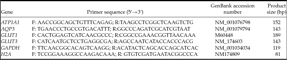
Quantitative real-time polymerase chain reaction (qPCR) amplifications were performed in a Master-Cycler EP Realplex4 S (Eppendorf AG; Hamburg, Germany). The quadruplicates of cDNA from each group were pooled prior to PCR experiments. Thereafter, the pooled cDNA were run in triplicate for target and reference genes (Table 1). Target genes were Na/K ATPase isoform 1 (ATP1A1), aquaporin 3 (AQP3), glucose transporter-1 (GLUT1) and glucose transporter-3 (GLUT3). These genes were chosen to evaluate osmotic regulation, blastocoele formation and energy metabolism of the embryos. Reference genes were glyceraldehyde 3-phosphate dehydrogenase (GAPDH) and member Z of the H2A histone family (H2A). Each reaction consisted of 20 μl total volume containing 10 μl 2× Power SYBR Green PCR Master Mix (Applied Biosystems; Foster City, CA, USA), 0.3 μM of each primer and 0.5 μl cDNA (equivalent to 0.075 embryo). The qPCR protocol consisted of an initial incubation at 95°C for 10 min, followed by 40 cycles of an amplification program of 95°C for 15 s, 55°C for 15 s and 60°C for 30 s. Fluorescence data were acquired during the 72°C extension step. Threshold, quantification cycle (Cq), and melting temperature (Tm) values were automatically determined by Realplex 2.2 software (Eppendorf AG), using default parameters. To determine the linearity (R2) and the efficiency (E) of the PCR amplifications, standard curves were generated for each gene using serial dilutions of a cDNA preparation from 20 (for GAPDH, H2A and ATP1A1) or 40 (for AQP3, GLUT1 and GLUT3) hatched blastocysts, with all other conditions being identical. Specificity of each reaction was achieved by performing the melting procedure (55–95°C, starting fluorescence acquisition at 55°C and taking measurements at 10 s intervals until the temperature reached 95°C) after every qPCR run. Samples with RNA but with no reverse transcriptase were used as negative controls.
Oligonucleotides used for qPCR analysis of gene expression in in vitro fertilized bovine embryos

F, forward primer; R, reverse primer.
Data analysis
The statistical analysis of cleavage and blastocyst rates was assessed by the non-parametric Fisher's exact test, whereas the average number of nuclei per blastocyst was compared using one-way analysis of variance (ANOVA). The relative quantification of gene expression was performed using the 2−ΔΔCt method (Livak & Schmittgen, Reference Livak and Schmittgen2001). Target gene expression was normalized against the geometric mean of GAPDH and H2A transcript levels (Vandesompele et al., Reference Vandesompele, Preter, Pattynm, Poppe, Roy, Paepe and Speleman2002). The differences in relative abundance of specific transcripts were compared using the Kruskal–Wallis test followed by the post-hoc Dunn's test. The corresponding qPCR efficiencies were calculated from the given slopes (S) of the standard curves, according to the equation: E = 10(−1/S) – 1. Linearity was expressed as the square of the Pearson correlation coefficient (R2). Tm data were expressed as mean ± standard deviation (SD) of three or more measurements. For all analysis, P-value < 0.05 was considered to be statistically significant using Prism 6.0 software (GraphPad Software Inc.; La Jolla, CA, USA).
Results
Embryo development
The present study assessed the possible effects of crotamine on the quality of IVF bovine embryos. Thus, we evaluated developmental competence up to the blastocyst stage. In addition, gene expression of selective quality genes from embryos exposed to crotamine in comparison with controls was quantitatively analyzed by qPCR.
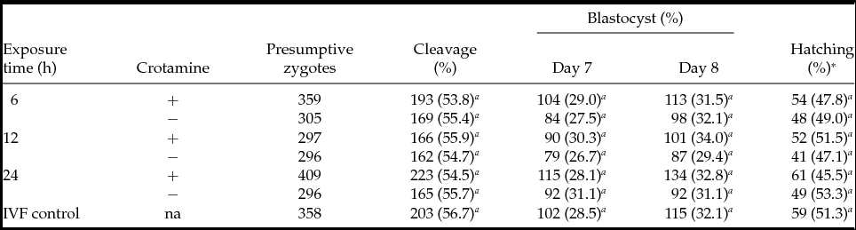
In Experiment 1, 2320 presumptive zygotes from 10 replicates produced 1281 (55.2%) cleaved embryos and 740 (31.9%) blastocysts. Cleavage and blastocyst rates were similar in zygotes exposed to 0.1 μM crotamine for 6, 12 or 24 h (P > 0.05) as well as to vehicle (0 μM crotamine) and IVF control groups (Table 2). Thus, exposing zygotes to 0.1 μM crotamine in the culture medium, even for as long as 24 h, did not affect cleavage and blastocyst production (P > 0.05). Additionally, embryo development appeared to be synchronized, as the blastocyst hatching rates at day 8 were statistically identical (P > 0.05) between the groups with or without crotamine for 6, 12 and 24 h or IVF control (Table 2).
Effect of exposure time with crotamine at 0.1 μM on development of in vitro fertilized bovine embryos

Experiment was replicated 10 times. a Values with different superscripts within the same column differ significantly (P < 0.05).
*Calculated from total number of blastocysts. na, not applicable.
Regarding Experiment 1, the exposure time to 0.1 μM crotamine did not affect the total number of nuclei per blastocyst when compared with the IVF control (P > 0.05). Expanded blastocysts had nuclei counts (mean ± SD) of 127.7 ± 54.1, 130.3 ± 37.6 and 132.7 ± 47.1 after 6, 12 or 24 h of crotamine exposure time, respectively, and 116.9 ± 28.9, 126.2 ± 32.7 and 133.4 ± 41.8 after 6, 12 or 24 h of vehicle exposure time, respectively. The IVF control produced expanded blastocysts with 116.3 ± 44.5 nuclei per embryo, similar (P > 0.05) to all exposure times of crotamine or vehicle solution. Likewise, hatched blastocysts, exposed or not to crotamine, respectively, had similar (P > 0.05) nuclei counts for the 6 h (175.4 ± 59.7 versus 220.8 ± 65.6), 12 h (151.0 ± 37.9 versus 179.5 ± 46.4) and 24 h (203.9 ± 92.2 versus 207.8 ± 71.2) treatments. In addition, all these nuclei counts were similar (P > 0.05) to that of the IVF control (210.8 ± 79.9).
Exposure to 150 mM NaCl up to 24 h did not affect bovine embryo development, when compared with the IVF control. Therefore, in Experiment 2, the control group was formed by embryos exposed to vehicle solution. In this assay, 1198 presumptive zygotes from 10 replicates produced 642 (53.6%) cleaved embryos and 312 (26.0%) blastocysts. Overall, concentrations as high as 10 μM crotamine did not affect the blastocyst rates evaluated at days 7 and 8 (Table 3). In addition, the hatching rates measured at day 8 were similar (P > 0.05) among different crotamine concentrations.
Effect of crotamine concentration on development of in vitro fertilized bovine embryos

Experiment was replicated 10 times. a,b Values with different superscripts within the same column differ significantly (P < 0.05).
*Calculated from total number of blastocysts.
Gene expression
Initially, to validate our qPCR conditions, standard curves prepared with serial dilutions of embryo cDNAs were plotted for all genes. These experiments give valuable information about the range of template concentrations that yielded adequate amplification efficiencies. In our analyses, when using the cDNA from 0.00016 to 1 embryo per reaction (varying according to the gene, Table 4), amplification reactions presented high linearity (R2 ≥ 0.98) and efficiency near to 1 (E from 0.99 to 1.05). These results indicate that differentially expressed mRNA species presented in this study can be analyzed using our qPCR conditions, as long as the template concentrations (such as 0.075 embryo per reaction) fall within the linear range (Dussault & Pouliot, Reference Dussault and Pouliot2006). Thus, qPCR amplifications were specific, once derivative melting curve of amplicons produced a single peak per gene. Finally, no amplification was observed for samples without reverse transcriptase enzyme (negative controls) in reaction tube.
Standard curve parameters for validation of qPCR amplifications in IVF bovine embryos

a Standard curves were constructed for target (ATP1A1, AQP3, GLUT1 and GLUT3) and reference (GAPDH and H2A) genes and the successfully amplified template ranges were presented.
b Derivative melting curves of target and reference gene amplicons produced the mean Tm values.
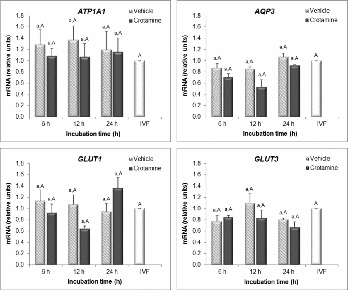
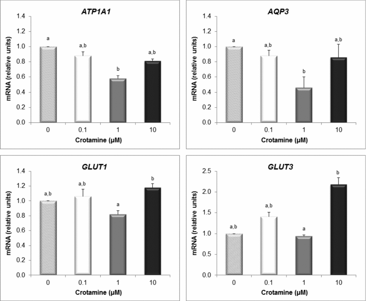
In general, ATP1A1, AQP3, GLUT1 and GLUT3 transcripts were detected successfully in all bovine embryos regardless of the exposure to crotamine. In Experiment 1, the presence of 0.1 μM crotamine in the culture medium did not affect gene expression of bovine embryo even up to 24 h (Fig. 1). Within each exposure time, mRNA levels were similar (P > 0.05) in embryos exposed or not to crotamine. Also, expression in each crotamine group did not differ (P > 0.05) from the IVF control. As expected, exposure up to 24 h with only vehicle solution did not alter (P > 0.05) the levels of ATP1A1, AQP3, GLUT1 or GLUT3 transcripts in bovine embryos when compared with IVF group. In Experiment 2 (Fig. 2), 0.1 and 10 μM crotamine concentrations for 24 h did not induce mRNA level changes when compared with the vehicle control. Remarkably, only at the concentration of 1 μM, crotamine decreased both ATP1A1 and AQP3 expression relative to vehicle control, whereas GLUT1 and GLUT3 transcript levels were similar (P > 0.05).
Effect of crotamine exposure time on gene expression of in vitro fertilized bovine embryos. Quantification of ATP1A1, AQP3, GLUT1 and GLUT3 transcripts was performed by qPCR using GAPDH and H2A as reference genes. The IVF group was used as calibrator. Comparisons were made between groups within each exposure time (a, b: P < 0.05) or for each group versus IVF (A, B: P < 0.05).

Effect of crotamine concentration on gene expression of in vitro fertilized embryos. Quantification of ATP1A1, AQP3, GLUT1 and GLUT3 transcripts were performed by qPCR using GAPDH and H2A as reference genes. The vehicle group (0 μM crotamine) was used as calibrator. Bars with different superscripts (a, b) are significantly different at P < 0.05.

Crotamine translocation
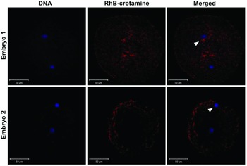
It was possible to visualize crotamine translocation through ZP of IVF bovine zygotes as soon as after 6 h exposure (Fig. 3). Moreover, partial overlapping of crotamine signal and DAPI nuclear DNA staining was observed. ZP removal after exposure and prior to fixation allowed us to differentiate membrane and intracellular labelling of embryos instead of possible ZP labelling.
Crotamine uptake by IVF bovine embryos after 6 h exposure to 10 μM RhB-labelled crotamine. Crotamine labelling (red) and nuclei counterstaining with DAPI (blue) was observed into fixed bovine zygote by fluorescence confocal microscopy. Partial overlapping of crotamine and DAPI fluorescence was seen (arrowhead).

Discussion
In the present work, the effect of crotamine on the in vitro development of bovine embryos and the quantitative expression of selected embryo quality genes were investigated. Immediately after fertilization, presumptive zygotes were cultured in the presence of 0.1–10 μM crotamine and for up to 24 h exposure. Overall, crotamine did not impair or disturb the embryo development under assayed conditions.
The presence of 0.1 μM crotamine in the culture medium even for 24 h neither affected the embryo production, as indicated by similar blastocyst rates among crotamine and control groups, nor morphological embryo quality, inferred by total nuclei counting. These data suggest that, under those conditions, crotamine has no negative effect on preimplantation development. It is possible that future applications of crotamine as CPP for animal transgenesis by embryo transfection may be limited to an exposure time of not longer than 24 h once the crotamine uptake by embryo seems to occur before 6 h of incubation. Kerkis et al. (Reference Kerkis, Kerkis, Rádis-Baptista, Oliveira, Vianna-Morgante, Pereira and Yamane2004) investigated in vitro the Cy3-crotamine uptake at 1 μM by human primary fibroblasts, lymphoblastic cells, murine embryonic stem and endothelial s-vec cells, monitoring the crotamine uptake after 5 min and 1, 3, 24, or 48 h of treatment by confocal microscopy. As indicated by this study, the cells internalized crotamine as fast as 5 min after its addition and the number of labelled cells reached a maximum after about 3 h of treatment. A similar interval for cellular uptake of fluorescently labelled crotamine was reported for highly proliferative CHO-K1 cells (Hayashi et al., Reference Hayashi, Nascimento, Kerkis, Oliveira, Oliveira, Pereira, Rádis-Baptista, Nader, Yamane, Kerkis and Tersariol2008). Conversely, when considering embryos, it is important to highlight that the barrier imposed by ZP could delay this time of internalization. However, crotamine uptake at 1 μM concentration was evidenced in compact mouse morulae after 24 h exposure (Kerkis et al., Reference Kerkis, Kerkis, Rádis-Baptista, Oliveira, Vianna-Morgante, Pereira and Yamane2004), indicating that this time was enough for crotamine to reach the inner cell mass of murine intact blastocysts.
The ZP is a glycoproteinaceous translucent matrix that surrounds the mammalian oocyte and plays a critical role in achieving fertilization (Gupta et al., Reference Gupta, Bhandari, Shrestha, Biswal, Palaniappan, Malhotra and Gupta2012). Another relevant aspect is the ZP glycoproteic composition, as crotamine–carbohydrate interaction plays a fundamental role in the first step of cell internalization, as reported extensively (reviewed in Rádis-Baptista & Kerkis, Reference Rádis-Baptista and Kerkis2011). In fact, it is known that some CPPs interact electrostatically with the extracellular matrix of the cell followed by endocytosis (Richard et al., Reference Richard, Melikov, Vives, Ramos, Verbeure, Gait, Chernomordik and Lebleu2003; Nascimento et al., Reference Nascimento, Hayashi, Kerkis, Oliveira, Oliveira, Rádis-Baptista, Nader, Yamane, Tersariol and Kerkis2007). More specifically, crotamine internalization seems to be mediated by heparan sulphate proteoglycans (HSPGs) in the uptake phase (Nascimento et al., Reference Nascimento, Hayashi, Kerkis, Oliveira, Oliveira, Rádis-Baptista, Nader, Yamane, Tersariol and Kerkis2007). In the context of the present study, we did not confirm if N- and O-glycans of bovine embryo ZP could interact with crotamine in the culture medium. Therefore, it is premature to infer whether the ZP itself could affect the number of crotamine molecules available for embryo cell uptake.
After 0.1 μM, we used 1 or 10 μM crotamine in the culture medium also for 24 h exposure with presumptive zygotes. The presence of crotamine concentrations greater than 0.1 μM seems to not affect the final production and quality of 8-day embryos. Nonetheless, levels of ATP1A1 and AQP3 transcripts detected in embryos exposed for 24 h to 1 μM crotamine were lower than in the control, but similar to 10 μM crotamine treatment. Interestingly, levels of ATP1A1 and AQP3 transcripts in embryos exposed to 0.1 μM and 10 μM crotamine for 24 h were similar to that of the control. Indeed, alterations in mRNA levels in 8-day embryos were not detected for GLUT1 and GLUT3 in any crotamine group. Additionally, in Experiment 1, embryos subjected to all exposure times with 0.1 μM crotamine had the same relative levels of all transcripts (ATP1A1, AQP3, GLUT1 and GLUT3) as the control groups. The expression level of these genes has been correlated with embryo quality (Camargo et al., Reference Camargo, Boite, Wohlres-Viana, Mota, Serapião, Sa, Viana and Nogueira2011; Kuzmany et al., Reference Kuzmany, Havlicek, Wrenzycki, Wilkening, Brem and Besenfelder2011) as the encoded proteins play important roles in the physiological process at preimplantation stage of embryo development (Rizos et al., Reference Rizos, Clemente, Bermejo-Alvarez, Fuente, Lonergan and Gutiérrez-Adán2008). The expression of ATP1A1 and AQP3 genes is reported to be involved in embryo osmotic regulation and blastocoele formation. ATP1A1 is the protein of the Na/K ATPase subunit responsible for generating an ionic gradient through the trophectoderm and subsequent water influx to the blastocoele (Watson et al., Reference Watson, Westhusin, De Sousa, Betts and Barcroft1999). AQP3 is an aquaglyceroporin that increases cellular permeability to water and other small solutes, such as glycerol, urea, purines and pyrimidines (Barcroft et al., Reference Barcroft, Offenberg, Thomsen and Watson2003). The GLUT proteins are described as being engaged in the energy metabolism of the mammalian embryos (Purcell & Moley, Reference Purcell and Moley2009). GLUT3 has been reported to play a central role in glucose absorption, while GLUT1 is responsible for intracellular glucose transport (Augustin et al., Reference Augustin, Pocar, Navarrete-Santos, Wrenzycki, Gandolfi, Niemann and Fischer2001).
In this context, the similarity of expression of GLUT1 and GLUT3 genes between embryos exposed to crotamine or vehicle solutions suggests that energy metabolism concerned with glucose uptake is not disturbed. Only embryos cultured in the presence of 1 μM crotamine for 24 h decreased the expression of osmotic-related genes (ATP1A1 and AQP3), while concentrations lower and higher than 1 μM did not affect these levels. It is possible that the 0.1 μM concentration is not able to alter the osmotic response of bovine embryos, whereas a crotamine concentration of 10 μM could promote a start-up compensatory embryo response at transcriptional level faster than that of 1 μM crotamine. This pattern of gene expression regulation on in vitro cultured bovine embryos has been reported previously in response to the presence of some substances in the culture medium (Camargo et al., Reference Camargo, Boite, Wohlres-Viana, Mota, Serapião, Sa, Viana and Nogueira2011). Nevertheless, these changes did not interfere with in vitro development of bovine embryos, as both blastocyst and hatching rates were similar among all groups.
In conclusion, crotamine application as a cell-penetrating peptide for bovine transgenesis and embryo transfection is apparently viable from the standpoint of its inherent capacity of translocate into intact IVF embryos and due to the lack of detectable toxicity. Moreover, as demonstrated in the current study, it is possible to use crotamine in the culture medium with concentrations ranging from 0.1–10 μM and exposure time from 6–24 h. Further studies could address the feasibility of crotamine as an effective carrier of DNA molecules into intact bovine embryos.
Financial support
This study was supported by CNPq (grant number 560613/2010–5). I.S.C. was supported by an MSc scholarship from FUNCAP. A.F.P., J.M.G.S-F and L.M.M. were supported by postdoctoral fellowships from CNPq and CAPES, respectively.
Conflict of interest
There are no conflicts of interest.
Acknowledgements
The authors would like to acknowledge Prof. Eduardo Brandt de Oliveira (University of São Paulo, Ribeirão Preto, Brazil) for providing crotamine. We are also thankful to Mr Claudio Andrade (inspection service of slaughterhouse, Morada Nova, Brazil) for supplying bovine ovaries.