Goat milk is believed by some consumers to offer nutritional benefits, and can thus attract a premium price (Haenlein, Reference Haenlein2004; Alférez et al., Reference Alférez, Rivas, Díaz-Castro, Hijano, Nestares, Moreno, Campos, Serrano-Reina and López-Aliaga2015). Goat milk products appear to be potentially less allergenic than cow milk (Agamy, Reference Agamy2007; Claeys et al., Reference Claeys, Cardoen, Daube, De Block, Dewettinck and Dierick2013) and can, therefore, have particular benefit for those people (including children) who suffer from allergenic intolerance of cow milk (Ballabio et al., Reference Ballabio, Chessa, Rignanese, Gigliotti, Pagnacco and Terracciano2011). Several techniques can be used for detection of goat milk contamination or adulteration, including polyacrylamide gel electrophoresis, capillary isoelectric focusing, liquid chromatography and ELISA (Ke et al., Reference Ke, Zhang, Lai, Chen, Zhang, Jiang, Mo and Ren2017). However, these methods have difficulty in quantifying adulteration percentage and are both laborious and time consuming. Based upon these considerations, we aimed to develop a competitive lateral flow immunoassay (LFIA) strip for simple, rapid, specific and sensitive detection of cow milk casein for quantitatively measuring the percentage of cow's whole milk contamination or adulteration in goat milk.
Materials and methods
Mice, milk and chemicals
Three Balb/c mice were obtained for immunization from experimental animal center, Southern Medical University, Guangzhou, China. Animal care was in accordance with national and institutional policies for animal health and well-being and the experiments were approved by Southern Medical University (SMU) Animal Care and Use Committee (Permit numbers: NFYY-2008-043 and NFYY-2010-076). Mouse surgery was performed under anesthesia. Goat and cow milk and five different kinds of milk or milk substitute (camel milk brand A, camel milk brand B, soymilk brand A, soymilk brand B and soybean milk) were obtained from domestic manufacturers. Freund's complete adjuvant, Freund's incomplete adjuvant were purchased from Sigma-Aldrich (St. Louis, MO, USA). Polyethylene glycol 4000 was supplied by Roche (Basel, Switzerland).
Production of monoclonal antibodies
Each BALB/c mouse was immunized primarily by intraperitoneal injection with 100 µl of 1 mg/ml cow casein (Sigma, 95% purity) emulsified with an equivalent volume of Freund's complete adjuvant. The booster injections with Freund's incomplete adjuvant were given twice by tail vein injection at two week intervals. Three days after the last injection, the mouse was sacrificed. Spleen cells were collected from the immunized mouse and fused with SP2/0 cells using 50% polyethylene glycol 4000. The cells were distributed into 96-well culture plates (Costar, USA) in 1640 medium supplemented with 1% HAT (Gibco, USA). After 11 d, the supernatants from the hybridoma growing wells were screened using an indirect ELISA. The positive hybridomas were sub-cloned three times by the limiting dilution method, and the reactive monoclonal antibodies were characterized by ELISA (Qiu et al., Reference Qiu, Wang, Wu, Zhang, Wang, Qiao, Chen, Gao, Allain and Li2012; Li et al., Reference Li, Hu, Chen, Luo, He, Wang, Allain and Li2017).
Preparation of LFIA strip
Colloidal gold particles (GNPs) with a mean diameter of 40 nm were produced by reduction of gold chloride with 1% sodium citrate as described previously (Zhang et al., Reference Zhang, Wang, Yang, Yang, Xing, Li, Zhao, Chai and Guo2006). The colloidal gold particles were labeled with mAbs specific to cow milk casein according to the method of Yokota (Yokota, Reference Yokota and Fujimori1992). The cow casein (2 mg/ml, Sigma) and goat anti-mouse IgG (1 mg/ml) were applied to the nitrocellulose (NC) membrane by dispenser as the test (T) and control (C) lines, respectively. The strips were assembled as described previously (Zhang et al., Reference Zhang, Wang, Zhi, Bao, Yang, Qu, Luo, Li, Guo, Wang, Yang, Xing, Chai, Shi and Liu2008). The sample pad, blotted NC membrane and absorption pad were assembled on the plastic backing support board sequentially with a 1–2 mm overlap and covered by color film at both ends. The master card was cut into 4 mm width strips using a CM 4000 Cutter (Bio-Dot). Strips were then sealed in a plastic bag in the presence of desiccant gel and stored at 4 °C.
Procedure and principle of LFIA strip
An aliquot of 200 µl of goat milk sample or cow milk reference standard solution (cow milk diluted in goat milk) was added into reaction buffer (5 µl of 5 M Tris-HCl pH 7.5, 10 µl of 20% Tween-20, 3 µl of colloidal gold labeled anti-casein mAb) and reacted at room temperature for 3 min. The solution was then added onto the sample pad, and migrated toward the absorbent pad. The results could be seen after 3 min. When cow casein was absent in the goat milk sample, the detection reagent would be trapped by capture reagent to form an easily visible T line. On the contrary, when cow casein was present in the sample, it will competitively bind to the immobilized capture reagent with the limited amount of detection reagent. The more cow casein present in the sample, the weaker color of test line appeared. If sufficient cow casein is present in the sample, it will completely block the reaction with the capture reagent, thus, there would be no visible T line on the nitrocellulose membrane. The C line was always visible, which indicated that the test strip and procedure worked well. If no colored capture lines or only T line was observed, the testing procedure was likely to be improper or invalid, under which circumstance the test should be repeated using a new strip.
Results
Characterization of monoclonal antibody to cow milk casein
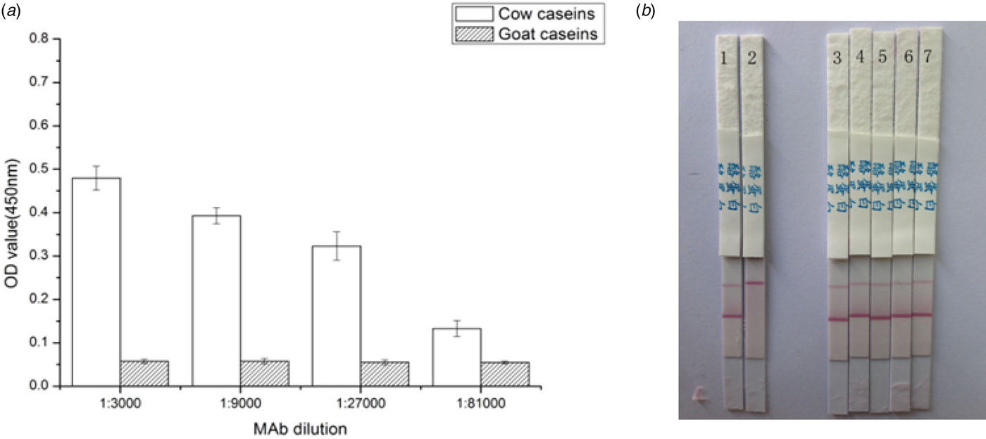
A total of 12 mAbs reactive with cow milk casein were selected from screening of hybridomas by ELISA. Clone 1G6 was selected and used for the LFIA strip. The mAb was purified from the ascites fluid. The titer of the purified ascites fluid was 5.12 × 10⁻⁵. The 1G6 clone was IgG1 and had high affinity constants (K a) of 4.5 × 10¹⁰ l/mol, while this mAb had negligible cross-reactivity with goat milk (<0.01%) (Fig. 1a).
Reactivity of monoclonal antibody specific to goat milk. (a) MAb1G6 cross-reactivity and affinity measured by ELISA. OD values were obtained by a micro-plate reader under 450 nm. Cow or goat caseins were diluted in the sodium bicarbonate coating buffer (pH 9.6) (Li et al., Reference Li, Hu, Chen, Luo, He, Wang, Allain and Li2017). (b) Specificity of LFIA strip with anti-casein gold conjugates. 1, negative control without cow caseins; 2, positive control with cow caseins; 3, camel milk brand A; 4, camel milk brand B; 5, soymilk brand A; 6, soymilk brand B; 7, soybean milk. All tests were performed in triplicate.

Specificity of LFIA strip for cow milk
The specificity of this assay was evaluated by reacting with two other milk solutions (camel milk brand A, camel milk brand B) and three milk substitutes (soymilk brand A, soymilk brand B and soybean milk). As presented in Fig. 1b, no cross-reaction was observed from testing of these 5 milk/milk substitutes at concentration of 2% in goat milk, which indicated a reasonably high specificity of the assay to cow milk casein.
Sensitivity of LFIA strip for cow milk
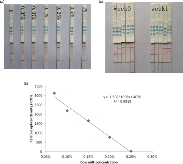
To test the sensitivity of this assay, a reference standard of cow milk was serially diluted at a final concentration of 0, 0.05, 0.07, 0.1, 0.25, 0.5 and 1% in goat milk. As shown in Fig. 2a, the color indicative of intensity of T line was stronger or similar than that of C line for negative and low concentration samples, which became obviously equivalent to 0.07%, or weaker than 0.1%, and then the T line disappeared when cow milk concentration reached 0.25%. Therefore, the visual detection limitation of this LFIA strip was 0.07% for cow milk added in goat milk. By measuring the T lines of strips with a HF3500 membrane strip reader (Huaan Magnech, Beijing), the scanned relative optical density (ROD) decreased as the cow milk concentration increased in these reference standard samples. A sigmoidal dose-response curve was plotted according to the concentrations of cow milk reference standard and the ROD (Fig. 2b), which gave a good linearity with the range between 0.07 and 0.25% of cow milk in goat milk.
Characterization of LFIA strip. (a) Sensitivity for detection of cow milk in goat milk. (b) Linear regression equation derived using the ROD data of HF3500 membrane strip reader. (c) Stability for detection of cow milk casein. The strips were tested after 1 week post production, in which cow milk was detected in a concentration of 0, 0.05, 0.1, 0.2 and 0.5% (from left to right), respectively. All tests were performed in triplicate.

Stability of LFIA strips for cow milk
The stability of the prepared LFIA strips was tested after being stored in different conditions for various times. No significant difference of color intensity was shown for those LFIA strips newly prepared or stored 1 week at 37 °C (Fig. 2c), indicating these LFIA strips were stable for up to 1 week at 37 °C.
Testing of cow milk adulteration from commercial goat milk brand samples
Firstly the artificial samples were made by adding 0.02–1% cow milk into goat milk, and then were detected using LFIA strips in comparison with R-Biopharm ELISA kit (R-Biopharm, Germany). Using LFIA, cow milk concentrations of 0.02 and 0.05% were not detected, but 0.07, 0.1 and 1% were all detected. Using ELISA, only the highest two concentrations were detected with signal cutoffs of 1.25 and 2.97 Then, 10 different manufactured goat milk brand samples purchased from markets around the country were investigated using both the LFIA and the ELISA. The same 3 (out of 10) goat milk samples tested positive for cow milk adulteration by both tests. These results demonstrated that the LFIA could be used for detection of cow milk contamination or adulteration in goat milk.
Discussion
Goat milk is considered a healthy food and high-quality product by the dairy industry because of its biochemical, nutritional and organoleptic characteristics. In addition, the characteristics of goat milk components allow to have highly priced dairy products, such as cheeses, yogurts, and a wide variety of other fermented products (Alférez et al., Reference Alférez, Rivas, Díaz-Castro, Hijano, Nestares, Moreno, Campos, Serrano-Reina and López-Aliaga2015; Moreno-Fernández et al., Reference Moreno-Fernández, Díaz-Castro, Alférez, Hijano, Nestares and López-Aliaga2016). In this study, an analytical method was developed for detecting and quantifying the percentage of cow milk in goat milk, which was useful for both consumer and dairy industry. Using this rapid lateral flow assay strip, the presence or absence of cow milk contamination/adulteration in goat milk can be tested without any further expensive equipment or professional laboratory technicians compared with commonly used methods like liquid chromatography or ELISA. The LFIA assay requires just one simple procedure to be completed by the operator and the result is visible within 6 min. The visual limitation of detection (LOD) achieves up to 0.07%, which is also more sensitive than the commercial ELISA kit (LOD: 0.1%). Currently over 700 LFIA strips have been tested in field trials across China.
In conclusion, the LFIA strip is rapid, user friendly and cost effective for detecting and quantifying goat milk contaminated or adulterated by cow milk, with no need of sample preparation and having excellent sensitivity, specificity and reproducibility. The developed assay could be used for verification of milk samples on site.
Acknowledgements
This work was supported by the grants from the National Natural Science Foundation of China (No. 31500134 and 31372443), the Guangzhou Pearl River S&T Nova Program (No. 201506010075), the Guangzhou major project of industry-university-research cooperation and collaborative innovation (No. 201508020061 and 201704020083), the Innovative R&D Team Introduction Program of Guangdong (No. 2014ZT05S123) and the Guangdong Provincial S&T Project (No. 2014A020214003 and 2012A080203003).
Financial support
None.