Introduction
Myliobatiformes, commonly known as stingrays, is a monophyletic order comprising more than 221 species (29 genera in ten families) with worldwide distribution (Dunn et al., Reference Dunn, McEachran and Honeycutt2003; Nelson, Reference Nelson2006). Several species of stingrays are used for human consumption and others, especially potamotrygonids, are highly appreciated by aquarists. Despite their great diversity and economic importance, less than 29% of stingray species have been studied for the presence of monogenean parasites (Chero et al., Reference Chero, Cruces, Iannacone, Sáez, Sánchez, Minaya and Alvariño2016, Reference Chero, Cruces, Sáez, Camargo, Santos and Luque2018a, Reference Chero, Cruces, Sáez and Luqueb, Reference Chero, Cruces, Sáez, Santos and Luque2020; Kohn et al., Reference Kohn, Justo, Cohen, Eiras, Velloso and Pereira2016; Lopes et al., Reference Lopes, Mainenti, Sanches, Knoff and Gomes2016; Merlo-Serna & García-Prieto, Reference Merlo-Serna and García-Prieto2016; Mendoza-Garfias et al., Reference Mendoza-Garfias, García-Prieto and Pérez-Ponce de León2017; Pollerspöck & Straube, Reference Pollerspöck and Straube2021).
The global fauna of monogeneans infecting stingrays is remarkably diverse and includes 147 species belonging to 33 genera in six families – namely, Acanthocotylidae Monticelli, 1903 (1 sp.), Capsalidae Baird, 1853 (23 spp.), Hexabothriidae Price, 1942 (9 spp.), Loimoidae Price, 1936 (1 sp.), Monocotylidae Taschenberg, 1879 (111 spp.) and Udonellidae Taschenberg, 1879 (2 spp.) (Chisholm & Kritsky, Reference Chisholm and Kritsky2020; Kritsky & Chisholm, Reference Kritsky and Chisholm2020; Pollerspöck & Straube, Reference Pollerspöck and Straube2021). More than half of these genera (19) and about 91 species are parasites of dasyatids, the most diverse group within the Myliobatiformes (Last et al., Reference Last, Naylor and Manjaji-Matsumoto2016). The monocotylid Monocotyle ijimae Goto (1894) was the first monogenean species described from a dasyatid host and was found in the mouth of the common stingray, Dasyatis pastinaca (Linnaeus, 1758), in Japan. Thirty-five monogenean species have been described from dasyatids in the last two decades and four new genera have been proposed – namely, Hypanocotyle Chero, Cruces, Sáez, Camargo, Santos & Luque, 2018; Listrocephalos Bullard, Payne & Braswell, 2004; Peruanocotyle Chero, Cruces, Sáez & Luque, 2018; and Septesinus Chisholm, 2013 (Cao et al., Reference Cao, Ding, Zhang and Liu2010; Vaughan & Chisholm, Reference Vaughan and Chisholm2010; Chisholm & Whittington, Reference Chisholm and Whittington2012; Santos et al., Reference Santos, Santos, Cunha and Chisholm2012; Chisholm, Reference Chisholm2013; Chero et al., Reference Chero, Cruces, Iannacone, Sáez, Sánchez, Minaya and Alvariño2016, Reference Chero, Cruces, Sáez, Camargo, Santos and Luque2018a, Reference Chero, Cruces, Sáez and Luqueb, Reference Chero, Cruces, Sáez, Santos and Luque2020; Kitamura & Ogawa, Reference Kitamura and Ogawa2019).
Loimoidae Price, 1936 includes three accepted genera, with species that infect marine elasmobranchs – namely, Loimopapillosum Hargis, 1955 (one species described from dasyatids in the Atlantic Ocean); Loimos MacCallum, 1917 (five species from carcharhinids in the Atlantic and Pacific Oceans); and Loimosina Manter, 1944 (two species described from sphyrnids in Central America and one species in Brazil) (Kohn et al., Reference Kohn, Justo, Cohen, Eiras, Velloso and Pereira2016; Lopes et al., Reference Lopes, Mainenti, Sanches, Knoff and Gomes2016; Mendoza-Garfias et al., Reference Mendoza-Garfias, García-Prieto and Pérez-Ponce de León2017; Pollerspöck & Straube, Reference Pollerspöck and Straube2021). Boeger & Kritsky (Reference Boeger and Kritsky1993) provided a phylogenetic analysis of the monogeneans at the family level based on cladistic analysis and clustered Loimoidae and Monocotylidae as sister taxa within the order Monocotylidea. This hypothesis was also considered by Chisholm et al. (Reference Chisholm, Wheeler and Beverley-Burton1995), who used Loimoidae as the outgroup in their cladistic analysis of relationships within Monocotylidae. Boeger et al. (Reference Boeger, Kritsky, Domingues and Bueno-Silva2014) assessed the phylogenetic position of Loimoidae within the monogeneans based on analyses of partial sequences of 18S and 28S ribosomal DNA (rDNA) of Loimosina sp. They found no phylogenetic support for the family and suggested that Loimoidae represents a subordinate taxon within Monocotylidae.
During a parasitological survey of Peruvian marine fishes carried out from July 2018 through November 2019, several monogenean specimens were found on the gill filaments of the diamond stingray Hypanus dipterurus (Jordan & Gilbert, 1880) from Puerto Pizarro, northern Peru. Detailed morphological examination revealed that the parasites represented a new species of Loimopapillosum, which is described based on morphological and molecular analysis. Additionally, nucleotide sequences (28S and 18S rDNA) obtained of the new species were used to evaluate its phylogenetic position.
Materials and methods
Specimen collection and morphological analyses
Stingrays were collected during July 2018 through November 2019 from the coastal zone of Puerto Pizarro, Tumbes, Peru (3°29′S, 80°24′W), using gillnets, euthanized immediately after capture and dissected. The gills were excised and placed in Petri dishes with sea water and examined for monogeneans with the aid of a stereomicroscope. Monogeneans were collected alive, washed in seawater and fixed in hot 4% formalin under slight coverslip pressure. They were then stained with Semichon's carmine, clarified in Eugenol (4-allyl-2-ethoxyphenol) and mounted in Canada balsam. Other specimens were mounted directly in Gray and Wess medium for study of sclerotized structures (Humason, Reference Humason1979). Specimens were examined using a compound Olympus™ BX51 light photomicroscope equipped with Nomarski Differential Interference Contrast optics (Olympus Corporation, Tokyo, Japan) and drawings were made with the aid of a drawing tube. Unless stated otherwise, measurements are in micrometres, representing straight-line distances between extreme points of the structures measured and are expressed as the range followed by the mean and number (n) of structures measured in parentheses. Body length represents the length of the body proper including the haptor. Scientific names, including taxonomic authorities and dates, for fishes follow Eschmeyer et al. (Reference Eschmeyer, Fricke and van der Laan2019). Nomenclature and classification of Myliobatiformes, Dasyatidae and Hypanus species follow Last et al. (Reference Last, Naylor and Manjaji-Matsumoto2016). Prevalence and mean intensity of infection were calculated according to Bush et al. (Reference Bush, Lafferty, Lotz and Shostak1997). Type specimens were deposited in the Helminthological Collection in Museum of Natural History at the San Marcos University (MUSM), Lima, Peru. Specimens of Loimopapillosum dasyatis Hargis, 1955 (USNM 1338744, holotype and paratypes) deposited in the Helminthological Collection of the National Museum of Natural History, Smithsonian Institution (USNM), USA, were examined. High-resolution imagens of the anterior end of Loimosina parawilsoni Bravo-Hollis, 1970 (CNHE 153, holotype) and of the anterior end and haptor of Loimos winteri Caballero & Bravo-Hollis, 1961 (CNHE 86, holotype) from the Nacional Collection of Helminths, Institute of Biology, National Autonomous University of Mexico (CNHE), Mexico, available at http://unibio.unam.mx were also examined.
DNA extraction, polymerase chain reaction (PCR) amplification and DNA sequencing
Two monogeneans were fixed directly in absolute Ethanol alcohol (EtOH) for molecular analyses. Genomic DNA was extracted using QIAamp DNA Mini Kit (Quiagen, Hilden, Germany), according to the manufacturer's recommendations. The partial 28S rDNA was amplified by PCR using the primers C1 (5′-ACCCG CTGAA TTTAA GCAT-3′) and D2 (5′-TGGTC CGTGT TTCAA GAC-3′) (according to Chisholm et al., Reference Chisholm, Morgan, Adlard and Whittington2001). For the partial 18S rDNA region the primers used were L7 (5′-TGATT TGTCT GGTTT ATTCC GAT-3′) and IR5 (5′-TACGG AAACC TTGTT ACGAC-3′) (Sinnappah et al., Reference Sinnappah, Lim, Rohde, Tinsley, Combes and Verneau2001). PCR assays were carried out in a total volume of 15 μl containing 7.5 μl of 2× GoTaq® Colorless Master Mix (Promega, Madison (Wisconsin), United States), 1.5 μl of each primer with final concentration at 0.5 μM, 2.5 μl of complementary DNA sample and ultrapure water to complete, using cycling parameters as previously described by these authors. The PCR products were analysed by electrophoresis in 1.5% agarose in Tris-borate Ethylenediamine tetraacetic acid (EDTA) gels, stained with SyberGreen (Invitrogen, Eugene, Oregon, USA) and photographed under ultraviolet transillumination. Amplified PCR products were purified with ExoSap-IT (USB® Products Affymetrix Inc., Cleveland, Ohio, USA). DNA cycle sequencing reactions were performed using BigDye Terminator v.3.1 (Applied Biosystems, Foster City, California, USA) and automated sequencing was done using the Sequencing Platform at the Fundação Oswaldo Cruz-PDTIS/FIOCRUZ in Brazil. Sequences of both strands were generated, edited and aligned by using the MEGA version 7.0 software (Kumar et al., Reference Kumar, Stecher and Tamura2016). Sequences were compared to others available in the GenBank database using the BLAST program from the National Center for Biotechnology Information (NCBI) server (http://www.ncbi.nlm.nih.gov/BLAST) (Altschul et al., Reference Altschul, Gish, Miller, Myers and Lipman1990).
Molecular analyses
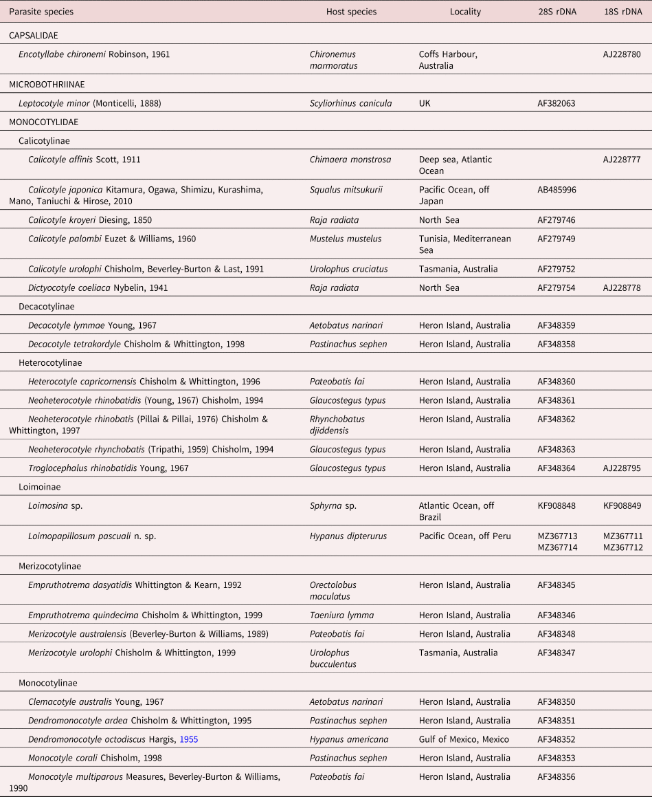
Sequences were aligned with Clustal W in MEGA 7.0 (Thompson et al., Reference Thompson, Higgins and Gibson1994). The best model found by JModelTest2 (Posada & Crandall, Reference Posada and Crandall1998), selected with the corrected information criterion, was GTR + I + G for the 28S and K80 + G for 18S. The best model was implemented in MrBayes 3.2.7a (Ronquist et al., Reference Ronquist, Huelsenbeck and Teslenko2011) for Bayesian Inference (BI) analysis. BI analysis was performed using Metropolis-coupled Markov chain Monte Carlo analyses, which were run with four chains and 5,000,000 generations, and were sampled every 1000 generations. The burn-in period discarded 25% generations. Posterior clade probabilities were used to assess nodal support. Tree topology represented on the 50% majority-rule consensus trees. The software we used was implemented on the CIPRES Science Gateway V.3.3 (http://www.phylo.org/) (Miller et al., Reference Miller, Pfeiffer and Schwartz2011). The sequences from GenBank that were used for the phylogenetic analysis are listed in table 1 as well the sequences for outgroups.
List of the species of Monogenea and outgroups used in the phylogenetic analyses of Loimopapillosum pascuali n. sp. (Loimoinae) using sequences of the partial 28S and 18S rDNA genes.

Results
Systematics
Order: Monocotylidea Lebedev, 1988
Monocotylidae Taschenberg, 1879
Loimoinae Price, 1936
Loimopapillosum pascuali n. sp.
Description
Based on 25 adult specimens. Body elongate, fusiform, 2.16–3.41 (2.70; n = 15) mm long, 0.32–0.44 (0.35; n = 14) mm wide at level of testis; slightly constricted at level of pharynx (fig. 1a). Tegument smooth. Peduncle slightly broad, elongate. Haptor differentiated from body proper, 264–380 (311; n = 12) in diameter, broadly ellipsoid, concavo-convex, undivided, with strong muscular ring, armed with two anchors and 14 similar hooks on muscular digitiform papillae; marginal membrane present. Anchor 30–31 (30.5; n = 4) long, deep and superficial roots distinct; deep root short, spatulate distally, constricted at base; superficial root rounded, thick, with distal knobs; slightly curved shaft and recurved point; point reaching level of tip of superficial root (fig. 1b). Hooks 9–11 (10; n = 7) long, each with short and curved point, protruding thumb and uniform shank; filamentous hook loop extending to near level of proximal end of shank (fig. 1c). Cephalic region slightly broad; four pairs of head organs (fig. 1a); cephalic glands posterolateral to pharynx. Eyespots absent. Mouth ventral, subterminal, posterior to head organs. Pharynx muscular, pyriform, 102–125 (113; n = 11) long, 92–120 (100; n = 11) in greatest width, comprising short anterior part and long posterior part with 7–8 transversal muscular packets. Oesophagus short. Intestinal bifurcation anterior to common genital aperture; intestinal caeca unbranched, not confluent posteriorly, extending up to posterior end of body proper. Common genital pore unarmed. Testis oval, 284–362 (332; n = 6) long, 102–141 (128; n = 6) wide, limited to intercaecal field, well separated from posterior end of body. Vas deferens slightly sinuous, runs anteriorly dorsal to left transverse vitelline duct, looping over ejaculatory bulb, expanding as seminal vesicle and entering at ejaculatory bulb. Ejaculatory bulb muscular, small, subspherical, 32–37 (34; n = 6) long, 24–29 (26; n = 6) wide. Male copulatory organ 39–55 (48; n = 12) long, sclerotized, funnel-shaped with asymmetric base (fig. 1d). Accessory piece absent. Ovary pretesticular, elongate, looping dorsoventrally right intestinal cecum (fig. 1e), 299–345 (323; n = 6) long, 128–201 (174; n = 6) wide. Oötype and Mehlis’ glands not observed. Vaginal pore unarmed, opening ventrally on right side. Vagina weakly sclerotized, club-shaped, lying slightly obliquely, anterior to ovary; proximal portion of vagina connected to seminal receptacle by narrow duct (fig. 1e); seminal receptacle 49–58 (54; n = 3) long, 43–49 (46; n = 3) wide. Vitelline follicles dense, extending from posterior level of intestinal bifurcation to posterior end of trunk, absent in regions of reproductive organs (fig. 1e). Egg oval (fig. 1f), 98–149 (134; n = 4) long, 86–117 (104; n = 4) wide; single terminal filament, short, 30–45 (38; n = 2) long, with small button at distal end.
Loimopapillosum pascuali n. sp. from the gill filaments of the diamond stingray Hypanus dipterurus, holotype: (a) whole specimen (ventral view); (b) anchor; (c) hook; (d) male copulatory organ; (e) reproductive system; (f) egg. Abbreviations: eb, ejaculatory bulb; mco, male copulatory organ; ov, ovary; sr, seminal receptacle; sv, seminal vesicle; tvd, transverse vitelline duct; te, testis; va, vagina; vd, vas deferens.

Taxonomic summary
-
Type host. Hypanus dipterurus (Jordan et Gilbert) (Myliobatiformes: Dasyatidae), diamond stingray.
-
Site of infection. Gill filaments.
-
Type locality. Puerto Pizarro, Tumbes Region, Peru (3°29′S, 80°24′W).
-
Prevalence. Two of three hosts infected (67%) with a total of 52 worms.
-
Mean intensity of infection. 26 monogeneans per infected host (range 25–27).
-
Specimens deposited. Holotype, MUSM 4660; 24 paratypes, MUSM 4661a-x.
-
Representative DNA sequences. Sequences were deposited in GenBank under the accession numbers MZ367711 and MZ367712 for the partial 18S rDNA with 489 bp, and MZ367713 and MZ367714 for the partial 28S rDNA with 914 bp.
-
Etymology. The new species is named in honour of Pascual Chero Sullon, father of the first author.
Remarks
The collected specimens belong to Loimopapillosum because of their haptor, which is a concavo–convex disk, undivided, armed with two anchors and 14 hooks located on digitiform papillae (Hargis, Reference Hargis1955). Loimopapillosum pascuali n. sp. can be easily distinguished from L. dasyatis Hargis, 1955 mainly by the male copulatory organ, which is funnel-shaped with an asymmetric base (almost incomplete retort-shaped in L. dasyatis). In addition, L. pascuali n. sp. differs from L. dasyatis by the position of the male copulatory organ (MCO), which is posterior to the beginning of the vitelline follicles (at the level of the vitelline follicles in L. dasyatis). The new species also differs from L. dasyatis by having the superficial root of the anchor with distal knobs, the deep root of the anchor with a constriction at its base, a single testis (two testes in L. dasyatis) and by the number of head organs (four in L. pascuali n. sp. vs. five in L. dasyatis). Loimopapillosum pascuali n. sp. is the second species described in the genus and represents the first record of Loimopapillosum species from South America.
Genetic characterization
For the first partial 18S rDNA sequence of L. pascuali n. sp., the BLAST results indicated 95% identity with Troglocephalus rhinobatidis (AJ228795); Loimosina sp. (KF908849) with 95% identity; and Dictyocotyle coeliaca (AJ228778) with 92% identity. For the first partial 28S rDNA sequence, the most similar sequences included those of Decacotyle lymmae (AF348359) with an 86% identity, and Heterocotyle capricornensis (AF348360) with 85% identity.
Phylogenetic analyses of Monocotylidae
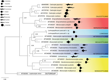
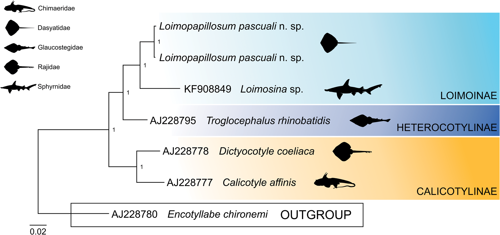
BI trees based on partial 28S and 18S rDNA show that Loimoidae (represented in this study by L. pascuali n. sp. and Loimosina sp.) is included within the Monocotylidae Taschenberg, 1879 (figs 2 and 3). Therefore, we propose the reestablishment of Loimoinae as a subfamily of Monocotylidae.
Phylogenetic tree based on 28S region for Loimopapillosum pascuali n. sp. (Monocotylidae: Loimoinae) inferred by Bayesian inference (BI) to show their relationships with other species of Monocotylidae. Numbers along branches indicate the bootstrap values obtained from the posterior probability of BI. GenBank accession numbers are shown, and the scale bar indicates the nucleotide mutations per site.

Phylogenetic tree based on 18S region for Loimopapillosum pascuali n. sp. (Monocotylidae: Loimoinae) inferred by Bayesian inference (BI) to show their relationships with other species of Monocotylidae. Numbers along branches indicate the bootstrap values obtained from the posterior probability of BI. GenBank accession numbers are shown, and the scale bar indicates the nucleotide mutations per site.

The phylogenetic tree based on the partial 28S rDNA shows two main clades (fig. 2). The first clade, with strong statistical support (0.97), contains species of Calicotylinae and Merizocotylinae. Calicotylinae, represented by four species of Calicotyle which infect carcharhiniforms, myliobatiforms and rajiforms – namely, C. urolophi, C. kroyeri, C. japonica and C. palombi – together with D. coeliaca, appear to form a sister clade of Merizocotylinae, represented by Empruthotrema dasyatidae and Empruthotrema quindecima, together with Merizocotyle urolophi and Merizocotyle australensis, showing good statistical support (1). The second clade, with statistical support of 1, is polytomic and comprises four subfamilies (Loimoinae, Heterocotylinae, Decacotylinae and Monocotylinae) with nine genera, divided into two main subclades, with a strong statistical support (0.98 and 1, respectively). The first subclade contains L. pascuali n. sp. (Loimoinae) and H. capricornensis (Heterocotylinae), both gill parasites of dasyatids, together with D. lymmae and Decacotyle tetrakordyle (Decacotylinae), both occurring in stingrays (aetobatids in the former species and dasyatids in the latter). Members of the Decacotylinae form a well-supported monophyletic group, but Heterocotylinae and Loimoinae appear as a non-monophyletic group. It is interesting to note the low statistical support (0.54) among these subfamilies. Another clade of Heterocotylinae, represented by species that infect rhinopristiforms (Glaucostegidae and Rhinidae) – namely, three Neoheterocotyle species and T. rhinobatidis, and Loimoinae with Loimosina sp. occurring in a hammerhead shark (Sphyrnidae) – presented strong statistical support (1). The last subclade contained members of the Monocotylinae infecting myliobatiform stingrays, represented by two Dendromonocotyle species, Dendromonocotyle octodiscus and Dendromonocotyle ardea, clustering together with Clemacotyle australis and two Monocotyle species, Monocotyle corali and Monocotyle multiparous, forming a well-supported monophyletic group.
The phylogenetic reconstruction based on the partial sequence of the 18S rDNA shows that our consensus sequence of L. pascuali n. sp. is grouped with Loimosina sp., forming a clade of the subfamily Loimoinae (fig. 3). This clade is the sister group of T. rhinobatidis, a member of the subfamily Heterocotylinae. A well-supported second clade is formed by Calicotylinae species, represented by Calicotyle affinis and D. coeliaca.
Discussion
Historically, representatives of Loimoinae Price, 1936 were included in Monocotylidae, order Capsaloidea Price, 1936. Bychowsky (Reference Bychowsky and Hargis1957) elevated Loimoinae to the family level, and it was included in Dactylogyridea Bychowsky, 1937. Lebedev (Reference Lebedev1988) recognized the family status but reassigned it to Monocotylidea Lebedev, 1988. According to Bychowsky (Reference Bychowsky and Hargis1957), Loimoidae include two genera, Loimos and Loimosina, which are characterized by having cephalic pits, an aseptate haptor with or without dorsal half-rounded sclerotized rib and a lobulated ovary not looping the right intestinal cecum. Hargis (Reference Hargis1955) proposed a third genus, Loimopapillosum, to accommodate L. dasyatis from the gills of the southern stingray, Hypanus americanus (Hildebrand & Schroeder, 1928) (type host) and the bluntnose stingray, Hypanus say (Lesueur, 1817) in Alligator Harbor, Florida, USA, and amended the diagnosis of the family. This species is characterized, besides other features, by having an ovary that loops the right intestinal cecum (also observed in the new species described here), a character considered apomorphic among the Monocotylidae (Chisholm et al., Reference Chisholm, Wheeler and Beverley-Burton1995), suggesting a phylogenetic relationship with the monocotylids.
Two Monocotylidae species have been described having an ovary with the same characteristic as Loimos and Loimosina (not looping the right intestinal caeca): Mehracotyle insolita Neifar, Euzet & Ben Hassine, 2002, a member of the Dasybatotreminae, from the gills of the Blackchin guitarfish Glaucostegus cemiculus (Geoffroy Saint-Hilaire, 1817) (Glaucostegidae) in Tunisia, and Anoplocotyloides papillatus (Doran, Reference Doran1953) Young, 1967, also a member of Dasybatotreminae, infecting the gills of rhinobatid fishes (Doran, Reference Doran1953; Bravo-Hollis, Reference Bravo-Hollis1969; Neifar et al., Reference Neifar, Euzet and Ben Hassine2002), exhibiting a phylogenetic relationship with Loimos and Loimosina species. This relationship is also supported by the presence of cephalic pits in M. insolita and A. papillatus and the aforementioned species (Boeger et al., Reference Boeger, Kritsky, Domingues and Bueno-Silva2014).
The phylogenetic position of Loimoidae was assessed by Boeger et al. (Reference Boeger, Kritsky, Domingues and Bueno-Silva2014) using molecular data, but only a single species, Loimosina sp., a monogenean from a hammerhead shark in the Neotropical region, was included in this analysis. As a result, they found that Loimoidae (represented by Loimosina sp.) fell within a major monocotylid clade including species of Heterocotylinae, Decacotylinae and Monocotylinae, suggesting that Loimoidae represents a subordinate taxon of the Monocotylidae. In their phylogenetic analyses, they found that Loimosina sp. formed a terminal clade with T. rhinobatidis and Neoheterocotyle rhinobatis.
In the present work, our partial sequences of the 28S rDNA of L. pascuali n. sp. from the Neotropical region are grouped in a clade with H. capricornensis, a member of the Heterocotylinae, from the Australian region. These two species are gill parasites of Myliobatiformes stingrays (Dasyatidae) and share some morphological characteristics – that is, an ovary that loops the right intestinal cecum, presence of head organs and small anchors with the deep roots larger and narrower than the superficial roots. This clade (L. pascuali + H. capricornensis) together with D. lymmae and D. tetrakordyle represents the sister group of a clade containing four Heterocotylinae species from the Australian region (three Neoheterocotyle species and T. rhinobatidis that infect Rhinopristiformes shovelnose rays), together with Loimosina sp. (Loimoinae). Apparently, the Heterocotylinae (here represented by H. capricornensis, three Neoheterocotyle species and T. rhinobatidis) and the Loimoinae (here represented by Loimosina sp. and L. pascuali n. sp.) are non-monophyletic groups, indicating that relationships are still widely misunderstood in these two subfamilies.
Apparently, Loimosina sp. and L. pascuali n. sp. (both in Loimoinae) are not phylogenetically related species. While it is true that both species exhibit strong morphological similarity, there are marked morphological differences between species of both genera that can support this hypothesis – that is, species of Loimopapillosum have an ovary that loops the right intestinal cecum (as opposed to an intercaecal ovary in Loimosina spp.), a compact ovary (as opposed to a lobulated ovary in species of Loimosina), a non-lobulated testis (as opposed to a lobulated testis in Loimosina spp.) and head organs (as opposed to cephalic pits in Loimosina spp.).
On the other hand, our sequences of the 18S rDNA of the new species are grouped with Loimosina sp. (KF908849) forming a clade of the subfamily Loimoinae. However, this result may be due to the low number of sequences used in the phylogenetic analysis. Probably adding more sequences of monocotylid species, mainly members of the Heterocotylinae and Decacotylinae, will enable the phylogenetic relationship among Loimosina sp. and L. pascuali n. sp. to be better resolved.
Finally, our results based on partial 28S and 18S rDNA confirm the rejection of Loimoidae Price, 1936 (represented in this study by L. pascuali n. sp. and Loimosina sp.) and its reincorporation as a subfamily of Monocotylidae, as previously suggested (Boeger et al., Reference Boeger, Kritsky, Domingues and Bueno-Silva2014). Unfortunately, there are no available genetic data for any species of Loimos to test its phylogenetic relationship with Loimosina sp. and L. pascuali n. sp.
Loimopapillosum now includes two species that parasitize marine stingrays: L. dasyatis, the type species described from the gills of the two different hosts, the southern stingray, H. americanus (type host) and the bluntnose stingray, H. say in the Gulf of Mexico (USA). Loimopapillosum pascuali n. sp., with a funnel-shaped MCO with asymmetric base, is the first species to be described from the Pacific Ocean. Therefore, L. dasyatis can be readily differentiated from L. pascuali n. sp. not only by morphological characters but also by the geographic distribution of its hosts. Hypanus dipterurus, host of L. pascuali n. sp., is typically a subtropical species with a distribution range restricted to the Eastern Pacific (from southern California, USA, to Peru), while H. americanus and H. say, hosts of L. dasyatis, are endemic to the western Atlantic (Dunn et al., Reference Dunn, McEachran and Honeycutt2003; Nelson, Reference Nelson2006; Last et al., Reference Last, Naylor and Manjaji-Matsumoto2016). Unfortunately, molecular data for L. dasyatis are not available in GenBank, which could help us to elucidate the phylogenetic relationships among these two taxa.
Forty-two species of monogeneans have been reported infecting marine chondrichthyans from South America (Santos et al., Reference Santos, Santos, Cunha and Chisholm2012; Irigoitia et al., Reference Irigoitia, Cantatore, Delpiani, Incorvaia, Lanfranchi and Timi2014, Reference Irigoitia, Chisholm and Timi2016, Reference Irigoitia, Braicovich, Rossin, Canel, Levy, Farber and Timi2019; Kohn et al., Reference Kohn, Justo, Cohen, Eiras, Velloso and Pereira2016; Luque et al., Reference Luque, Cruces, Chero, Paschoal, Alves, Da Silva, Sánchez and Iannacone2016; Chero et al., Reference Chero, Cruces, Sáez, Camargo, Santos and Luque2018a, Reference Chero, Cruces, Sáez and Luqueb, Reference Chero, Cruces, Sáez, Camargo, Santos and Luque2019, Reference Chero, Cruces, Sáez, Santos and Luque2020; Ñacari et al., Reference Ñacari, Sepulveda, Escribano and Oliva2018, Reference Ñacari, Sepulveda, Escribano and Oliva2019, Reference Ñacari, Sepulveda, Droguet, Escribano and Oliva2020). Of these, 24 species belong to the Monocotylidae and only three species belong to the Loimoinae: Loimos scitulus Burhnheim, 1972 from the Atlantic sharpnose shark, Rhizoprionodon terraenovae (Richardson, 1836) in Brazil; Loimos scoliodoni (Manter, 1938) from the sharptooth smooth-hound, Mustelus dorsalis Gill, 1864 in Peru; and Loimosina sp. from a hammerhead shark in Brazil. Loimopapillosum pascuali n. sp. is added to the 42 previously known species infecting marine chondrichthyans from South America and represents the second species of Loimoinae infecting chondrichthyans from Peru.
Acknowledgements
Our thanks to Anna Phillips (USNM) for allowing us access to specimens under their care. We also thank Leslie A. Chisholm (South Australian Museum) and Luis García-Prieto (Nacional Collection of Helminths, Institute of Biology, National Autonomous University of Mexico) for providing literature on monocotylids. Our thanks also to Ivette Cuellar, Eva G. Huancachoque and Cynthia E. Rodríguez, who helped in the collection of fishes, all from the National University Federico Villarreal (UNFV).
Financial support
Jhon D. Chero and Celso L. Cruces were supported by a student fellowship from the Coordenação de Aperfeiçoamento de Pessoal do Ensino Superior, Brazil (CAPES) (Finance Code 001). José L. Luque and Cláudia P. Santos were supported by Researcher fellowships from the Conselho Nacional de Desenvolvimento Científico e Tecnológico, Brazil (CNPq).
Conflicts of interest
None.
Ethical standards
The authors declare that all applicable international, national and/or institutional guidelines for the care and use of animals were followed.