Introduction
We present a patient who presented at our emergency department after self-inflicted penetrating laryngeal trauma. After initial evaluation, it became clear there was severe destruction of the thyroid cartilage. The patient was brought to the operating theatre for tracheostomy and laryngeal reconstruction. Because of the extent of the injury, internal fixation was used for laryngeal reconstruction.
Ethical considerations
We received written informed consent from our patient for the publication of this case report.
Case report
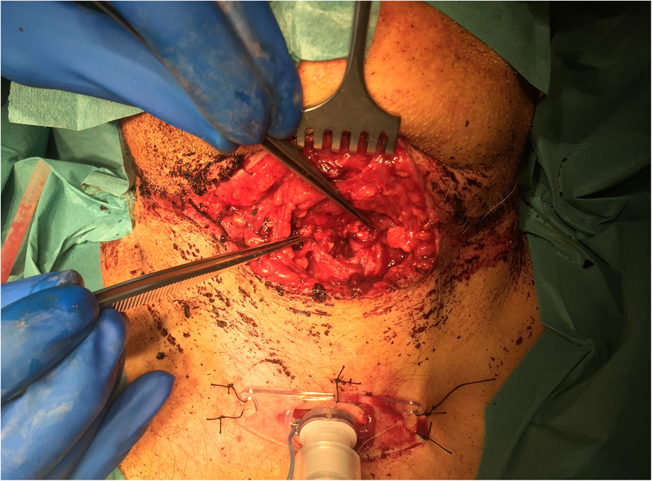
A 50-year-old male patient with a past psychiatric history of schizophrenia was brought to our emergency department by ambulance after self-inflicted penetrating laryngeal trauma by the use of scissors. He was conscious and alert upon arrival. On the midline of thyroid level, there was a horizontal 10 cm frayed laceration, with involvement of the thyroid cartilage (Figure 1). There was normal breathing without signs of stridor, with airflow both through the mouth and through the neck laceration. There was evident breathiness of the voice, but otherwise no dyspnoea or tachypnoea, and his vital signs were normal. After initial evaluation according to the Advanced Trauma Life Support® protocol, there was no suspicion of other injuries.
External view of the larynx. The forceps are holding the left and right lamina of the thyroid cartilage.

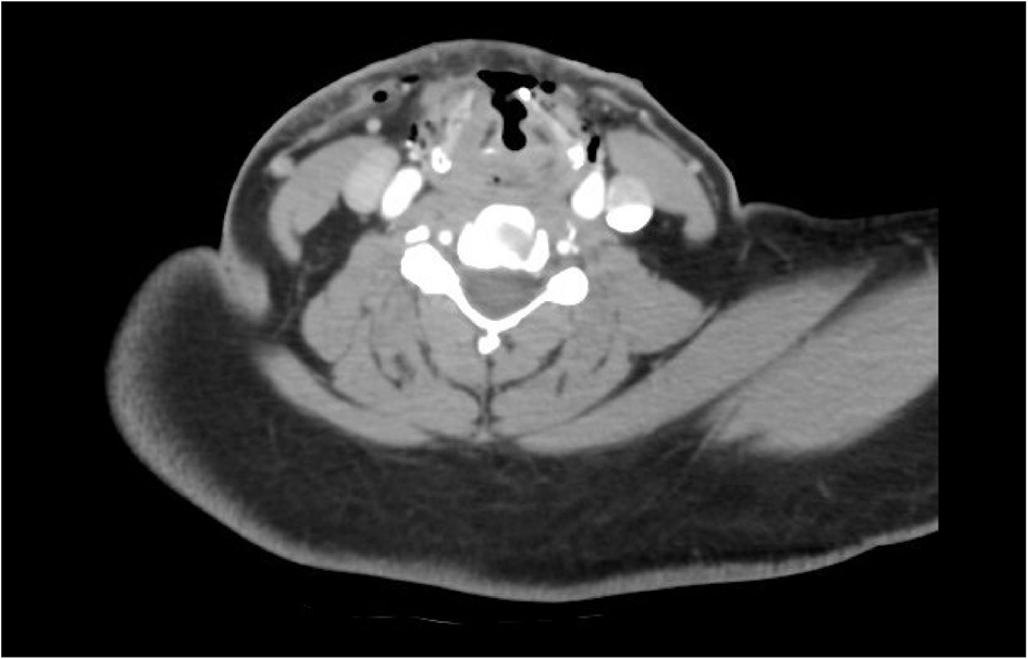
On flexible laryngoscopy, there was an adequate breathing lumen, with discrete swelling at the supraglottic level and of the vocal folds. Mobility of the vocal folds could not be properly assessed. In the absence of an acute compromised airway, a computed tomography (CT) angiography of the neck was performed before going to the operating theatre. This revealed a severe penetrating injury running through the thyroid cartilage above the level of the vocal folds. There was a loose fragment at the aryepiglottic fold on the right side. The cervical vasculature was intact. The tract of the recurrent laryngeal nerve appeared not to be involved (Figure 2).
Pre-operative computed tomography angiography of the neck, revealing the extent of injury, laryngeal swelling, subcutaneous emphysema and a fractured element at the right aryepiglottic fold.

Orotracheal intubation was performed without difficulty, followed by a tracheostomy through a separate incision caudal of the injury, at the level of the fourth tracheal ring. Inspection of the neck injury revealed multiple lesions, without continuation to the vascular compartment. The thyroid cartilage at the level of the false vocal cords was fractured and elements were missing, so there was a direct view into the larynx with identification of the vocal folds and epiglottis. The injury did not extend completely laterally or posteriorly of the larynx.
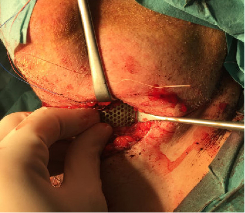
Because of the lack of structural support associated with the destruction of the anterior part of the thyroid cartilage, there was collapse of the false vocal cords. In order to restore airway patency, the decision was made to reconstruct the thyroid cartilage with a titanium mesh. Firstly, holes were drilled through the intact part of the thyroid caudally, through which the false vocal cords were sutured. The epiglottis was sutured through the anterior part of the thyroid. Secondly, the titanium mesh plate was used and fixated to the thyroid by three screws on both sides (Figure 3). The sutures of the false vocal cords and epiglottis were fixated to the mesh plate. Afterwards, an endolaryngeal stent was placed because of mucosal involvement. For the stent, an endotracheal tube size 8 was cut to the preferred size, and correct placement was confirmed through laryngoscopy. One suture was used to fixate the stent to the mesh plate. The wound was closed in two layers. The patient was transferred to the intensive care unit and could rapidly be weaned.
Pre-operative view of the mesh plate in situ.

Eleven days after surgery, the endolaryngeal stent was removed under general anaesthesia, and the patient was able to ingest thin fluid without aspiration. On day 14, the cannula was switched to an uncuffed cannula, and capping of the cannula could be commenced. The patient's voice was slightly breathy. On day 16, decannulation was successful and his oral intake was normal.
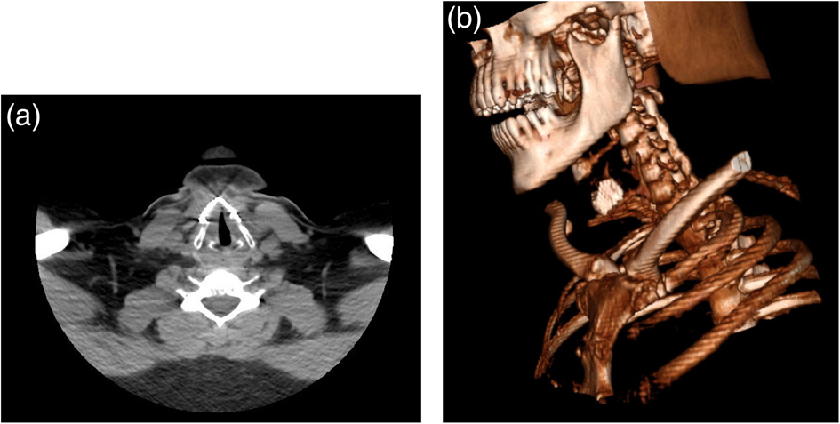
A CT scan of the neck was performed three weeks after surgery (Figure 4a and b). The mesh adequately restored the skeletal framework. On flexible laryngoscopy, there was supraglottic swelling and normal adduction of the vocal folds, which confirmed that the recurrent nerve was intact. Most importantly, the patient was free of complaints. He had a normal intake, and a subjectively good voice quality that was maintained thirty-six months post-surgery.
(a) Axial computed tomography scan and (b) three-dimensional reconstruction of the neck three weeks after surgery, revealing some intralaryngeal swelling without signs of infection.

Discussion
Acute laryngeal trauma may be obvious and severe, as in the patient presented here, but can be occult such that there is no physical evidence of the injury at initial presentation. Acute laryngeal trauma accounts for less than 1 per cent of all cases seen at major trauma centres. It may present with a variety of symptoms, such as hoarseness, pain or tenderness, haemoptysis, dysphagia, subcutaneous emphysema, impaired respiration, and hematoma.Reference Schaefer1
Predating World War II, there were little data on how to manage laryngeal trauma because of the low incidence and high fatality rate. Experience regarding the classification and treatment possibilities saw an increase after the 1950s.Reference Lynch2 Early recognition and treatment of laryngeal trauma is vital to restore proper laryngeal function, in order to prevent laryngeal stenosis and tracheostomy dependency, and achieve good voice quality and swallow function.Reference Maran, Murray, Stell and Tucker3
The initial management of acute laryngeal trauma is the establishment of a secure airway. In our patient, there was no acute respiratory distress, which allowed thorough evaluation of the airway. We opted for oral intubation as there was good visualisation of the airway on flexible laryngoscopy. Intubation via the wound was not undertaken to avoid further traumatising the already damaged larynx. Intubation through the open wound would otherwise have been a viable option.Reference Hwang, McGinness, Choroomi and Jacobson4,Reference Moonsamy, Sachdeva and Morse5 A CT scan is required to further assess the extent of the injury prior to treatment.Reference Becker, Leuchter, Platon, Becker, Dulguerov and Varoquaux6 We also performed angiography to exclude vessel involvement.
Laryngeal mucosa involvement can lead to formation of synechia and adhesions, which may alter the shape of the glottis and decrease laryngeal lumen. It has been established that endolaryngeal stent placement helps to counteract these complications.Reference Schaefer7 The availability and modifiable nature of an endotracheal tube made it an easy and effective option to use as a stent in our patient. One suture was used for easy post-operative removal in the operating theatre.
A mesh plate was used to restore the laryngeal skeletal framework. The plate could be modified to the preferred angle, thereby serving as a replacement for the missing skeletal elements and restoring the proper anatomical position of the lamina. Furthermore, the plate served as the base to fixate the epiglottis and false vocal cords.
The use of biodegradable and metal plates has been described previously.Reference Schaefer7 Internal fixation by either system leads to cartilaginous union of the fragments, whereas fixation by suture wire alone tends to heal by fibrous union, and may not provide sufficient support to tolerate the strength exerted by the extralaryngeal and intralaryngeal musculature.Reference de Mello-Filho and Carrau8
• Internal and external laryngeal reconstruction with a mesh plate has good functional outcomes, even when a large part of the larynx is missing
• Titanium mesh plates can be used as an alternative to miniplates to reconstruct the external part of the larynx
• The internal parts of the larynx can easily be fixed to the mesh plate for greater structural support
• One suture was used to fix the endolaryngeal stent, to facilitate easy removal and avoid manipulation
• Use of an endotracheal tube as an endolaryngeal stent is recommended, as it is widely available and easy to modify when there is risk of adhesion formation
Mesh plates have the advantage of being able to be customised to various shapes and sizes.Reference Sato, Nito, Ueha, Goto and Yamasoba9 However, the soft nature of cartilage could potentially result in insufficient support for fixation materials. It has been suggested that the use of drill holes smaller than screw sizes can increase the plate-tissue friction for optimisation of support;Reference de Mello-Filho and Carrau8,Reference Plant and Pinczower10 however, we did not see mesh plate displacement or associated discomfort in our patient.
Conclusion
Immediate surgical reconstruction of the larynx in penetrating laryngeal trauma can lead to satisfactory results. A longer follow-up duration will be needed to determine whether this reconstruction stands the test of time or whether other means of reconstruction will be needed.
Competing interests
None declared