Introduction
Primates, like other mammals, are subject to selection pressures (e.g. sperm competition, cryptic post-copulatory female choice, sexual coercion, climatological factors; see review in Martinez and Garcia, Reference Martinez and Garcia2020) resulting in inter-specific and intra-specific variability in sperm shapes. Analyzing this variability with morphological sperm measurements is becoming increasingly relevant, especially in the context of animal health and fertility monitoring (García-Vázquez et al., Reference García-Vázquez, Gadea, Matás and Holt2016; Oliveira et al., Reference Oliveira, Santos, Leão, Brito, Lima, Sampaio and Domingues2016; Piña-Aguilar et al., Reference Piña-Aguilar, López-Saucedo, Ruiz-Galaz, de Jesús Barroso-Padilla, Gallegos-Rivas, González-Ortega and Gutiérrez-Gutiérrez2016). Indeed, the worldwide decline of many primate populations (Estrada et al., Reference Estrada, Garber, Rylands, Roos, Fernandez-Duque and Di Fiore2017) could lead to an increase in the use of medically assisted reproduction techniques (ART) to conserve the genetic material, detect inbreeding and potentially address primate fertility issues (Andrabi and Maxwell, Reference Andrabi and Maxwell2007) in the framework of ex situ conservation. For instance, evaluation of sperm morphology makes it possible to determine an individual sperm quality when standards exist, as morphology is closely linked to successful fertilization (García-Vázquez et al., Reference García-Vázquez, Gadea, Matás and Holt2016). Although most studies on primate semen parameters are primarily conducted on laboratory model species (de Villiers, Reference de Villiers2018), there is a growing collection of published literature for endangered and critically endangered species (Martin et al., Reference Martin, Gould and Warner1975; Thomson et al., Reference Thomson, Iliff-Sizemore, Gliessman and Wolf1992; Robson et al., Reference Robson, Rouse and Pettigrew1997; Steinberg et al., Reference Steinberg, Nieves, Ascunce, Palermo and Mudry2009; Nakazato et al., Reference Nakazato, Yoshizawa, Isobe, Kusakabe, Kuraishi, Hattori and Kai2015; Sampaio et al., Reference Sampaio, Oliveira, Leão, Caldas-Bussiere, Queiroz, Paim and Domingues2017; Leão et al., Reference Leão, Sampaio, Sousa, Moura, Oskam, Santos and Domingues2020).
Here we provide, for the first time, sperm morphometric and morphologic data from De Brazza’s monkey (Cercopithecus neglectus), moustached monkey (Cercopithecus cephus), Guinea baboon (Papio papio), and the critically endangered Roloway monkey (Cercopithecus roloway), as well as supplementary data for L’Hoest’s monkey (Allochrocebus lhoesti), mandrill (Mandrillus sphinx) and olive baboon (Papio anubis). These data are highly valuable, especially for the Roloway monkey, which is listed as one of the 25 most endangered primate species in the world (Koné et al., Reference Koné, Oates, Dempsey, Gonedelé, McGraw and Wiafe2019).
Materials and methods
Animals
All samples were obtained from French zoos, EAZA (European Association of Zoos and Aquaria) members, assuring the best veterinary care practices for the animals. Testes from A. lhoesti, C. roloway, C. cephus and C. neglectus were collected from adult males of proven fertility (registered offspring) who died from accident or disease not affecting spermatogenesis. Testes from M. sphinx, P. anubis and P. papio were collected from sexually mature males at the time of castration. Testicles were sent to the laboratory for sperm cryoconservation and creation of a primate sperm biobank. Morphological analyses were carried out as part of the sperm evaluation before freezing.
Sperm collection and preparation
Testis were kept at 4°C and processed within 48 h after castration or death discovery. Sperm extraction was carried out by dissecting the tail of the epididymis using needles in a Petri dish with 2 ml Sperm Preparation Medium (SPM, Origio). Spermatozoa were allowed to swim for 10 min at 37°C, washed twice in SPM (by centrifugation at 500 g, 5 min), displayed over a slide, dried at room temperature, and then fixed in ethanol 70° for Harris–Schorr staining. Simple and easy to perform, this staining gives good results in routine human sperm evaluation (World Health Organization, 2010).
Sperm morphometric analysis
All cells measurements were realized on a Nikon Eclipse 80i microscope equipped with a Nikon DS-Ri1 camera with NIS-Elements D (version 3.1.) software. At least 100 cells were measured for each individual. Measured parameters were head length (L), head width (W), head perimeter (P), head area (A), midpiece length (MP) and total flagellum length (Flag). Calculated parameters were head volume (V = 4/3π(L/2)2(W/2)), head ellipticity (ξ = L/W), head elongation (Σ = (L − W)/(L + W)), head roughness (Ro = 4π(A/P²)) and head regularity (Re = π((L*W)/(4*A)). Given the small sample size within each species (only one individual), we were unable to statistically investigate differences in morphometric measurements between species.
Sperm morphologic analysis
Morphology was assessed by a trained biologist (GM) with a counting of at least 400 spermatozoa per slide using a magnification of ×1000. Analyses were performed according to World Health Organization (WHO) recommendations for human sperm analysis based on a modified Kruger classification (Cooper et al., Reference Cooper, Noonan, Von Eckardstein, Auger, Baker, Behre and Vogelsong2010).
Results
Morphometry
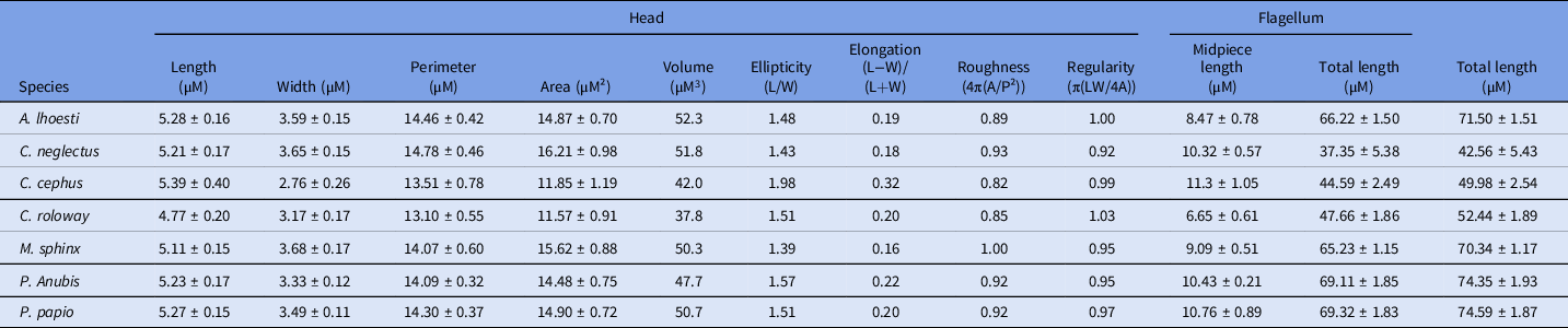
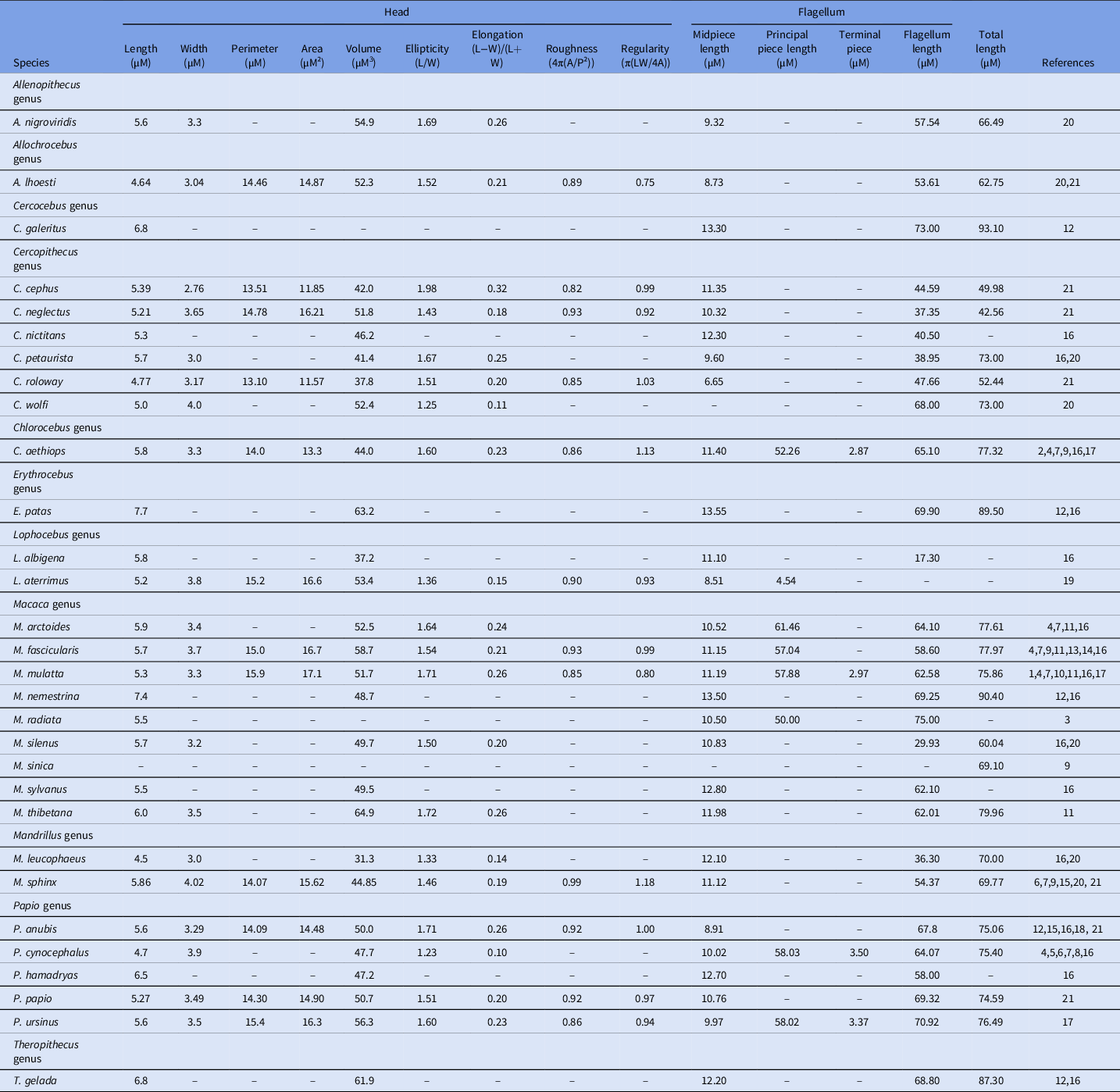
Our morphometric data are presented in Table 1 and alongside the literature in Table 2 with means (±SD) calculated for each genus. All individuals displayed an oval-shaped sperm head, with C. cephus displaying the most elongated and narrowest head within all known spermatozoa species (head width 2.76 ± 0.26, ellipticity 1.98 and elongation 0.32). Regarding total sperm length, P. papio, P. anubis, M. sphinx and A. lhoesti were measured at around 70 µM whereas C. neglectus, C. cephus and C. roloway lengths were around 50 µM (see Table 2 for further details). Moreover, C. roloway spermatozoa were characterized by a notable morphological particularity, i.e. the shortest intermediate piece among Cercopithecinae (6.65 ± 0.61 µM).
Sperm morphometric data in our sample of seven Cercopithecinae species

Sperm morphometric data of Cercopithecinae

All measurements are presented uncritically and as mean when multiple sources.
References: 1Mastroianni and Manson, Reference Mastroianni and Manson1963; 2Bedford, Reference Bedford1967; 3Flećhon and Hafez, Reference Flećhon and Hafez1975; 4Martin et al., Reference Martin, Gould and Warner1975; 5Flećhon et al., Reference Flećhon, Kraemer and Hafez1976; 6Gould and Martin, Reference Gould and Martin1978; 7Gould, Reference Gould1980; 8Carrick and Hughes, Reference Carrick and Hughes1982; 9Cummins and Woodall, Reference Cummins and Woodall1985; 10Bedford and Hoskins, Reference Bedford, Hoskins and Lamming1990;11Shang et al., Reference Shang, Ji, Yang, Chen and Zou1993; 12Gage, Reference Gage1998; 13Gago et al., Reference Gago, Perez-Sanchez, Yeung, Tablado, Cooper and Soler1998; 14Gago et al., Reference Gago, Pérez-Sánchez, Yeung, Tablado, Cooper and Soler1999; 15Cseh et al., Reference Cseh, Chan, Corselli and Bailey2000; 16Anderson et al., Reference Anderson, Nyholt and Dixson2005; 172011 Maree, Reference Maree2011; 18Nyachieo et al., Reference Nyachieo, Spiessens, Chai, Kiulia, Mwenda and D’Hooghe2012; 19Gadea et al., Reference Gadea, Toledano-Díaz, Navarro-Serna, Jiménez-Movilla, Soriano, Matás and Santiago-Moreno2019; 20San Diego Zoo, 2019; 21This study.
Morphology
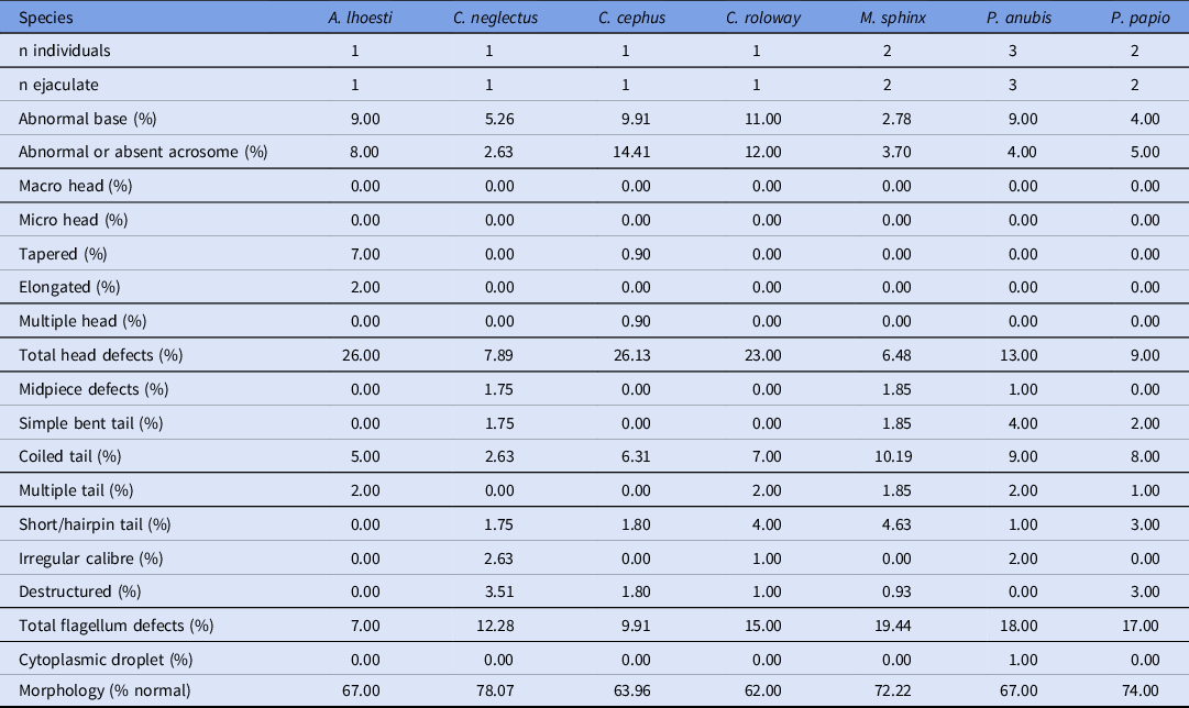
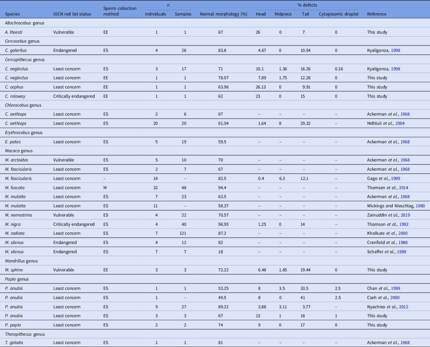
Our morphologic data are presented in Table 3 and alongside the literature in Table 4. The proportion of morphologically normal spermatozoa in the ejaculate is homogeneous for the seven species examined (mean ± SD of 69.18 ± 5.77%, range 62.00–78.07%) and across the literature (72.19 ± 12.17%, range 50–96.95%). Conversely, the localization of the morphological abnormalities is heterogeneous from one species to another and mainly concentrated on the tail (15.70 ± 6.52%, range 7–29.32%) or head (10.53 ± 9.83%, range 0.4–26.13%) but few on the midpiece (1.78 ± 2.80%, range 0–8%). At the species level, the anomalies are mostly present on the tail, with the exception of A. lhoesti, C. cephus and C. roloway, for which mostly present were head anomalies (26.00, 26.13 and 23.00% respectively). The presence of cytoplasmic droplets should be noted for P. anubis and C. neglectus but in very low proportion (2.00 and 0.08%, respectively).
Sperm morphologic data

Sperm morphologic data of Cercopithecinae

All measurements are presented uncritically and as mean when multiple sources. EE: epididymal extraction, ES: electrostimulation, M: masturbation.
Discussion
This study contributes to increase our knowledge of primate sperm morphometry and morphology by providing original data for four primate species scarcely studied and supplementary data for three other species. Our data are consistent with the general morphometric and morphologic proportions reported in the literature for primates (Tables 2 and 4).
All the species measured here had an oval-shaped sperm head, like most primates, the only exceptions being Lorisidae and Lemuroidea taxa (Gould and Martin, Reference Gould and Martin1978). It should be noted that C. cephus displays a specific head pattern with a narrow head. Some species of the Cercopithecus genus tend to have shorter spermatozoa with an average length of 58.20 ± 13.99 µM (Macaca: 75.85 ± 9.41; Papio: 69.89 ± 0.33), with C. neglectus, C. cephus and C. roloway being the shortest representatives of all Cercopithecinae. Yet, due to the small sample sizes inherent in such studies, we report only suggestive trends that may illuminate further studies with more individuals in order to determine if our current findings can be generalized to these different species. Therefore, these morphometric differences between species deserve more attention and future studies combining sperm morphometrics on additional specimens with data on mating systems and reproductive ecology are needed.
This study brings to 48 the number of primate species for which we have sperm morphological data. Our normal-form sperm rates (mean 69.18%, range 62.00–78.07%) are consistent with those observed in the literature for Cercopithecinae (mean 73.44%, range 49.50–96.95%). Although preliminary, these data could help to assess the reproductive potential of individuals, as an abnormal decrease in the number of normal-form sperm is often associated with infertility (Gould et al., Reference Gould, Young, Smithwick and Phythyon1993; Piña-Aguilar et al., Reference Piña-Aguilar, López-Saucedo, Ruiz-Galaz, de Jesús Barroso-Padilla, Gallegos-Rivas, González-Ortega and Gutiérrez-Gutiérrez2016) when it occurs during a reproductive period (Seier et al., Reference Seier, Conradie, Oettle and Fincham1993; Hernández-López et al., Reference Hernández-López, Parra, Cerda-Molina, Pérez-Bolaños, Sánchez and Mondragón-Ceballos2002).
Interestingly, the distribution of morphological abnormalities appears heterogeneous, with some species mainly presenting head anomalies (A. lhoesti, C. cephus, C. roloway) and others flagellum anomalies (C. neglectus, M. sphinx, P. anubis, P. papio) with sometimes strong ratios (3.7 times more head abnormalities than flagellum for A. lhoesti and 3 times more flagellum than head abnormalities for M. sphinx). The causes of this differential positioning are not known and non-human primate spermocytograms in the literature are rarely detailed, making further analysis impossible without additional data collection.
Sperm morphological abnormalities increasing with the inbreeding degree (Gomendio et al., Reference Gomendio, Cassinello and Roldan2000), the data on sperm parameters presented here can also be of major importance for ART as valuable indicators of inbreeding depression. The increase in matings between genetically related individuals due to shrinking habitats and declining populations in the wild or space and management constrains in captivity can lead to a reduction in genetic diversity. The negative impact of inbreeding on reproductive fitness is also well known and the avoidance of inbreeding is one of the priorities in the conservation and captive breeding of endangered species (Hedrick and Kalinowski, Reference Hedrick and Kalinowski2000). Therefore, assisted breeding techniques may be useful in the future to ensure assisted selective genetic propagation, avoid inbreeding and thus prevent the decline of the general reproductive ability of the captive species (Pukazhenthi et al., Reference Pukazhenthi, Comizzoli, Travis and Wildt2005).
In conclusion, all data on primate reproduction are critical in the current context of global population declines, especially for endangered species. Beyond the improvement of our general knowledge on primate sperm, we provide here essential data for potential future implementation of ex situ conservation actions and monitoring of animal health and fertility.
Acknowledgements
The authors would like to deeply thank Marlène Gandula, Charlotte Guyot and Jean-Pascal Hograindleur for their technical assistance.
Ethical approval
Not applicable.
Financial support
None.
Conflict of interest
The authors have declared that no competing interests exist.
Author contributions
Study design: GM, CA, RL; sample collection and processing: GM, CF-B, SL, AL, AL, BM, TP, BQ, SB, CA, CC; data analysis and interpretation: GM, CG, RL; manuscript drafting: GM, CG, RL. All authors read, critically revised and approved the final manuscript.