


Significant outcomes
Some schizophrenia cases show cognitive decline over the course of illness without neuropathological changes matching the clinical symptoms.
Limitations
Our evaluation of only three cases may have reduced the power of the study, and the findings may not be generalisable to other populations.
Introduction
Schizophrenia is a major mental illness affecting approximately 1% of the population, and its etiology remains unknown. Besides, the biological background of the various symptoms associated with schizophrenia remains unclear, and the environmental background factors influencing the diversity of symptoms are also unknown. Schizophrenic patients often demonstrate a cognitive decline and/or dementia symptoms as they age over their long disease course. A cohort study reported that patients with schizophrenia were at an increased risk of developing dementia (Cai & Huang, Reference Cai and Huang2018). However, it was conversely reported that the prevalence of Alzheimer’s disease (AD) in schizophrenia did not differ markedly from the normal control group neuropathologically (Arnold & Trojanowski, Reference Arnold and Trojanowski1996b; Niizato et al., Reference Niizato, Arai, Kuroki, Kase, Iritani and Ikeda1998). It is difficult to determine whether the cognitive decline is due to the disease itself or a neurodegenerative disease based on the clinical symptoms alone.
The overlap between the so-called negative symptoms of schizophrenia and the symptoms of dementia can also be difficult to distinguish (van Os & Kapur, Reference van Os and Kapur2009; Lang et al., Reference Lang, Kosters, Lang, Becker and Jager2013), and more precise neuroimaging or neuropathological investigations may ultimately be required. In neuroimaging research on schizophrenia, morphological changes (volume reduction) in the brain over time have been reported with reproducibility, so examinations of the brain histopathology are indispensable for elucidation (Van Haren et al., Reference Van Haren, Cahn, Hulshoff Pol and Kahn2013; Veijola et al., Reference Veijola, Guo, Moilanen, Jaaskelainen, Miettunen, Kyllonen, Haapea, Huhtaniska, Alaraisanen, Maki, Kiviniemi, Nikkinen, Starck, Remes, Tanskanen, Tervonen, Wink, Kehagia, Suckling, Kobayashi, Barnett, Barnes, Koponen, Jones, Isohanni and Murray2014; Guo et al., Reference Guo, Huhtaniska, Miettunen, Jaaskelainen, Kiviniemi, Nikkinen, Moilanen, Haapea, Maki, Jones, Veijola, Isohanni and Murray2015).
Schizophrenia was originally proposed as a disease entity by Kraepelin around 100 years ago and initially described as ‘Dementia Praecox’ (early-onset dementia) (Hoenig, Reference Hoenig1983). In German psychiatry at that time, the pathophysiology of neuropsychiatric disorders was mainly explored using neuropathological approaches. Through such research activities, a number of disease entities, such as AD, were established. However, concerning so-called endogenous mental illnesses, no significant findings were able to be detected by neuropathological techniques (Plum, Reference Plum1972). Despite subsequent biological research efforts, the pathogenesis of schizophrenia has yet to be fully elucidated. Its etiology is thought to be multifactorial, including genetic, biological and environmental factors. The neurodevelopmental hypothesis has been proposed as an etiology of this disease (Murray et al., Reference Murray, Bhavsar, Tripoli and Howes2017), and it is believed that some central nervous system (CNS) vulnerability is responsible (Ladea & Prelipceanu, Reference Ladea and Prelipceanu2009).
On the other hands, the cognitive dysfunction aspect of schizophrenia has been considered an important target site for the treatment and prognosis recently (Kahn & Keefe, Reference Kahn and Keefe2013; Vita et al., Reference Vita, Deste, De Peri, Barlati, Poli, Cesana and Sacchetti2013). Elucidating the pathogenesis of this disease will require examining the neuropathological background of the dementia symptoms observed in the long-term course of schizophrenia, in order to consider the pathogenesis of cognitive impairment and the prognosis of the disease in later life. This is because most patients suffer some kind of deficit in their higher mental function over their lifetime.
Aim of the study
We conducted a neuropathological examination of three schizophrenia patients affected in early adulthood who developed severe cognitive decline and dementia symptoms late in life and investigated the background of the brain pathology of the dementia symptoms in these older schizophrenia patients.
Materials and methods
Three schizophrenic patients who met the diagnostic criteria for Diagnostic and Statistical Manual of Mental Disorders 5th edition (DSM-5) (American Psychiatric Association, 2013) had been diagnosed before 30 years old and developed severe cognitive decline or dementia syndrome later in life were enrolled. Detailed medical histories of three cases were retrospectively investigated from medical records.
Following their passing, an autopsy was conducted, and their brains were immediately removed and fixed in formalin for the appropriate periods. The samples were embedded in paraffin and cut into 8-μm-thick sections and then subjected to Hematoxylin–Eosin, Gallyas–Braak (GB), Klüver–Barrera (KB) and immunohistochemical staining with the following respective agents: 1) phosphorylated tau (AT8; monoclonal, at 1:1000; Innogenetics, Ghent, Belgium), 2) β-amyloid (monoclonal, at 1:100; DAKO, Glostrup, Denmark), 3) phosphorylated 43-kDa TAR DNA-binding protein (pTDP-43 ser409/410; polyclonal, at 1:2500; CosmoBio, Tokyo, Japan) and 4) phosphorylated α-synuclein (monoclonal, at 1:3000; Wako Pure Chemical Industries, Osaka, Japan). We observed these stained specimens under a microscope.
We evaluated the frequency of neurofibrillary tangles (NFTs) and senile plaques (SPs) according to the Braak stage (Braak & Braak, Reference Braak and Braak1991; Braak et al., Reference Braak, Alafuzoff, Arzberger, Kretzschmar and Del Tredici2006), Consortium to Establish a Registry for AD (CERAD) reference (Mirra et al., Reference Mirra, Heyman, McKeel, Sumi, Crain, Brownlee, Vogel, Hughes, van Belle and Berg1991) and Thal amyloid deposition stage classification (Thal et al., Reference Thal, Rub, Orantes and Braak2002). The appearance and frequency of Lewy bodies (LBs) were evaluated according to the pathological diagnostic criteria described in the third report of the dementia with Lewy bodies (DLB) consortium and Braak Parkinson’s disease (PD) stage (Braak et al., Reference Braak, Del Tredici, Rub, de Vos, Jansen Steur and Braak2003; McKeith et al., Reference McKeith, Dickson, Lowe, Emre, O’Brien, Feldman, Cummings, Duda, Lippa, Perry, Aarsland, Arai, Ballard, Boeve, Burn, Costa, Del Ser, Dubois, Galasko, Gauthier, Goetz, Gomez-Tortosa, Halliday, Hansen, Hardy, Iwatsubo, Kalaria, Kaufer, Kenny, Korczyn, Kosaka, Lee, Lees, Litvan, Londos, Lopez, Minoshima, Mizuno, Molina, Mukaetova-Ladinska, Pasquier, Perry, Schulz, Trojanowski and Yamada2005), and the appearance of argyrophilic grains were in accordance with Saito’s staging (Saito et al., Reference Saito, Ruberu, Sawabe, Arai, Tanaka, Kakuta, Yamanouchi and Murayama2004), throughout clinicopathological conferences (CPC) with multiple neuropathologists and psychiatrists.
Results
Case 1
A 75-year-old man at death had a 45-year history of schizophrenia.
After graduating from junior high school, the patient worked at construction companies. He had an identical twin brother who was also diagnosed with schizophrenia.
At 30 years old, he was brought under police protection following a suicide attempt, and after returning home, he became anxious and delusional and exhibited strange behaviour, such as monologuing and laughing, so he was admitted to a psychiatric hospital. He was subsequently repeatedly hospitalised and released, and thereafter underwent long periods of hospitalisation in later life. During his hospitalisation, he showed remarkable symptoms/behaviours, such as hallucinations, epilepsy, sensory hallucinations (insects erupting), insomnia, violence, self-injury and drug refusal.
In his late 60s, auditory hallucinations only infrequently occurred, but active visual hallucinations were often observed, and he became angry and violent toward other patients, resorting to self-injury and occasionally needing placement in a protected room. He also showed parkinsonism, including aspiration and propulsion and gait disorder at this time. Since around 72 years old, he showed decreased speech as well as disorientation and incontinence. His unstable walking caused his body to tilt, and he was unable to maintain his posture, resulting in increased falls. At 74 years old, his cognitive function was further reduced, and his conversations became superficial and simple. His contact with other patients also decreased, and he no longer complained of hallucinations of delusions. At 75 years old, he died of repeated episodes of aspiration pneumonia.
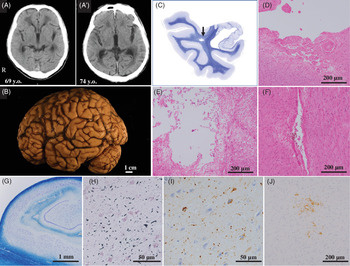
Head computed tomography (CT) (taken three times: 69 [Fig. 1(A)], 73 and 74 years old [Fig. 1(A)]) indicated slight cortex atrophy and enlargement of the ventricle chronologically.
Clinicopathological findings of Case 1. A, A’. Brain CT at 6 years before death (A) and 1 year before death (A’). Slight cortical atrophy progressed over time but not significantly. B. The general appearance of the left cerebral hemisphere showed unremarkable changes. C. Loupe image of the temporal region, including anterior hippocampal formation of Klüver–Barrera (KB)-stained species. Lacunar infarction was seen in the white matter. D. Microscopic observation of the arrow in (C). Cystic encephalomalacia was observed. E. Small infarction of the pons. Many macrophages were accumulated. F. Old cerebral hemorrhaging in the putamen. G. Microscopic observation of the hippocampal area of the KB-stained species. Pyramidal neurons were preserved. H. Argyrophilic grains were observed in the ambient gyrus. I. Microscopic observation of the ambient gyrus of the AT-8-immunostained species. Tau-positive neurofibrillary tangles and neurites were observed. J. Microscopic observation of the striatal area of the occipital lobe of the Aβ-immunostained species. Diffuse-type senile plaques were observed. KB staining (C,G), HE staining (D–F), GB staining (H), AT8 immunostaining (I), Aβ11-28 immunostaining (J).

The brain weighed 1356 g at the autopsy [Fig. 1(B)]. On a macroscopic examination, slight cleft dilations of the frontal and temporal lobes, stiffness of the basilar artery and scattered soft spots in the white matter and the basal ganglia were detected. The neurons of the substantia nigra, locus coeruleus, cerebellum and nucleus of the cranial nerve responsible for the motor function were preserved. Arteriosclerosis and infarcts were occasionally observed in the cerebral white matter [Fig. 1(C, D)], basal ganglia, thalamus and pons [Fig. 1(E)]. Argyrophilic grains indicated Saito stage II [Fig. 1(H, I))]. NFTs indicated Braak NFT stage II, AT-8 NFT stage III. The low number of SPs indicated CERAD stage 0/A, Braak stage A, Thal phase 1 [Fig. 1(J)]. LBs, α-synuclein positive constructs and phosphorylated TDP-43 were not found.
The pathological diagnosis was multiple cerebral infarction and cerebral arteriosclerosis. We did not recognise any neurodegenerative diseases that were compatible with the neuropsychiatric symptoms and cognitive dysfunction.
Case 2
A 64-year-old woman at death had a 48-year history of schizophrenia.
After finishing junior high school, she obtained a job but was fired within 2 months. She was subsequently unable to get a regular job and remained unmarried. Her sister had a history as a patient in a mental institution.
At 16 years old, she showed insomnia, auditory hallucinations and violent behaviour and was admitted to a psychiatric hospital. She also presented with psychomotor excitement, monologuing and laughter despite being prescribed antipsychotic medication. She showed treatment-resistant schizophrenia for a long time. At 48 years old, she developed a disturbance of ego, and could not have insight of disease herself, demonstrating severe negative symptoms, including autism. At 63 years old, she had been living with her days and nights reversed and showed strange behaviours, including a tendency to undress her lower body. She refused guidance and assistance with meals, bathing and using the toilet but would not maintain her personal hygiene herself. She stuffed her mouth and food, increasing the risk of aspiration and suffocation, so she needed careful attention and observation. At 64 years old, she died of acute respiratory failure.
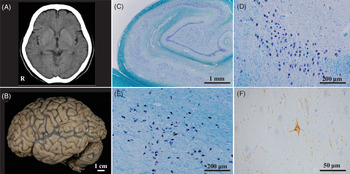
Head CT taken 2 days before her death indicated no significant atrophy and no abnormal findings [Fig. 2(A)].
Clinicopathological findings of Case 2. A. Brain CT at 2 days before death. Slight cortical atrophy was observed. B. The general appearance of the left cerebral hemisphere showed unremarkable changes. C. Microscopic observation of the hippocampal area. Pyramidal neurons were preserved. D. Microscopic observation of the locus coeruleus. The neurons were well preserved. E. Microscopic observation of the substantia nigra. The neurons were well preserved. F. Microscopic observation of the ambient gyrus. Neurofibrillary tangles were seldom observed. KB staining (C–E), AT8 immunostaining (F).

Her brain weighed 1080 g at the autopsy [Fig. 2(B)]. On a macroscopic observation, no marked abnormalities in the cerebrum, cerebellum, basal ganglia, substantia nigra, locus coeruleus, dorsal nucleus of vagus nerve were noted. A microscopic observation revealed Braak NFT stage I, SPs (-), LBs (-) and vascular lesions (-).
No lesions capable of causing sudden death were noted in the CNS, and there were no abnormal brain organic findings. There was also no evidence of neurodegenerative diseases that might explain her cognitive impairment.
Case 3
A 69-year-old woman at death had a 40-year history of schizophrenia.
After graduating from a vocational school, she worked at a hospital. She married at 26 years old and had two children, living as a housewife after marriage. Her eldest son developed schizophrenia.
At 29 years old, she developed schizophrenia, presenting with hallucinations and a delusional state. She was admitted to psychiatric hospitals multiple times before 38 years of age. However, her condition, including her hallucinatory delusions, subsequently subsided with some remnant negative symptoms, such as a decline in her personality level and the existence of thought disorders, and she was able to live a stable life at home.
Around 64 years old, she showed dementia syndrome, including amnesia, wandering and urinary incontinence and was again admitted to a psychiatric hospital. She then showed gradual cognitive decline, presenting with wandering, pica, ranting, violence under resistance for care, undressing and inappropriate urinating. At this point, she was unable to recall the names of common items or how to use them, and her revised Hasegawa dementia scale (HDS-R) (Jeong et al., Reference Jeong, Kim, Lee, Lee, Park, Choi, Choe, Do, Ryang, Roh, Park, Choi and Woo2007) score was 7/30. At 69 years old, she died of acute respiratory failure.
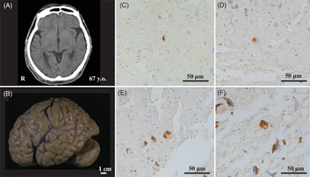
Head CT (taken 2 years before death) showed slight cortex atrophy but no areas of abnormal density [Fig. 3(A)].
Clinicopathological findings of Case 3. A. Brain CT at 2 years before death. Slight cortical atrophy was observed. B. General appearance of the left side. C. Neurofibrillary tangles and neuropil threads were detected in the transentorhinal cortex. Lewy bodies were observed in the dorsal vagal nucleus (D), substantia nigra (E) and locus coeruleus (F). AT8 immunostaining (C), phosphorylated α-synuclein immunostaining (D–F).

Her brain weighed 1376 g at the autopsy. On a macroscopic observation, the cerebral cortex and white matter were preserved, and the basal ganglia and thalamus showed no significant changes. A microscopic observation revealed Braak NFT stage I [Fig. 3(C)], with no SPs. Neurons in hippocampal formation were preserved. Several brain stem-type LBs and Lewy neurites (LNs) were noted in the dorsal vagal nucleus [Fig. 3(D)], substantia nigra [Fig. 3(E)] and locus coeruleus [Fig. 3(F)]. A large number of LBs and LNs were observed in the medullary reticular formation and raphe nucleus. In the cerebrum, a few phosphorylated α-synuclein-positive structures were observed in the amygdala, and in the cingulate gyrus, several LBs and LNs were observed in the nucleus basalis of Meynert. [Fig. 3(C–F)]. She was diagnosed with LB disease, brain stem-type, Braak PD stage 3.
Discussion
In the present study, we performed clinical neuropathological investigations of three cases of schizophrenia that developed typically in early adulthood with cognitive dysfunction or dementia syndrome manifesting in late adulthood. The first case of this report showed reduced speech, disorientation and incontinence, among other symptoms, about 40 years after the onset of schizophrenia; however, only a preclinical AD pathology and mild multiple cerebral infarction were detected, which could not explain his clinical symptoms. In the second case, the patient developed schizophrenia in her teens and had difficulty achieving remission even with intensive antipsychotic drug treatment. From 60 years old, she needed nursing care, including clothing and personal hygiene. However, no significant neuropathological findings were detected at her autopsy. In the third patient, schizophrenia developed in her 20s, and remission was achieved with treatment, but in her 60s, she developed symptoms such as amnesia, wandering and urinary incontinence. In addition, she showed psychiatric symptoms such as violence or/and abusive language against her care giver. Pathologically, mild brain stem-type LB disease was diagnosed, and these findings could not explain her clinical symptoms of dementia.
In all cases, the neuropathological findings failed to explain the clinical symptoms of cognitive decline empirically and reasonably. Dementia syndrome or severe cognitive decline in elderly schizophrenic patients might be due to schizophrenic unrevealed pathogenesis itself, and not due to comorbid dementia occurred secondly such as AD.
How brain degenerative changes influence the symptoms of schizophrenia
From a clinicopathological investigative perspective, we should consider that the brain degenerative changes might influence the clinical symptoms of elderly schizophrenic patients at the bedside.
In Case 1, argyrophilic grains were detected in this schizophrenia patient. The characteristic clinical symptoms of argyrophilic grain disease (AGD) are behavioural and psychological symptoms, including stubbornness, irritability, ideas of persecution and personality change, and the AGD pathology tends to progress slowly (Togo et al., Reference Togo, Isojima, Akstu, Suzuki, Uchikado, Katsuze, Iseki, Kosaka and Hirayasu2005). Indeed, Case 1 had demonstrated violent behaviour in her 60s, a symptom that might have indicated the pathology of AGD. However, the other schizophrenia patients typically showed hostility, poor rapport and/or suspiciousness; it is thus much more difficult to determine whether these clinical symptoms were due to AGD pathology or schizophrenia itself. Furthermore, in Case 1, the neuropathological degree of AGD was estimated to be stage II. Such mild AGD pathology would be expected at preclinical stage at the bedside. In addition, the AGD pathology may also affect the clinical symptoms of residual schizophrenia to some degree.
In Case 3, LB pathology was observed mainly in the brainstem, and the pathological diagnosis was LB disease, brain stem-type, Braak PD stage 3. This degree pathology is expected to be accompanied by mild cognitive impairment (not dementia), if any, and the patient’s disease progression remained at the prodromal stage, showing no psychotic symptoms, including hallucinations and delusions (Kon et al., Reference Kon, Tomiyama and Wakabayashi2020). Indeed, she did not exhibit any hallucinations or delusion in her older years at all. However, Case 3 had been indicated to have severe level dementia on the dementia screening test administered in her mid-60s, a result that did not correlate with her neuropathological findings. Notably, she showed urinary incontinence, with respiratory failure as the cause of death. These symptoms suggest the hypofunction of the autonomic nervous system, leading to the relevance to brain stem-type LB disease.
When neurodegenerative changes develop in the brains of elderly schizophrenia (even mild changes), it becomes difficult to determine the origin of the symptoms. And it is assumed that we have difficulty with judging which treatment is rational for the patients, treatment for schizophrenia or treatment for neurodegenerative diseases.
Neuropathology of schizophrenia
Dementia Praecox is a concept underlying schizophrenia proposed by Emil Kraepelin (1856−1926) (Kendler & Engstrom, Reference Kendler and Engstrom2018). Kraepelin had considered that the pathogenesis of this disease involved organic factors in the brain and elucidated the pathogenesis of mental illness through neuropathological techniques. However, no reproducible or significant brain pathological findings have been detected in patients with this disease. Subsequently, neuropathological research to elucidate the pathology of schizophrenia was conducted less and less frequently, and the notion that ‘schizophrenia is a graveyard for neuropathologists’ began to be bandied about in ridicule (Plum, Reference Plum1972).
However, around the 1980s, marked advances in molecular biological research began to identify many risk-related genes for this disease, and many of these risk genes are reportedly involved in the formation of the CNS (Giusti-Rodriguez & Sullivan, Reference Giusti-Rodriguez and Sullivan2013). The reduction in the brain volume of this disease throughout the disease course has been reported with reproducibility (Guo et al., Reference Guo, Huhtaniska, Miettunen, Jaaskelainen, Kiviniemi, Nikkinen, Moilanen, Haapea, Maki, Jones, Veijola, Isohanni and Murray2015). Such research results have once again drawn focus to what is occurring in the schizophrenic brain tissue. Several neuropathological findings have been reported in the postmortem brains of schizophrenic patients with reference to the neuropathological examinations of animal models (Schmitt & Falkai, Reference Schmitt and Falkai2014). However, no useful diagnostic reproducible pathological findings are presently available for schizophrenia.
The biological pathogenesis of schizophrenia and neuropathology
Genomic studies have suggested some form of neurodevelopmental disorder in patients with this disease (Rund, Reference Rund2018). As another potential pathogenesis, the brain volume has been suspected to decrease over time after the onset, and remission has been shown to worsen gradually, eventually leading to mental devastation.
Given these phenomena, the pathogenesis of this disease is suspected to involve neurodegeneration (Lieberman et al., Reference Lieberman, Chakos, Wu, Alvir, Hoffman, Robinson and Bilder2001; Perez-Neri et al., Reference Perez-Neri, Ramirez-Bermudez, Montes and Rios2006), and the two aspects of neurodevelopmental disorder and neurodegenerative disorder are predicted to exist in this disease. The pathogenesis of dementia syndrome in elderly cases of schizophrenia is thought to be associated with some failure in the protective function of CNS based on the initial fragility that was observed during the developmental stage of this disease, and such fragility tends to increase as the patients continue to increase in age (Pino et al., Reference Pino, Guilera, Gomez-Benito, Najas-Garcia, Rufian and Rojo2014; Eyler & Jeste, Reference Eyler and Jeste2018). On reviewing the reported cases again, Cases 1 and 3 showed cognitive decline even with preclinical degenerative findings. In Case 2, however, few degenerative neuropathological findings were found. The fragility of the brain of schizophrenia patients is believed to accelerate the impact of aging on the body, manifesting as clinical symptoms. However, while this fragility seems to be undoubtedly a fundamental component of the disease, the details of this vulnerability remain to be clarified.
Neurocognition and schizophrenia
There have been some reports that an anti-dementia drug (acetylcholinesterase inhibitor) was effective for managing cognitive decline in schizophrenia patients (Sarter, Reference Sarter1994; Sarter et al., Reference Sarter, Lustig and Taylor2012). The schizophrenic brain does not demonstrate the same loss of cholinergic neurons found in AD patients, but the activation of cholinergic neurons at an early age may indicate an improved cognitive function. Another anti-dementia drug, memantine, is an N-Methyl-D-aspartate (NMDA) receptor antagonist. Memantine is a low-affinity antagonist of the NMDA receptor (NMDAR) that exerts neuroprotective effects in AD (Kishi et al., Reference Kishi, Matsunaga, Oya, Nomura, Ikuta and Iwata2017), and in schizophrenia patients, these drugs are thought to be effective by their ability to enhance or maintain the NMDAR function (Perez-Neri et al., Reference Perez-Neri, Ramirez-Bermudez, Montes and Rios2006). Schizophrenia has reportedly become milder overall, particularly concerning the severity of psychiatric symptoms, and its prognosis has improved since the development of relevant drugs in the 1950s (Der et al., Reference Der, Gupta and Murray1990). The spread of drug treatment with a focus on neuroprotective aspects may have altered the course and prognosis of this disease (Li & Xu, Reference Li and Xu2007).
The brain pathology of dementia and schizophrenia in the elderly might therefore have the same pathogenesis from the point of attention deficit (Sarter, Reference Sarter1994). In Case 2, clinically severe cognitive decline was observed, but no corresponding pathological findings were detected. Such case reports of elderly schizophrenia patients have been considered since the era of Kraepelin (Arnold & Trojanowski, Reference Arnold and Trojanowski1996a). The present study indicates that there are some schizophrenia cases that show cognitive decline over the course of their illness while lacking any neuropathological changes matching the clinical symptoms. This suggests that some degenerative changes might be happening without the association of any known brain pathology in cases of this disease. However, not all cases of schizophrenia show a decline in the cognitive function over time, so such patients might be members of a subgroup of this disease. In any case, in order to clarify the fragility of the brain and the more fundamental etiology of schizophrenia, recent advances in genomic research and neuroimaging have clarified many points regarding the brain pathology, thus allowing us to have a better understanding of the diseased brain.
In this report, the decline of cognitive function observed in elderly schizophrenia is partially thought to due to disease specific pathogenesis or/and the vulnerability to aging rather than complication of neurodegenerative disease. It is hoped that future cases will continue to be accumulated and studied in order to clarify the pathogenesis of schizophrenia anyway.
Authors’ contributions
Each author contributed to the article. The study was conceptualized by SI, HF, KI and KK. The clinical data of the sample were evaluated by AM, YT, HS and CH. The neuropathological findings were investigated by MH and MY. The manuscript was written by AM, MH and SI. All authors have approved the final script.
Financial support
This study was partially supported by Japan Agency for Medical Research and Development (AMED) under grant 20dm0107108h005, 20dm017105. Japan Society for the Promotion of Science (JSPS) KAKENHI Grant Numbers JP17K10294 (2017-2020), JP19K17059 (2019-2021).
Statement of interest
The authors declare no conflict of interest in association with the present study.
Ethical standards
The authors assert that all procedures in this work comply with the ethical standards of the relevant national and institutional committees on human experimentation and with the 1975 Declaration of Helsinki, as revised in 2008. This study was approved by the Nagoya University School of Medicine Ethical Review Board.