INTRODUTION
Recent environmental issues have changed the current social context and call for urgency in the conservation, recuperation and management of marine coastal environments. These regions have been exploited haphazardly, which has resulted in the reduction of species and the irreversible loss of biological diversity (Ghilardi et al., Reference Ghilardi, Filho and Berchez2008).
Among the different biological compartments, marine benthos plays a vital role, both as a receiver of energy from the pelagic environment and as a supplier of energy for organisms that forage near the bottom, such as fish, crustaceans and other invertebrates (Amaral & Rossi-Wongtschowski, Reference Amaral and Rossi-Wongtschowski2004). Many benthic invertebrate species, or other species associated with the seafloor, have direct economic importance, as is the case with crustaceans and molluscs, or also indirect economic importance, since they are often the main food item for demersal fish species of economic interest (e.g. some polychaetes and echinoderms).
In this context, the study of biological benthic diversity and marine environments remains the basis for the formulation of ecological and preservation programs, including the monitoring of ecosystems and environmental impact assessment, as well as procedures for their conservation. Hence, it is essential to investigate these species in order to conduct any further study and/or planning towards the conservation of estuary regions (Almeida et al., Reference Almeida, Guerrazzi and Coelho2007). The situation is even worse regarding places in Northern and North-eastern Brazil, where many locations and environments have not yet been surveyed faunistically (Lana et al., Reference Lana, Camargo, Brogim and Isaac1996).
Estuaries are some of the most productive ecosystems on the planet (Duxbury & Duxbury, Reference Duxbury and Duxbury1996). They stand out as complex environments that serve as a shelter for many organisms and they constitute a suitable place for the reproduction, feeding and nursery of many species of fish and invertebrates (Mesquita et al., Reference Mesquita, Costa, Pereira and Magalhães2006; Fiori, Reference Fiori2007).
Despite their significant ecological and economic importance, little is known about the environmental and biological characteristics of Brazilian estuaries. A literature review on the faunal and taxonomic studies conducted in these environments has shown that researchers mainly investigate specific groups, such as crustaceans (Severino-Rodrigues et al., Reference Severino-Rodrigues, Pita and Graça-Lopes2001; Almeida et al., Reference Almeida, Guerrazzi and Coelho2007), bivalves (Fiori, Reference Fiori2007), gastropods (Vasconcelos et al., Reference Vasconcelos, Santos and Trindade2004; Fiori, Reference Fiori2007; Ourives, Reference Ourives2007), nematodes (Fiori, Reference Fiori2007), polychaetes (Vasconcelos et al., Reference Vasconcelos, Santos and Trindade2004; Fiori, Reference Fiori2007), leeches (Fiori, Reference Fiori2007), cnidarians (Mesquita et al., Reference Mesquita, Costa, Pereira and Magalhães2006; Fiori, Reference Fiori2007) and fishes (Ramos & Vieira, Reference Ramos and Vieira2001; Loebmann & Vieira, Reference Loebmann and Vieira2005; Falcão et al., Reference Falção, Sarpédonti, Spach, Otero, Queiroz and Santos2006).
Echinoderms, especially ophiuroids, despite their important ecological role in marine benthic communities, are not among the most studied groups. Nonetheless, the importance of ophiuroids in these environments is evidenced by their abundance and wide distribution (Hendler, Reference Hendler1996). Studies conducted in the United States and France indicate the importance of these organisms in the characterization and stability of the environments they inhabit; and these studies also emphasize their use as bioindicators of polluted ecosystems (Thorson, Reference Thorson1957; Barnard & Ziesenhenne, Reference Barnard and Ziesenhenne1961; Harmelin et al., Reference Harmelin, Bouchon and Hong1981).
Studies on the ophiuroids of Brazil are still insufficient for us to possess a full knowledge of the group. It is worth highlighting the studies conducted by Tommasi (Reference Tommasi1970, Reference Tommasi1999), Alves & Cerqueira (Reference Alves and Cerqueira2000), Manso (Reference Manso2004), Gondim et al. (Reference Gondim, Lacouth, Alonso and Manso2008), Lima & Fernandes (Reference Lima and Fernandes2009) and Lima et al. (Reference Lima, Correia, Sovierzoski and Manso2011), in the North and North-east of Brazil, and Borges et al. (Reference Borges, Monteiro and Amaral2002), Borges & Amaral (Reference Borges, Amaral, Amaral, Rizzo and Arruda2005, Reference Borges and Amaral2007) and Borges & Campos (Reference Borges and Campos2011), in the South and South-east of Brazil.
The estuary of Camamu bay, located in the Brazilian State of Bahia, is an ecosystem of great ecological and economic significance for the local population, due to fishing and tourism-related activities. However, anthropogenic pollutants have affected this ecosystem (Guerrazzi, personal communication). Consequently, faunal inventories are of great importance, since few studies have been conducted in the region, with the exception of crustaceans studied by Almeida et al. (Reference Almeida, Guerrazzi and Coelho2007), gastropods by Ourives et al. (Reference Ourives, Guerrazzi and Simone2011) and echinoderms by Manso (Reference Manso2004).
Therefore, the purpose of our study is to survey the Ophiuroidea fauna of the estuary of Camamu Bay, in order to redescribe and illustrate the species we recorded, so as to identify the local biodiversity, as well as to enable monitoring programs and wildlife management. Additionally, we will comment on the occurrence of the species found in the study area, including any published records.
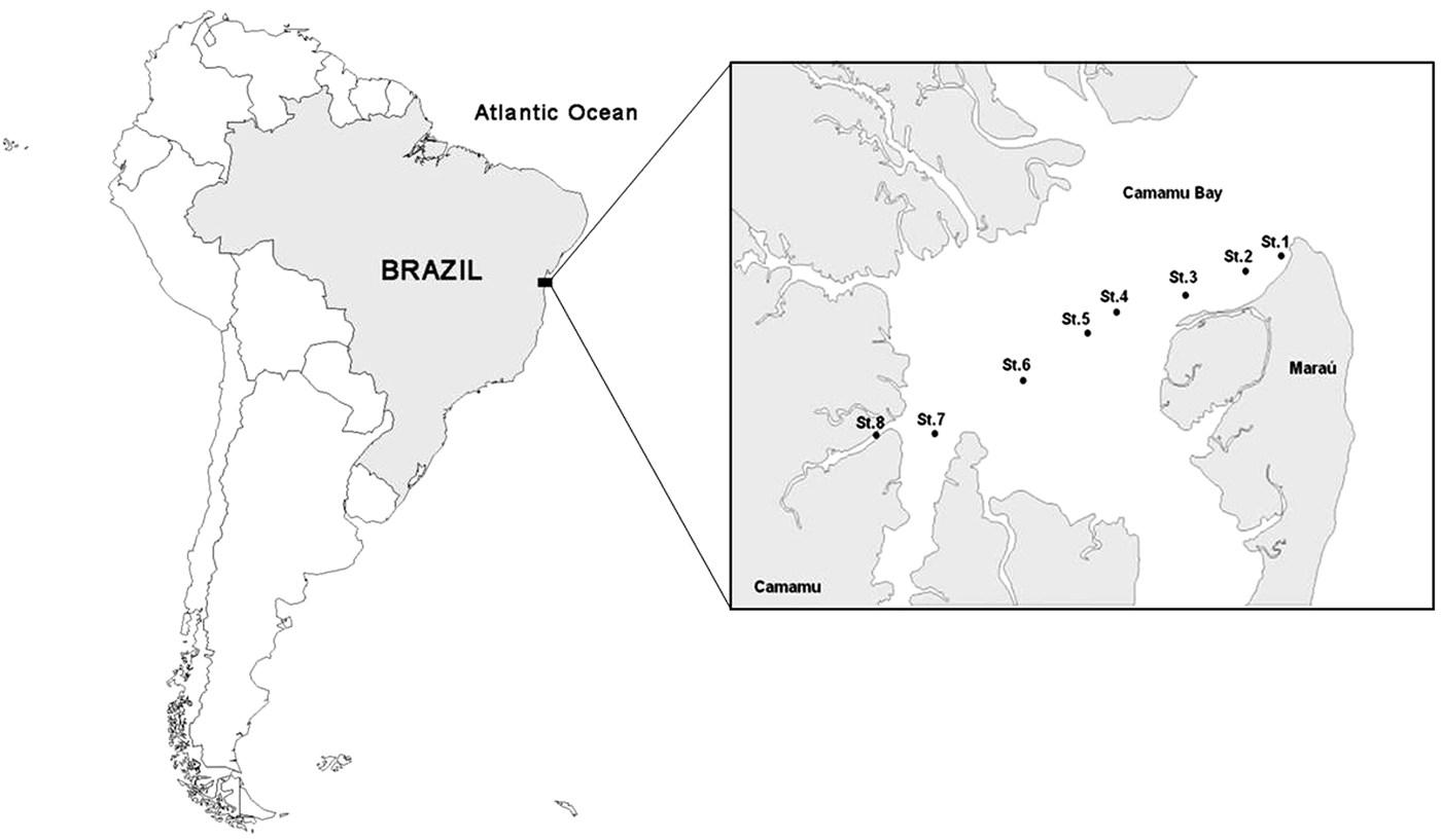
STUDY AREA
Camamu Bay is an estuary located in the compartment structure defined as the Camamu Basin (Manso & Souza-Lima, Reference Manso and Souza-Lima2007) and is located on the central coast of Bahia, between 13°50′–14°06′S and 38°57′–39°4′W (Figure 1). This bay is the third largest bay in Brazil after Todos os Santos Bay (Bahia) and Guanabara Bay (Rio de Janeiro) (Oliveira et al., Reference Oliveira, Queiroz, Damasceno, Santos and Freitas1998, Reference Oliveira, Queiroz and Argôlo2002).
Map of the study area in Camamu Bay, Bahia, Brazil. Detail showing the eight sampling stations (St. 1–St. 8). Source: Ourives et al. (Reference Ourives, Guerrazzi and Simone2011), with modifications.

Camamu Bay has a roughly circular shape with an internal area of 85 km2. It contains many islands in its interior and has an extensive estuary area bordered by mangroves, salt marshes, rocky shores and the Atlantic Forest, forming natural pools and reefs along the coast, which are still well preserved and also little studied. Another peculiar characteristic of the area is the great opening of the mouth, which allows a massive influx of sea water in comparison to the current flow of the rivers, giving the bay a favourable environment for marine life (Oliveira et al., Reference Oliveira, Queiroz, Damasceno, Santos and Freitas1998, Reference Oliveira, Queiroz and Argôlo2002; Almeida et al., Reference Almeida, Guerrazzi and Coelho2007; Hatje et al., Reference Hatje, Barros, Magalhães, Riatto, Amorim, Figueiredo, Spano and Cirano2008).
The humidity is high, with an annual average of approximately 75–85% relative humidity, lacking a dry season. The average annual temperature is 24°C, with minor variations over the year, with a maximum of 26°C and a minimum of 20°C.
MATERIAL AND METHODS
We collected samples during 18 months in the estuary of the Marau River, Camamu Bay (Bahia), at eight fixed stations (Figure 1). The stations were established approximately 2.5 km apart, and their coordinates were determined with a GPS device (Global Position System, 12X-L, Garmin). The samples were obtained between latitude 13°53′04″–13°56′24″S and longitude 38°57′06″–39°05′04″W.
The benthic organisms were sampled between isobaths from 1.5 to 15.5 m, using a fishing boat (trawler type) with a trawl door system, a mesh of 3 cm (measured internodes) and a mouth of 4 m. At each station, the drag lasted approximately 10 min, at a constant speed of 2.5 km/h.
We also collected sediment samples, using a Van Veen dredge for granulometric parameter estimates of sediment and organic matter from the substrate.
The samples of biological material from the trawling were initially screened onboard the vessel and fixed in 70% alcohol.
Brittle stars were identified with the help of specific references and identification keys (Hendler et al., Reference Hendler, Miller, Pawson and Kier1995; Borges et al., Reference Borges, Monteiro and Amaral2002; Borges & Amaral, Reference Borges, Amaral, Amaral, Rizzo and Arruda2005; Pomory, Reference Pomory2007), and counted. Species names are according to Stöhr et al. (Reference Stöhr, O'Hara and Thuy2014) and the terminology adopted from Stöhr et al. (Reference Stöhr, O'Hara and Thuy2012). Most of the material was deposited in the collection of Echinodermata at the Laboratory of Ecology at the State University of the Southwest of Bahia, Campus Jequié, labelled with the code UESBOFR. Duplicates were deposited in the collection of Ophiuroidea at the Museum of Zoology at the University of Campinas, labelled with the code ZUEC OPH.
RESULTS
We analysed and identified 6282 individuals belonging to five families in the order Ophiurida (Müller & Troschel, 1840) and 11 species.
Checklist of brittle stars from Camamu Bay, Bahia, Brazil
Order OPHIUROIDEA Gray, 1840
Suborder OPHIURIDA Müller & Troschel, 1940
Family AMPHIURIDAE Ljungman, 1867
Amphipholis januarii Ljungman, 1866
Amphipholis squamata (Delle Chiaje, 1829)
Microphiopholis atra (Stimpson, 1852)
Ophiophragmus filograneus (Lyman, 1875)
Ophiostigma isocanthum Say, 1825
Family OPHIACTIDAE Matsumoto, 1915
Ophiactis lymani Ljungman, 1871
Ophiactis savignyi (Müller & Troschel, 1842)
Family OPHIOCOMIDAE Ljungman, 1867
Ophiocoma echinata (Lamarck, 1816)
Family OPHIODERMATIDAE Ljungman, 1867
Ophioderma cinerea Müller & Troschel, 1842
Ophioderma januarii Lütken, 1856
Family OPHIOTRICHIDAE Ljungman, 1867
Ophiothrix (Ophiothrix) angulata (Say, 1825)
Key to the families of brittle stars from Camamu Bay, Bahia, Brazil
1. Presence of cluster of dental papillae on the apex of the jaw…………3
Without cluster of dental papillae…………2
2. One pair of infradental oral papillae on the apex of the jawAmphiuridae
Only one apical papilla on the apex of the jaw…………4
3. A continuous series of lateral oral papillae…………Ophiocomidae
Without lateral oral papillae…………Ophiotrichidae
4. Disc covered with granules; numerous oral papillae in continuous series…………Ophiodermatidae
Disc covered by scales and spines. Presence of a diastema separating the lateral oral papillae from the apical papilla.…………Ophiactidae
Key to the members of the family Amphiuridae registered from Camamu Bay, Bahia, Brazil
1. Distal oral papilla of similar size as the other lateral papillae.…………Ophiophragmus filograneus (Figure 4)
Larger, opercular, distal oral papilla, partly or fully closing the oral slit…………2
2. Disc covered by scales; two perpendicular tentacle scales; distal oral papilla partly closing the oral slit…………3
— Disc covered by small papillae; two parallel tentacle scales; distal oral papilla fully closing the oral slit…………Ophiostigma isocanthum (Figure 5)
3. Small and delicate disc…………4
— Larger disc (up to 1 cm), covered with smaller scales; perpendicular marginal scales, forming a fringe…………Microphiopholis atra (Figure 3)
4. Disc covered with small scales; three or four arm spines, the second and third with two hyaline denticles at the tip. Narrow and long radial shields…………Amphipholis januarii (Figure 2A–C)
— Disc with larger scales; three arm spines with tip tapering. Radial shields slightly longer than broad…………Amphipholis squamata (Figure 2D–F)
Amphipholis januarii: A – dorsal view; B – ventral view; C – ventral view of the arm. Amphipholis squamata: D – dorsal view; E – ventral view; F – ventral view of the arm. Scale bars (A, B, D, E, F) = 0.5 mm; (C) = 0.2 mm.
Microphiopholis atra: A – dorsal view; B – ventral view; C – oral view. Scale bars (A, B) = 1 mm; (C) = 0.5 mm.
Ophiophragmus filograneus: A – dorsal view; B – dorsal view with detail of the radial shields; C – ventral view; D – oral view. Scale bars (A, C) = 0.5 mm; (B) = 0.2 mm; (D) = 0.1 mm.
Ophiostigma isocanthum: A – dorsal view; B – ventral view; C – oral view. Scale bars (A, B) = 1 mm; (C) = 0.2 mm.
Ophiactis lymani: A – dorsal view; B – ventral view; C – oral view. Ophiactis savignyi: D – dorsal view; E – ventral view; F – oral view. Scale bars (A, B, C, F) = 0.5 mm; (D, E) = 1 mm.
Key to the members of the family Ophiactidae identified in Camamu Bay, Bahia, Brazil
1. Six arms; disc covered with small and strong overlapping scales, with some spines scattered over the dorsal disc; much larger radial shields. One or two lateral oral papillae…………Ophiactis savignyi (Figure 6D–F)
— Six arms; disc covered by larger scales slightly overlapped; few delicate spines usually restricted to the disc margin, easily lost. One oral papilla…………Ophiactis lymani (Figure 6A–C)
Key to the members of the family ophiodermatidae identified in Camamu Bay, Bahia, Brazil
1. Robust, greenish disc; radial shields not visible, covered by granules…………Ophioderma januarii (Figure 8D–F)
— Robust, brown disc; oval radial shields visible not covered by granules…………Ophioderma cinerea (Figure 8A–C)
SYSTEMATICS
Family AMPHIURIDAE Ljungman, 1867
Genus Amphipholis Ljungman, 1866
Amphipholis januarii Ljungman, 1866
(Figure 2A–C)
EXAMINED MATERIAL
24ex.: St. 2, 13°53′21″S 38°57′49″W, ix.9.2006 (UESB OFR 19, 3 ex.; ZUEC OPH 989, 2 ex.); St. 4, 13°54′06″S 39°00′22″W, iii.24.2005 (UESB OFR 483, 1 ex.; UESB OFR 494, 1ex.); St. 5, 13°54′14″S 39°00′34″W, iv.24.2004 (UESB OFR 193, 1ex.; UESB OFR 336, 1 ex.; UESB OFR 337, 1 ex.; UESB OFR 338, 1 ex.; UESB OFR 376, 1 ex.), viii.28.2004 (UESB OFR 217, 1 ex.), viii.29.2004 (UESB OFR 449, 1 ex.), ix.25.2004 (ZUEC OPH 999, 1 ex.), x.31.2004 (UESB OFR 64, 1ex.; UESB OFR 175, 1ex.; UESB OFR 396, 3 ex.; ZUEC OPH 982, 1 ex.), viii.7.2005 (UESB OFR 438, 1 ex.), ix.8.2005 (UESB OFR 290, 1 ex.), St. 6, 13°55′21″S 39°02′13″W, ix.11.2006 (ZUEC OPH 973, 1 ex.).
DESCRIPTION
Disc diameter: 2.0–6.0 mm. Dorsal disc covered with small and imbricated scales. Primary scales evident. Radial shields narrow and long, separated proximally by one or two scales (Figure 2A). Ventral interradius covered by scales similar to the dorsal surface. Oral shields slightly longer than wide, diamond-shaped, with rounded edges and small latero-posterior indentations. Adoral shields distally extended and separated proximally. Two oral papillae on each side of jaw angle, the distal one twice as wide as the proximal. A pair of elongated infradental papillae, widely separated from each other (Figure 2B). Long arms, approximately ten times the diameter of the disc. Dorsal arm plates slightly wider than long (Figure 2A); ventral arm plates pentagonal, sub-elliptical, with rounded edges. Two tentacle scales, the larger supported on the ventral arm plate and the smaller on the lateral arm plate (Figure 2C). Four elongated arm spines, reduced to three at the end of the arms. In segments with four spines, the second ventralmost with one or two lateral terminal denticles and smaller denticles at all edges. At segments with three spines, the middle one presents such denticles (Figure 2C). Spines at the proximal segments and on the distal arms, lack denticles.
REMARKS
Amphipholis januarii is a characteristic inhabitant of soft bottoms, sand and mud, but is also found on algae, under rocks and rubble, and is associated with sponges and corals, among others (Hendler et al., Reference Hendler, Miller, Pawson and Kier1995; Borges & Amaral, Reference Borges, Amaral, Amaral, Rizzo and Arruda2005; Pomory, Reference Pomory2007). This species is commonly sampled in live substrate, together with other brittle stars, such as Amphipholis squamata, Ophiactis savignyi and Ophiothrix (O.) angulata. In Brazil, the species has been recorded in the states of Rio de Janeiro (type locality), São Paulo, Pará, Ceará, Paraíba, Alagoas and Bahia (Tommasi, Reference Tommasi1970; Albuquerque, Reference Albuquerque1986; Borges, Reference Borges2006; Gondim et al., Reference Gondim, Lacouth, Alonso and Manso2008; Manso et al., Reference Manso, Alves and Martins2008; Lima et al., Reference Lima, Correia, Sovierzoski and Manso2011). In this study, it was sampled in the phytal zone and under rocks.
GEOGRAPHICAL DISTRIBUTION
South Carolina to Florida, Texas, Mexico, Antilles, Cuba, Puerto Rico, Virgin Islands, Tobago, Barbados and Brazil (Hendler et al., Reference Hendler, Miller, Pawson and Kier1995; Borges & Amaral, Reference Borges, Amaral, Amaral, Rizzo and Arruda2005; Laguarda-Figueras et al., Reference Laguarda-Figueras, Hernández-Herrejón, Solís-Marín and Durán-González2009).
BATHYMETRIC DISTRIBUTION
1–311 m (Tommasi, Reference Tommasi1970; Hendler et al., Reference Hendler, Miller, Pawson and Kier1995; Borges, Reference Borges2006; Laguarda-Figueras et al., Reference Laguarda-Figueras, Hernández-Herrejón, Solís-Marín and Durán-González2009). In this study, it was sampled at depths ranging from 4.2 to 8.4 m.
RECORDS IN THE STATE OF BAHIA
Todos os Santos Bay, city of Porto Seguro and Salvador (Itapuã and Ondina beaches) (Magalhães et al., Reference Magalhães, Martins and Alves2005; Manso et al., Reference Manso, Alves and Martins2008).
RECORDS IN CAMAMU BAY
This is a new record of this species in the study area.
Amphipholis squamata (Delle Chiaje, 1829)
(Figure 2D–F)
EXAMINED MATERIAL
418 ex.: St. 1, 13°53′04″S 38°57′06″W, iv.24.2004 (UESB OFR 260, 2 ex.; UESB OFR 261, 3 ex.), viii.6.2005 (UESB OFR 309, 1 ex.); St. 2, 13°53′21″S 38°57′49″W, iv.24.2004 (UESB OFR 71, 1 ex.); ix.25.2004 (UESB OFR 07, 2 ex.); viii.6.2005 (UESB OFR 51, 1 ex.); St. 3, 13°54′25″S 38°59′14″W, iii.24.2004 (UESB OFR 23, 2 ex.); viii.28.2004 (UESB OFR 354, 17 ex.; UEBS OFR 392, 1 ex.; UESB OFR 414, 3 ex.; UESB OFR 425, 14 ex.; UESB OFR 454, 12 ex.; UESB OFR 470, 7 ex.; UESB OFR 496, 14 ex.); ix.7.2005 (ZUEC OPH 969, 1 ex.); ix.19.2000 (UESB OFR 205, 2 ex.); x.31.2004 (UESB OFR 313, 1 ex.); St. 4, 13°54′06″S 39°00′22″W, ix.13.2003 (UESB OFR186, 1 ex.); iii.24.2004 (UESB OFR 442, 1 ex.); iii.24.2005 (UESB OFR 368, 1 ex.; UESB OFR 482, 8 ex.); iv.24.2004 (UESB OFR 197, 1 ex.; UESB OFR 242, 4 ex.; UESB OFR 270, 1 ex.); ix.25.2004 (UESB OFR 406, 5 ex.); x.30.2004 (UESB OFR 302, 1 ex.; UESB OFR 311, 1 ex.; UESB OFR 366, 1 ex.; UESB OFR 421, 2 ex.); viii.6.2005 (UESB OFR 488, 10 ex.; UESB OFR 493, 14 ex.); ix.6.2005 (UESB OFR 452, 6 ex.); ix.7.2005 (UESB OFR 199, 1 ex.); ix.11.2005 (UESB OFR 150, 2 ex.); ix.9.2006 (UESB OFR 416, 2 ex.); St. 5, 13°54′14″S 39°00′34″W, iv.24.2004 (UESB OFR 230, 12 ex.; UESB OFR 268, 8 ex.; UESB OFR 336, 1 ex.; UESB OFR 339, 1 ex.; UESB OFR 378, 3 ex.; UESB OFR 379, 1 ex.; UESB OFR 381, 2 ex.; UESB OFR 395, 2 ex.; UESB OFR 477, 5 ex.); vii.28.2004 (UESB OFR 274, 5 ex.); vii.29.2004 (UESB OFR 156, 1 ex.); viii.25.2004 (UESB OFR 283, 1 ex.); viii.28.2004 (UESB OFR 318, 7 ex.); viii.29.2004 (UESB OFR 152, 13 ex.; UESB OFR 398, 1 ex.; UESB OFR 403, 5 ex.; UESB OFR 408, 18 ex.; UESB OFR 423, 6 ex.; UESB OFR 448, 5 ex.; UESB OFR 471, 4 ex.; UESB OFR 480, 3 ex.); ix.25.2004 (UESB OFR 216, 1 ex.; UESB OFR 273, 1 ex.; UESB OFR 275, 1 ex.; UESB OFR 287, 1 ex.; UESB OFR 328, 2 ex.; UESB OFR 329, 4 ex.; UESB OFR 388, 8 ex.; UESB OFR 410, 22 ex.); x.31.2004 (UESB OFR 183, 1 ex.; UESB OFR 389, 1 ex.); iii.25.2005 (UESB OFR 340, 14 ex.; UESB OFR 404, 5 ex.; UESB OFR 405, 1 ex.; UESB OFR 446, 13 ex.; UESB OFR 486, 1 ex.; ZUEC OPH 1006, 2 ex.); viii.7.2005 (UESB OFR 06, 13 ex.; UESB OFR 78, 2 ex.; UESB OFR 391, 1 ex.; UESB OFR 431, 4 ex.; UESB OFR 433, 13 ex.; UESB OFR 435, 5 ex.; UESB OFR 437, 13 ex.); ix.8.2005 (UESB OFR 413, 7 ex.; UESB OFR 473, 25 ex.); St. 6, 13°55′21″S 39°02′13″W, viii.29.2004 (UESB OFR 338, 1 ex.; x.31.2004 (UESB OFR 390, 1 ex.); ix.11.2006 (ZUEC OPH 972, 1 ex.); St. 7, 13°56′19″S 39°03′57″W, viii.7.2005 (UESB OFR 234, 1 ex.); St. 8, 13°56′24″S 39°05′04″W, ix.14.2005 (UESB OFR 294, 2 ex.).
DESCRIPTION
Disc diameter: 1.0–3.0 mm. Disc covered with irregular and imbricated scales. Radial shields slightly longer than wide, contiguous throughout, except at the proximal edge, where there is a small triangular scale (Figure 2D). Outside edge of the radial shields lightly curved and straight internally. Ventral interradius covered by imbricated scales, smaller than those of the dorsal surface. Oral shields diamond-shaped, with convex distal edge; adoral shields well developed, the distal end wing-like, enlarged and proximally united. Two oral papillae at each side of jaw angle, the distal one rectangular, enlarged and wider than the proximal one. A pair of elongated infradental papillae (Figure 2E). Bursal slits visible. Dorsal arm plates wider than long, fan-shaped with rounded edges; ventral arm plates pentagonal, with a small lateral notch at the tentacle pores (Figure 2F); lateral arm plates well-developed, meeting in the longitudinal mid-line, both dorsal and ventral. Two tentacle scales. Three pointed arm spines, almost as long as an arm segment (Figure 2F).
REMARKS
This species is known to dwell on several types of bottoms, such as sand, rock, rubble and different types of biological substrates such as sponges, bryozoans, corals, polychaete colonies and algae, mainly in the shallow zone, even though there are many records for 100 m depth. There are also records from mangroves and estuaries (Hendler et al., Reference Hendler, Miller, Pawson and Kier1995; Borges & Amaral, Reference Borges, Amaral, Amaral, Rizzo and Arruda2005; Pomory, Reference Pomory2007). In Brazil, it was reported in the states of Pará, Ceará, Paraíba, Alagoas, Bahia and São Paulo (Tommasi, Reference Tommasi1970; Albuquerque, Reference Albuquerque1986; Borges, Reference Borges2006; Gondim et al., Reference Gondim, Lacouth, Alonso and Manso2008; Manso et al., Reference Manso, Alves and Martins2008; Lima et al., Reference Lima, Correia, Sovierzoski and Manso2011). In this study, it was collected in seagrass and under rocks.
GEOGRAPHICAL DISTRIBUTION
Cosmopolitan in tropical and subtropical areas, absent only from the Polar Regions (Hendler et al., Reference Hendler, Miller, Pawson and Kier1995; Borges, Reference Borges2006). However, this is most likely a species complex (Boissin et al., Reference Boissin, Féral and Chenuil2008).
BATHYMETRIC DISTRIBUTION
From 0 to 1962 m depth (considering the species complex) (Alvarado & Solís-Marín, Reference Alvarado and Solís-Marín2013). Borges et al. (Reference Borges, Monteiro and Amaral2002) sampled at 147 m and Borges (Reference Borges2006), at depths between 5 and 240 m. In this study, this species was collected at depths from 3.2 to 9.6 m.
RECORDS IN BAHIA
Todos os Santos Bay, city of Porto Seguro and Salvador (Itapuã and Ondina beaches) (Magalhães et al., Reference Magalhães, Martins and Alves2005; Manso et al., Reference Manso, Alves and Martins2008).
RECORDS IN CAMAMU BAY
This is a new record of this species in the study area.
Genus Microphiopholis Turner, 1985
Microphiopholis atra (Stimpson, 1852)
(Figure 3)
EXAMINED MATERIAL
1 ex.: St. 6, 13°55′21″S 39°02′13″W, iv.25.2004 (UESB OFR 370, 1 ex.).
DESCRIPTION
Disc diameter: 3.1–10.0 mm. Disc covered by small and imbricated scales. Approximately 20 scales between the centrodorsal and the edge of the disc. Primary scales evident. Radial shields twice as long as wide, with abradial edge convex, pairs separated proximally by two to four elongated scales. Scales near a pair of shields slightly bigger than the rest of the disc (Figure 3A). Ventral interradius covered with scales smaller than the dorsal ones and strongly imbricated (Figure 3B). Oral shields lozenge-shaped, longer than wide with the proximal and distal edges tapered. Rounded madreporite, with pores in the anterior margin, distally narrow. Adoral shields wing-like extended at their distal end, united and narrow proximally. Two oral papillae at each side of jaw angle, triangular, the distal one twice as wide as the proximal and more elongated. A pair of semi-rectangular infradental papillae, spaced from one another (Figure 3C). Dorsal arm plates two to three times wide as long, contiguous. Ventral arm plates sub-rectangular to pentagonal, with a small notch in the distal edge. Three thin arm spines. Two tentacle scales well-developed; one in the ventral plate and another on the lateral plate.
REMARKS
Microphiopholis atra is possibly a scavenging infaunal species, due to the large amount of sediment found in its stomach contents (Tommasi, Reference Tommasi1970; Borges, personal observation). It is a common specimen in the intertidal zone at approximately 100 m depth, and it occurs on soft bottoms such as sand, mud and puddles, often with other infaunal brittle stars (Borges & Amaral, Reference Borges, Amaral, Amaral, Rizzo and Arruda2005; Pomory, Reference Pomory2007). In Brazil, they have been reported in the states of Pará, Maranhão, Pernambuco, Bahia, Rio de Janeiro, São Paulo, Paraná and Santa Catarina (Tommasi, Reference Tommasi1970; Albuquerque, Reference Albuquerque1986; Borges, Reference Borges2006; Manso et al., Reference Manso, Alves and Martins2008). We collected the specimens on soft bottoms of puddles and mud, with rich organic matter.
GEOGRAPHICAL DISTRIBUTION
From Virginia, Gulf of Mexico and Puerto Rico to Brazil (Borges & Amaral, Reference Borges, Amaral, Amaral, Rizzo and Arruda2005).
BATHYMETRIC DISTRIBUTION
Intertidal zone – 100 m (Alvarado & Solís-Marín, Reference Alvarado and Solís-Marín2013). Tommasi (Reference Tommasi1970) records it at 20 m, and Borges (Reference Borges2006) between 13 and 24 m. In our study, they were sampled at 4.5 m depth.
RECORDS IN BAHIA
Salvador, Camamu Bay, Todos os Santos Bay and Aratu Bay (Tommasi, Reference Tommasi1970; Manso, Reference Manso2004; Magalhães et al., Reference Magalhães, Martins and Alves2005; Manso et al., Reference Manso, Alves and Martins2008).
PREVIOUS RECORDS IN CAMAMU BAY
Manso (Reference Manso2004).
Genus Ophiophragmus Lyman, 1865
Ophiophragmus filograneus (Lyman, 1875)
(Figure 4)
EXAMINED MATERIAL
1 ex.: St. 5, 13°54′14″S 39°00′34″W, ix.13.2003 (ZUEC OPH 1018, 1 ex.).
DESCRIPTION
Disc diameter: up to 9.0 mm. Disc with circular outline, covered by imbricated scales. Primary plates evident (Figure 4A). Radial shields twice as long as wide, proximally united; distal end separated by two or three scales; the proximal scale triangular and larger, and the posterior small and narrow. Disc edge delineated by small papillae (Figure 4A, B). Ventral interradius lacks scales, covered with fine papilliform granulation. Bursal slits visible (Figure 4C). Oral shields lozenge-shaped. Adoral shields sub-triangular, slightly wider at the distal end, united in the midline. Two oral papillae at each side of jaw angle, distal one slightly larger. A pair of sub-rectangular infradental papillae, separated from one another (Figure 4C, D). Dorsal arm plates trapezoid, wider than long, flabelliform, with a rounded distal edge. Ventral arm plates pentagonal. Lateral arm plates touch ventrally. Three sub-equal, conical arm spines; the median one with tiny denticles. Two tentacle scales, the inner one larger and more elongated.
REMARKS
Ophiophragmus filograneus is a species found on soft bottoms and seagrass (Hendler et al., Reference Hendler, Miller, Pawson and Kier1995). It has also been found in brackish water in Florida (USA). In Brazil, it was reported on sandy bottoms and gravel with calcareous algae in the states of Pará and Maranhão (Albuquerque, Reference Albuquerque1986). In our study, this species was sampled under rocks and associated with algae.
GEOGRAPHICAL DISTRIBUTION
Florida, Gulf of Mexico, Antilles and Brazil (Albuquerque, Reference Albuquerque1986; Hendler et al., Reference Hendler, Miller, Pawson and Kier1995; Stöhr et al., Reference Stöhr, O'Hara and Thuy2014).
BATHYMETRIC DISTRIBUTION
Species of shallow regions, recorded at approximately 80 m depth (Albuquerque, Reference Albuquerque1986; Hendler et al., Reference Hendler, Miller, Pawson and Kier1995). In the current study, we sampled it at 6.7 m.
RECORDS IN BAHIA
First records made by the current study.
RECORDS IN CAMAMU BAY
This is a new record of this species in the study area.
Genus Ophiostigma Lütken, 1856
Ophiostigma isocanthum (Say, 1825)
(Figure 5)
EXAMINED MATERIAL: 5476 ex.: St. 1, 13°53′04″S 38°57′06″W, iv.24.2004 (UESB OFR 09, 12 ex.; UESB OFR 13, 10 ex.; UESB OFR 24, 37 ex.; UESB OFR 190, 1 ex.; UESB OFR 191, 7 ex.; UESB OFR 192, 14 ex.); St. 2, 13°53′21″S 38°57′49″W, ix.19.2000 (UESB OFR 474, 2 ex.), ix.14.2003 (UESB OFR 164, 1 ex.); St. 3, 13°54′25″S 38°59′14″W, ix.19.2000 (UESB OFR 262, 1 ex.; UESB OFR 427, 19 ex.), iv.24.2004 (UESB OFR 129, 1 ex.; ZUECOPH 985, 1 ex.), viii.28.2004 (UESB OFR 38, 78 ex.; UESB OFR 39, 41 ex.; UESB OFR 250, 5 ex.; UESB OFR 251, 9 ex.; UESB OFR 252, 2 ex.; UESB OFR 253, 1 ex.; UESB OFR 254, 1 ex.; UESB OFR 255, 1 ex.; UESB OFR 355, 15 ex.; UESB OFR 357, 1 ex.; UESB OFR 358, 1 ex.; UESB OFR 359, 1 ex.; UESB OFR 360, 1 ex.; UESB OFR 362, 34 ex.; UESB OFR 363, 70 ex.; UESB OFR 453, 80 ex.; UESB OFR 469, 71 ex.; UESB OFR 315, 19 ex.; ZUEC OPH 991,1 ex.; ZUEC OPH 992,1 ex.); St. 4, 13°54′06″S 39°00′22″W, ix.10.2003 (UESB OFR 42, 3 ex.; ZUEC OPH 1010, 1 ex.), ix.13.2003 (UESB OFR 12, 1 ex.; UESB OFR 159, 4 ex.; UESB OFR 198, 2 ex.), iii.24.2004 (UESB OFR 441, 13 ex.; UESB OFR 481, 178 ex.), iv.24.2004 (UESB OFR 01,63 ex.; UESB OFR 26, 11 ex.; UESB OFR 27, 12 ex.; UESB OFR 115, 1 ex.; UESB OFR 349, 1 ex.; UESB OFR 350, 1 ex.; ZUEC OPH 976, 1 ex.), vi.13.2004 (UESB OFR 240, 1 ex.), ix.25.2004 (UESB OFR 31, 46 ex.), x.30.2004 (UESB OFR 33, 6 ex.; UESB OFR 54, 8 ex.; UESB OFR 55, 8 ex.; UESB OFR 66, 13 ex.; UESB OFR 98, 1 ex.; UESB OFR 303, 1 ex.; UESB OFR 321, 1 ex.; UESB OFR 495, 1 ex.; UESB OFR 323, 1 ex.; UESB OFR 324, 1 ex.; UESB OFR 364, 10 ex.; UESB OFR 365, 2 ex.; UESB OFR 383, 1 ex.; UESB OFR 420, 1 ex.; ZUEC OPH 968, 1 ex.; ZUEC OPH 978, 12 ex.; UESB OFR 304, 6 ex.; UESB OFR 307, 1 ex.), x.31.2004 (UESB OFR 170, 88 ex.; UESB OFR 325, 2 ex.), viii.6.2005 (UESB OFR 95, 1 ex.; UESB OFR 119, 1 ex.; UESB OFR 120, 1 ex.; UESB OFR 121, 1 ex.; UESB OFR 220, 2 ex.; UESB OFR 221, 1 ex.; UESB OFR 222, 2 ex.; UESB OFR 223, 6 ex.; UESB OFR 322, 7 ex.; UESB OFR 487, 119 ex.; UESB OFR 492, 295 ex.; UESB OFR11, 2 ex.), ix.6.2005 (UESB OFR 450, 124 ex.), ix.7.2005 (UESB OFR 218, 3 ex.), ix.9.2006 (UESB OFR 37, 10 ex.); St. 5, 13°54′14″S 39°00′34″W, ix.10.2003 (UESB OFR 202, 2 ex.), iii.25.2004 (UESB OFR 85, 23 ex.; UESB OFR 286, 1 ex.), iv.24.2004 (UESB OFR 117, 1 ex.; UESB OFR 48, 26 ex.; UESB OFR 03, 14 ex.; UESB OFR 20, 11 ex.; UESB OFR 227, 5 ex.; UESB OFR 228, 6 ex.; UESB OFR 335, 2 ex.; UESB OFR 375, 84 ex.; UESB OFR 380, 1 ex.; UESB OFR 458, ex. 63 ex.; UESB OFR 475, ex. 130 ex.; ZUEC OPH 979, ex. 19 ex.; ZUEC OPH 983, ex. 13 ex.), viii.28.2004 (UESB OFR 243, 7 ex.; UESB OFR 244, 1 ex.; UESB OFR 245, 6 ex.; UESB OFR 246, 2 ex.; UESB OFR 426, 1 ex.), viii.29.2004 (UESB OFR 104, 82 ex.; UESB OFR 110, 1 ex.; UESB OFR 112, 1 ex.; UESB OFR 113, 126 ex.; UESB OFR 153, 1 ex.; UESB OFR 154, 14 ex.; UESB OFR 157, 1 ex.; UESB OFR 52, 34 ex.; UESB OFR 35, 98 ex.; UESB OFR 158, 1 ex.; UESB OFR 317, 5 ex.; UESB OFR 319, 70 ex.; UESB OFR 320, 26 ex.; UESB OFR 316, 1 ex.; UESB OFR 463, 86 ex.; UESB OFR 447, 73 ex.; UESB OFR 394, 1 ex.; ZUEC OPH 977, 10 ex.; ZUEC OPH 987, 2 ex.; UESB OFR 478, 89 ex.; UESB OFR 479, 1 ex.; UESB OFR 484, 1 ex.), ix.25.2004 (UESB OFR 34, 341 ex.; UESB OFR 213, 65 ex.; UESB OFR 214, 9 ex.; UESB OFR 274, 1 ex.; UESB OFR 277, 1 ex.; UESB OFR 278, 14 ex.; UESB OFR 285, 15 ex.; UESB OFR 327, 1 ex.; UESB OFR 330, 46 ex.; UESB OFR 331, 15 ex.; UESB OFR 332, 2 ex.; UESB OFR 334, 17 ex.), ix.29.2004 (UESB OFR 276, 1 ex.), x.25.2004 (ZUEC OPH 993, 2 ex.), x.31.2004 (UESB OFR 108, 1 ex.; UESB OFR 68, 63 ex.; UESB OFR 69, 1 ex.; UESB OFR 74, 3 ex.; UESB OFR 165, 1 ex.; UESB OFR 166, 1 ex.; UESB OFR 167, 30 ex.; UESB OFR 168, 1 ex.; UESB OFR 169, 1 ex.; UESB OFR 171, 23 ex.; UESB OFR 172, 83 ex.; UESB OFR 173, 11 ex.; UESB OFR 176, 3 ex.; UESB OFR 177, 1 ex.; UESB OFR 182, 9 ex.; UESB OFR 184, 2 ex.; UESB OFR 305, 2 ex.; UESB OFR 312, 13 ex.; UESB OFR 460, 125 ex.; ZUEC OPH 981, 9 ex.; ZUEC OPH 1009, 2 ex.; ZUEC OPH 1016, 2 ex.), xii.19.2004 (UESB OFR 429, 37 ex.; ZUEC OPH 997, 1 ex.), iii.25.2005 (UESB OFR 84, 81 ex.; UESB OFR 86, 13 ex.; UESB OFR 194, 1 ex.; UESB OFR 343, 27 ex.; UESB OFR 344, 8 ex.; UESB OFR 347, 63 ex.; UESB OFR 443, 120 ex.; UESB OFR 445, 1 ex.; UESB OFR 459, 58 ex.; UESB OFR 485, 118 ex.; ZUEC OPH 1005, 1 ex.), vii.7.2005 (UESB OFR 36, 23 ex.), viii.7.2005 (UESB OFR 32, 44 ex.; UESB OFR 45, 93 ex.; UESB OFR 79, 1 ex.; UESB OFR 118, 1 ex.; UESB OFR 384, 1 ex.; UESB OFR 400, 1 ex.; UESB OFR 430, 226 ex.; UESB OFR 432, 146 ex.; UESB OFR 434, 116 ex.; UESB OFR 436, 133 ex.), ix.8.2005 (UESB OFR 57, 11 ex.; UESB OFR 465, 175 ex.; UESB OFR 466, 158 ex.), ix.11.2005 (UESB OFR 151, 1 ex.), ix.11.2006 (ZUEC OPH 975, 4 ex.), ix.25.2006 (UESB OFR 333, 1); St. 6, 13°55′21″S 39°02′13″W, ix.14.2003 (UESB OFR 163, 1 ex.; ZUEC OPH 965, 1 ex.), iv.25.2004 (UESB OFR 295, 2 ex.; UESB OFR 296, 1 ex.), vi.13.2004 (UESB OFR 263, 1 ex.), viii.29.2004 (UESB OFR 76, 1 ex.; ZUEC OPH 984, 1 ex.), x.31.2004 (UESB OFR 16, 2 ex.; UESB OFR 162, 1 ex.; UESB OFR 271, 1 ex.; UESB OFR 272, 1 ex.; UESB OFR 367, 1 ex.), xii.19.2004 (UESB OFR 114, 1 ex.), iii.25.2005 (UESB OFR 02, 3 ex.; UESB OFR 235, 5 ex.; UESB OFR 236, 4 ex.; UESB OFR 264, 4 ex.; UESB OFR 265, 1 ex.; UESB OFR 266, 2 ex.; UESB OFR 269, 1 ex.; UESB OFR 270, 1 ex.; UESB OFR 281, 1 ex.; UESB OFR 282, 1 ex.; UESB OFR 280, 1 ex.), iii.25.2006 (UESB OFR 267, 1 ex.); ix.11.2006 (ZUEC OPH 974, 2 ex.); St. 7, 13°56′19″S 39°03′57″W, vi.7.2000 (UESB OFR 204, 4 ex.), iv.24.2004 (ZUEC OPH 996, 24 ex.), viii.29.2004 (ZUEC OPH 994, 1 ex.), viii.7.2005 (UESB OFR 49, 5 ex.; UESB OFR 237, 1 ex.); St.8, 13°56′24″S 39°05′04″W, vi.7.2000 (UESB OFR 179, 1 ex.; UESB OFR 180, 1 ex.; UESB OFR 206, 2 ex.), ix.14.2003 (UESB OFR 301, 2 ex.; UESB OFR 373, 48 ex.), x.31.2004 (UESB OFR 17, 5 ex.).
DESCRIPTION
Disc diameter: up to 5.0 mm. Disc circular to pentagonal, covered by small blunt tubercles. Some large and blunt tubercles distributed in the interradius, usually near the radial shields (Figure 5A). Ventral interradius covered by short and blunt tubercles similar to the dorsal ones (Figure 5B). Radial shields small, united and little evident due to the tubercles on the disc. Oral shield lozenge-shaped, somewhat wider than long, narrow proximally; distal margin slightly rounded and lobed. Adoral shields big, sub-triangular, with rounded edges, united proximally. Two oral papillae on each side of jaw angle, distal operculate, large, occupying more than half the length of the jaw and closing oral slit. Proximal papillae small and squared. A pair of blunt infradental papillae, close to each other (Figure 5C). Long arms, at least four times the diameter of the disc. Dorsal arm plates flabelliform, separated by the lateral plates. Ventral arm plates pentagonal with slightly rounded distal edge. Lateral arm plates touching dorsally. Three small and rhombic arm spines that are smaller than the length of one arm segment, with tiny marginal denticles. Two elongated tentacle scales (Figure 5C).
REMARKS
Small and delicate species with long arms, common in environment with reefs, under rocks and on different types of biological substrates (algae, corals, sponges). Seagrass beds, under stones and rubble, and in branching coral and algae (Hendler et al., Reference Hendler, Miller, Pawson and Kier1995; Manso et al., Reference Manso, Alves and Martins2008; Gondim, Reference Gondim2009). In Brazil, Ophiostigma isocanthum was recorded in the states of Pará, Ceará, Paraíba, Pernambuco, Alagoas and Bahia (Albuquerque, Reference Albuquerque1986; Manso et al., Reference Manso, Alves and Martins2008; Gondim, Reference Gondim2009). In this study, it was found on sandy bottoms with mud and organic matter, as well as associated with algae and under rocks.
GEOGRAPHICAL DISTRIBUTION
Florida, North Carolina, México, Bermuda, Bahamas, Tortuga, Cuba, Jamaica, Puerto Rico, Virgin Islands, Leeward Islands, Barbados, Tobago, Curacao, Aruba, Costa Rica, Panama, Colombia, Venezuela, Antilles and Brazil (Albuquerque, Reference Albuquerque1986; Albuquerque & Guille, Reference Albuquerque and Guille1991; Hendler et al., Reference Hendler, Miller, Pawson and Kier1995; Laguarda-Figueras et al., Reference Laguarda-Figueras, Solís-Marín, Durán-González, Ahearn, Sánchez and Torres-Veja2005; Manso et al., Reference Manso, Alves and Martins2008; Gondim, Reference Gondim2009).
BATHYMETRIC DISTRIBUTION
1–223 m (Albuquerque, Reference Albuquerque1986; Laguarda-Figueras et al., Reference Laguarda-Figueras, Solís-Marín, Durán-González, Ahearn, Sánchez and Torres-Veja2005). Between 1.5 and 56 m (Manso et al., Reference Manso, Alves and Martins2008). In this study, species were collected at depths between 3.2 and 9.6 m.
RECORDS IN BAHIA
Abrolhos Archipelago, Todos os Santos Bay (Albuquerque & Guille, Reference Albuquerque and Guille1991; Manso et al., Reference Manso, Alves and Martins2008).
RECORDS IN CAMAMU BAY
This is a new record of this species in the study area.
Family OPHIACTIDAE Matsumoto, 1915
Genus Ophiactis Lütken, 1856
Ophiactis lymani Ljungman, 1872
(Figure 6A–C)
EXAMINED MATERIAL
41 ex.: St. 2, 13°53′21″S 38°57′49″W, ix.25.2004 (UESB OFR 399, 1 ex.), ix.9.2006 (ZUEC OPH 990, 1 ex.); St. 3, 13°54′25″S 38°59′14″W, vi.12.2004 (UESB OFR 181, 2 ex.), viii.28.2004 (UESB OFR 356, 1 ex.; UESB OFR 397, 1 ex.; UESB OFR 415, 1 ex.; UESB OFR 455, 1 ex.), ix.7.2005 (ZUEC OPH 971, 2 ex.); St. 4, 13°54′06″S 39°00′22″W, viii.6.2005 (UESB OFR 490, 4 ex.); St. 5, 13°54′14″S 39°00′34″W, ix.13.2003 (UESB OFR 116, 1 ex.), iii.25.2004 (UESB OFR 88, 1 ex.), iv.24.2004 (UESB OFR 377, 1 ex.; UESB OFR 476, 1 ex.; ZUEC OPH 980, 1 ex.), viii.28.2004 (UESB OFR 155, 2 ex.), viii.29.2004 (UESB OFR 409, 2 ex.; UESB OFR 418, 1 ex.; UESB OFR 419, 1 ex.; UESB OFR 422, 1 ex.; ZUEC OPH 986, 1 ex.), ix.25.2004 (UESB OFR 326, 1 ex.; UESB OFR 387, 4 ex.; UESB OFR 411, 1 ex.), x.31.2004 (UEBS OFR 393, 1 ex.), iii.25.2005 (UESB OFR 248, 1 ex.), viii.7.2005 (UESB OFR 369, 1 ex.; UESB OFR 440, 2 ex.), ix.8.2005 (UESB OFR 472, 2 ex.); St. 7, 13°56′19″S 39°03′57″W, viii.29.2004 (ZUEC OPH 995, 1 ex.).
DESCRIPTION
Disc diameter: from 1.0 to 5.0 mm. Disc sub-circular, covered with large and irregular scales. Radial shields are twice as long as wide and joined distally, along most of their length separated by a long scale (Figure 6A). Delicate spines sparse on disc. Ventral interradius covered by small scales with some spines that are smaller than the dorsal ones (Figure 6B). Oral shields sub-lozenge shaped and slightly narrower above the curved distal edge. Adoral shields are strongly wing-like, extended laterally, almost united proximally and truncated distally. One lateral oral papilla at each jaw edge. A rectangular apical papilla (Figure 6C). Bursal slits are large (Figure 6B). Usually hexamerous, occasionally pentamerous. Dorsal arm plates flabelliform (Figure 6A); ventral arm plates pentagonal; lateral arm plates robust, meeting on longitudinal mid-line dorsally and ventrally from the fourth or fifth segment. One tentacle scale. Three sub-equal, blunt arm spines, with marginal denticles. Distal segments with serrated, hooked spines.
REMARKS
Species with fission and regeneration capacity. Hexamerous individuals. Sometimes arms of different sizes, usually three smaller arms and half of the disc, indicating recent regeneration. They occur on sandy bottoms and are associated with different types of substrates such as sponges, algae, corals and polychaetes colonies (Borges & Amaral, Reference Borges, Amaral, Amaral, Rizzo and Arruda2005; Lima et al., Reference Lima, Correia, Sovierzoski and Manso2011). In Brazil, Ophiactis lymani has been registered in the states of Pará, Maranhão, Ceará, Paraíba, Pernambuco, Alagoas, Bahia and São Paulo (Tommasi, Reference Tommasi1970, Albuquerque, Reference Albuquerque1986; Borges et al., Reference Borges, Monteiro and Amaral2002; Borges & Amaral, Reference Borges, Amaral, Amaral, Rizzo and Arruda2005; Neves et al., Reference Neves, Lima and Pérez2007; Gondim et al., Reference Gondim, Lacouth, Alonso and Manso2008; Manso et al., Reference Manso, Alves and Martins2008; Lima et al., Reference Lima, Correia, Sovierzoski and Manso2011). In this study, it was sampled under rocks and associated with algae.
GEOGRAPHICAL DISTRIBUTION
Species with amphi-Atlantic distribution, recorded off the West coast of Africa (from Senegal to the Gulf of Ginea), Inhaca Island, Antilles and Brazil (Tommasi, Reference Tommasi1970; Borges et al., Reference Borges, Monteiro and Amaral2002; Borges & Amaral, Reference Borges, Amaral, Amaral, Rizzo and Arruda2005).
BATHYMETRIC DISTRIBUTION
The intertidal zone up to depths of 600 m (Alvarado & Solís-Marín, Reference Alvarado and Solís-Marín2013). In this study, they were sampled at depths between 3.2 and 9.6 m.
RECORDS IN BAHIA
Todos os Santos Bay (Magalhães et al., Reference Magalhães, Martins and Alves2005; Manso et al., Reference Manso, Alves and Martins2008).
RECORDS IN CAMAMU BAY
This is a new record of this species in the study area.
Ophiactis savignyi (Müller & Troschel, 1842)
(Figure 6D–F)
EXAMINED MATERIAL
35 ex.: St. 3, 13°54′25″S 38°59′14″W, viii.28.2004 (UESB OFR 314, 1 ex.; UESB OFR 353, 1 ex.; UESB OFR 424, 1 ex.; UESB OFR 461, ex. 1 ex.; ZUEC OPH 1017, ex. 2 ex.), ix.7.2005 (ZUEC OPH 970, 1 ex.); St. 4, 13°54′06″S 39°00′22″W, vii.12.2003 (UESB OFR 219, 1 ex.), x.30.2004 (ZUEC OPH 1007, 1 ex.), iii.24.2005 (UESB OFR 224, 1 ex.; UESB OFR 225, 2 ex.), iii.29.2005 (UESB OFR 210, 1 ex.), viii.6.2005 (UESB OFR 489, 1), ix.6.2005 (UESB OFR 451, 1 ex.); St. 5, 13°54′14″S 39°00′34″W, iv.24.2004 (UESB OFR 374, 1 ex.), viii.29.2004 (UESB OFR 101, 1 ex.; UESB OFR 407, 1 ex.; ZUEC OPH 962, 1 ex.), ix.31.2004 (ZUEC OPH 967, 1 ex.), x.31.2004 (UESB OFR 174, 1 ex.), iii.25.2005 (UESB OFR 87, 1 ex.; UESB OFR 195, 2 ex.; UESB OFR 209, 1 ex.; UESB OFR 249, 3 ex.; ZUEC OPH 1003, 2 ex.), viii.7.2005 (UESB OFR 99, 1 ex.; UESB OFR 107, 1 ex.), ix.8.2005 (UESB OFR 200, 1 ex.; UESB OFR 229, 1 ex.); St. 6, 13°55′21″S 39°02′13″W, vi.13.2004 (UESB OFR 257, 1 ex.).
DESCRIPTION
Disc diameter: 0.5–8.5 mm. Disc covered with imbricated, irregular scales; those around the radial shields are larger. Some small spines on the disc, more numerous at the edges. Radial shields large and triangular, length about one third of the diameter of the disc. They are united distally and proximally separated by approximately two scales (Figure 6D). Ventral interradius covered with smaller scales with sparse spines (Figure 6E). Oral shields sub-lozenge shaped, slightly wider than long. Adoral shields wider distally and separated proximally. Two spatulate oral papillae at each side of a jaw, sometimes only one. One rectangular apical papilla (Figure 6F). Bursal slits are large. Hexamerous species, occasionally pentamerous, five or six arms (Figure 6D, E). Dorsal arm plates sub-rectangular; ventral arm plates octagonal. Five to six arm spines, with marginal denticles, the ventral one smaller. One large tentacle scale.
REMARKS
The species is usually found in high densities, associated with different types of biological substrates. It has a high degree of fission and is commonly found with arms and parts of the disc in regeneration. Its ability for sexual and asexual reproduction may explain its abundance and wide distribution, since it is one of the most common species in fauna associated with biological substrates such as algae, sponges, bryozoan colonies, polychaete tubes, coral reefs and seagrass. It also occurs on sandy bottoms, mud, among rocks and mangroves (Borges & Amaral, Reference Borges, Amaral, Amaral, Rizzo and Arruda2005; Hendler et al., Reference Hendler, Miller, Pawson and Kier1995). In Brazil, it has been registered in the states of Amapá, Pará, Maranhão, Ceará, Paraíba, Pernambuco, Alagoas, Bahia, Rio de Janeiro and São Paulo (Tommasi, Reference Tommasi1970; Albuquerque, Reference Albuquerque1986; Albuquerque & Guille, Reference Albuquerque and Guille1991, Borges et al., Reference Borges, Monteiro and Amaral2002; Borges & Amaral, Reference Borges, Amaral, Amaral, Rizzo and Arruda2005; Neves et al., Reference Neves, Lima and Pérez2007; Gondim et al., Reference Gondim, Lacouth, Alonso and Manso2008; Manso et al., Reference Manso, Alves and Martins2008; Lima et al., Reference Lima, Correia, Sovierzoski and Manso2011). In this study, we registered them on sandy bottoms with mud and organic matter, under rocks and associated with algae.
GEOGRAPHICAL DISTRIBUTION
Species considered to be circum-tropical and circum-subtropical. Records of occurrences in the Mediterranean Sea and the Red Sea, Philippines, Zanzibar, Madagascar, Reunion, Mauritius, Mozambique, Sandwich Island, Tortuga, Africa (west, south and the Gulf of Guinea), Sea of Japan, Australia (west), Hawaii, California Bay, Florida, South Carolina (USA), Gulf of Mexico, Bermuda, the Caribbean, West Indies, Bahamas, Jamaica, Puerto Rico, Mexico, Panama and Brazil (Tommasi, Reference Tommasi1970; Hendler et al., Reference Hendler, Miller, Pawson and Kier1995; Borges et al., Reference Borges, Monteiro and Amaral2002).
BATHYMETRIC DISTRIBUTION
Common species in shallow waters, but already recorded at depths of up to approximately 520 m (Alvarado & Solís-Marín, Reference Alvarado and Solís-Marín2013). In this study, it was sampled at depths between 4.5 and 9.6 m.
RECORDS IN BAHIA
Porto Seguro, Salvador (Itapuã, Pituba, Ondina and Ribeira beaches), Todos os Santos Bay, Aratu Bay, Abrolhos Archipelago (Magalhães et al., Reference Magalhães, Martins and Alves2005; Manso et al., Reference Manso, Alves and Martins2008).
RECORDS IN CAMAMU BAY
This is a new record of this species in the study area.
Family OPHIOCOMIDAE Ljungman, 1867
Genus Ophiocoma L. Agassiz, 1835
Ophiocoma echinata (Lamarck, 1816)
(Figure 7)
Ophiocoma echinata: A – dorsal view; B – ventral view; C – oral view. Scale bars (A, B) = 2 mm; (C) = 1 mm.
EXAMINED MATERIAL
1 ex.: St. 2, 13°53′21″S 38°57′49″W, viii.29.2004 (UESB OFR 348, 1 ex.).
DESCRIPTION
Disc diameter: 15 mm. Disc pentagonal, covered with spherical granules, a little denser at the edge. Radial shields not contiguous, covered by granules (Figure 7A). Ventral interradius also covered by granules, wedge-shaped from the disc edge inwards, leaving the proximal part bare. Bursal slits elongated (Figure 7B). Oral shield longer than wide and sub-quadrangular. Adoral shields small and sub-triangular, proximally separated. Four elongated oral papillae at each side of jaw angle (Figure 7B). A cluster of dental papillae at the apex of the jaw, at the same level as the lateral papillae (Figure 7C). Dorsal arm plates wider than long, fan-shaped, with curved distal edge (Figure 7A). First ventral arm plate sub-pentagonal; subsequent ones slightly hexagonal. First tentacle pore with two well-developed, oval tentacle scales. Following segments with one tentacle scale (Figure 7C). Four blunt arm spines, the dorsal-most being the largest.
REMARKS
Species is considered to be large, with records on unconsolidated bottoms (especially sand with gravel) in reef and mangrove areas, with calcareous algae, corals and sponges, among others. It is particularly abundant under rocks (Hendler et al., Reference Hendler, Miller, Pawson and Kier1995; Manso et al., Reference Manso, Alves and Martins2008; Gondim, Reference Gondim2009). In Brazil, there are records in the states of Ceará, Paraíba, Pernambuco, Alagoas and Bahia (Tommasi, Reference Tommasi1970, Albuquerque, Reference Albuquerque1986; Manso et al., Reference Manso, Alves and Martins2008; Gondim, Reference Gondim2009; Lima et al., Reference Lima, Correia, Sovierzoski and Manso2011). In this study, Ophiocoma echinata was recorded associated with algae.
GEOGRAPHICAL DISTRIBUTION
Florida (USA), Bermuda, Bahamas, Barbados, Antilles, Panama, Curacao, Venezuela, Haiti, Puerto Rico, Cuba, Mexico, Jamaica and Brazil (Tommasi, Reference Tommasi1970; Albuquerque & Guille, Reference Albuquerque and Guille1991; Hendler et al., Reference Hendler, Miller, Pawson and Kier1995).
BATHYMETRIC DISTRIBUTION
Intertidal at 183 m deep (Hendler et al., Reference Hendler, Miller, Pawson and Kier1995; Laguarda-Figueras et al., Reference Laguarda-Figueras, Solís-Marín, Durán-González, Ahearn, Sánchez and Torres-Veja2005). In this study, it was sampled at a depth of 4.2 m.
RECORDS IN BAHIA
Porto Seguro, Salvador (Itapuã, Pituba, Amaralina and Ondina beaches), Todos os Santos Bay (Tommasi, Reference Tommasi1970; Magalhães et al., Reference Magalhães, Martins and Alves2005; Manso et al., Reference Manso, Alves and Martins2008).
RECORDS IN CAMAMU BAY
This is a new record of this species in the study area.
Family OPHIODERMATIDAE (Ljungman, 1867)
Genus Ophioderma Müller & Troschel, 1840
Ophioderma cinerea Müller & Troschel, 1842
(Figure 8A–C)
EXAMINED MATERIAL: 7 ex.: St. 2, 13°53′21″S 38°57′49″W, ix.19.2000 (UESB OFR 189, 1 ex.); St. 6, 13°55′21″S 39°02′13″W, iii.14.2003 (UESB OFR 385, 1 ex), iv.25.2004 (UESB OFR 239, 1 ex.; UESB OFR 291, ex. 3 ex.), vi.13.2004 (UESB OFR 256, 1 ex.).
Ophioderma cinerea: A – dorsal view; B – ventral view; C – oral view. Ophioderma januarii: D – dorsal view; E – ventral view; F – oral view. Scale bars (A, B, D, E) = 2 mm; (C) = 1 mm; (F) = 1.5 mm.
DESCRIPTION
Disc diameter: 2.0–16.0 mm. Disc covered with small granules, except on the radial shields, which are visible, oval and separated from each other (Figure 8A). Ventral interradius and jaw covered by granules similar to dorsal ones (Figure 8B, C). Sub-triangular oral shields with a slightly rounded proximal margin and a slightly straight distal margin, slightly convex in middle part. Eight to nine oral papillae on each side of a jaw, proximal one the narrowest, and the two distal-most larger and more rounded; there is a triangular distal oral papilla. A pair of long and robust apical papillae, similar to the lateral ones (Figure 8C). Two bursal slits in each interradial margin (Figure 8B). Dorsal arm plates sub-divided into irregular pieces. Ventral arm plates as wide as long, with rounded distal margin. Eight compressed, short arm spines; the ventral being the largest and partially covered by the outer tentacle scale. Two well-developed and spatulate tentacle scales, located on the lateral plate, the inner one long and narrow, the outer one small and sub-triangular (Figure 8C).
REMARKS
A large and robust species, member of the epifauna on unconsolidated bottoms, rocky shores, reefs and/or in rock cracks. It is also found associated with sponges, corals and coralline algae, as well as between macroalgae and mangrove (Albuquerque, Reference Albuquerque1986; Hendler et al., Reference Hendler, Miller, Pawson and Kier1995; Borges & Amaral, Reference Borges, Amaral, Amaral, Rizzo and Arruda2005; Borges, Reference Borges2006; Lima et al., Reference Lima, Correia, Sovierzoski and Manso2011). In Brazil, it has been registered from the states of Ceará (North) to São Paulo (South-east), including the Fernando de Noronha archipelago (Tommasi, Reference Tommasi1970, Albuquerque, Reference Albuquerque1986; Gondim, Reference Gondim2009; Lima et al., Reference Lima, Correia, Sovierzoski and Manso2011). In this study, Ophioderma cinerea was sampled on soft bottoms, mud with sand and organic matter.
GEOGRAPHICAL DISTRIBUTION
Florida (USA), Mexico, Central America, Brazil. Also in the East Atlantic of Senegal to Angola (Tommasi, Reference Tommasi1970; Hendler et al., Reference Hendler, Miller, Pawson and Kier1995; Borges, Reference Borges2006).
BATHYMETRIC DISTRIBUTION
Species is most common in intertidal and shallow waters (Tommasi, Reference Tommasi1970; Hendler et al., Reference Hendler, Miller, Pawson and Kier1995). In this study, it was sampled at depths between 4.2 and 4.5 m.
RECORDS IN BAHIA
Todos os Santos Bay, city of Porto Seguro, Abrolhos Archipelago, city of Salvador (Itapuã, Pituba, Amaralina and Ondina beaches), Medo/Itaparica Islands (Tommasi, Reference Tommasi1970; Magalhães et al., Reference Magalhães, Martins and Alves2005; Tanure & Cerqueira, Reference Tanure and Cerqueira2007).
RECORDS IN CAMAMU BAY
This is a new record of this species in the study area.
Ophioderma januarii Lütken, 1856
(Figure 8D–F)
EXAMINED MATERIAL
66 ex.: St. 3, 13°54′25″S 38°59′14″W, ix.19.2000 (ZUEC OPH 958, 1 ex.), viii.28.2004 (UESB OFR 351, 1 ex.; UESB OFR 352, 1 ex.); St. 4, 13°54′06″S 39°00′22″W, iv.24.2004 (UESB OFR 187, 2 ex.), x.30.2004 (ZUEC OPH 988, 1 ex.), iii.24.2005 (UESB OFR 226, 1 ex.), viii.6.2005 (ZUEC OPH 956, 3 ex.; ZUEC OPH 960, 2 ex.); St. 5, 13°54′14″S 39°00′34″W, iv.24.2004 (UESB OFR 188, 1 ex.; UESB OFR 232, 1 ex.), viii.29.2004 (UESB OFR 70, 1 ex.; UESB OFR 97, 1 ex.; UESB OFR 345, 1 ex.; UESB OFR 402, 1 ex.; UESB OFR 417, 1 ex.; UESB OFR 464, 1 ex.), ix.25.2004 (ZUEC OPH 961, 1 ex.), x.31.2004 (UESB OFR 47, 3 ex.), xii.19.2004 (ZUEC OPH 998, 1 ex.), iii.25.2005 (UESB OFR 29, 13 ex.; UESB OFR 211, 1 ex.; UESB OFR 258, 4 ex.; UESB OFR 259, 1 ex.), viii.7.2005 (UESB OFR 4, 2 ex.; UESB OFR 135, 1 ex.; UESB OFR 178, 2 ex.; UESB OFR 439, 1 ex.; ZUEC OPH 1004, 1 ex.), ix.8.2005 (UESB OFR 201, 2 ex.; UESB OFR 310, 3 ex.; UESB OFR 467, 1 ex.), ix.25.2009 (UESB OFR 105, 1 ex.); St. 6, 13°55′21″S 39°02′13″W, iv.15.2004 (UESB OFR 293, 1 ex.), iii.25.2005 (UESB OFR 123, 1 ex.); St. 8, 13°56′24″S 39°05′04″W, ix.14.2003 (UESB OFR 14, 6 ex.).
DESCRIPTION
Disc diameter: 7.0–26.0 mm. Disc and radial shields covered by granules (Figure 8D). Radial shields not-visible. Radial region of the disc with a recess bearing three or four dorsal arm plates, lined by small scales without granules. Oral shields rounded triangular (Figure 8E, F). Adoral shields small, short, proximally separated. Jaw also covered by granules. Seven to eight oral papillae tapered and sub-equal; in line with these, there is a triangular distal oral papilla. A pair of apical papillae, similar to the lateral ones (Figure 8F). Two bursal slits along each arm, the proximal one wider and shorter than the distal one (Figure 8E). Dorsal arm plate wider than long, rectangular (Figure 8D). Basal ventral arm plates slightly wider than long; subsequent ones as wide as long and octagonal. Eight to ten short, compressed arm spines. Two well-developed and rounded tentacle scales, situated at the lateral plate (Figure 8E, F).
REMARKS
A large and robust species, common in infralittoral habitats, at depths of up to 120 m. A member of the epifauna, it lives mainly on sandy bottoms, mud, rubble and shell fragments. In Brazil, Ophioderma januarii has already been recorded in the states of Rio de Janeiro (type locality), São Paulo, Amapá, Pará, Sergipe, Bahia and the South Region (Tommasi, Reference Tommasi1970, Albuquerque, Reference Albuquerque1986; Borges & Amaral, Reference Borges, Amaral, Amaral, Rizzo and Arruda2005). It is important to note the similarity between Ophioderma januarii and Ophioderma brevispina, the latter being a more common species in the North and North-east of Brazil. According to Costa & Costa (Reference Costa and Costa1962) and Albuquerque (Reference Albuquerque1986), what separates the two species is the aspect of the arm; in Ophioderma januarii it tapers towards the end, whereas in Ophioderma brevispina the diameter is maintained throughout the arm. In this study, species were sampled on sandy bottoms with mud and organic matter, as well as under rocks and seaweed.
GEOGRAPHICAL DISTRIBUTION
Antilles and Brazil (Tommasi, Reference Tommasi1970; Albuquerque, Reference Albuquerque1986; Borges, Reference Borges2006).
BATHYMETRIC DISTRIBUTION
Intertidal zone at 1500 m depth (Alvarado & Solís-Marín, Reference Alvarado and Solís-Marín2013). In this work, species were sampled at depths between 3.5 and 9.6 m.
RECORDS IN BAHIA
Aratu Bay and Abrolhos Archipelago (Tommasi, Reference Tommasi1970; Magalhães et al., Reference Magalhães, Martins and Alves2005).
RECORDS IN BAY CAMAMU
This is a new record of this species in the study area.
Family OPHIOTRICHIDAE Ljungman, 1867
Genus Ophiothrix Müller & Troschel, 1840
Ophiothrix (Ophiothrix) angulata (Say, 1825)
(Figure 9)
EXAMINED MATERIAL: 58 ex.: St. 1, 13°53′04″S 38°57′06″W, vii.12.2003 (UESB OFR 185, 1 ex.), x.30.2004 (UESB OFR 342, 1 ex.), viii.6.2005 (UESB OFR 309, 1 ex.), ix.9.2006 (ZUEC OPH 1014, 1 ex.); St. 2, 13°53′21″S 38°57′49″W, ix.19.2000 (UESB OFR 462, 1 ex.), ix.13.2003 (UESB OFR 59, 1 ex.), iv.24.2004 (UESB OFR 386, 1 ex.); St. 3, 13°54′25″S 38°59′14″W, iv.28.2004 (UESB OFR 56, 1 ex.), ix.28.2004 (UESB OFR 62, 2 ex.; UESB OFR 63, 1 ex.; UESB OFR 215, 1 ex.), ix.13.2005 (UESB OFR 341, 1 ex.); St. 4, 13°54′06″S 39°00′22″W, ix.13.2003 (UESB OFR 289, 1 ex.), iv.24.2004 (UESB OFR 109, 1 ex.; UESB OFR 142, 1 ex.; UESB OFR 196, 3 ex.), ix.25.2004 (UESB OFR 28, 1 ex.), x.30.2004 (UESB OFR 306, 1 ex.), iii.24.2005 (UESB OFR 292, 1 ex.), viii.6.2005 (UESB OFR 491, 1 ex.; ZUEC OPH 966, 1 ex.; ZUEC OPH 1000, 1 ex.; ZUEC OPH 1001, 1 ex.; ZUEC OPH 1013, 1 ex.); St. 5, 13°54′14″S 39°00′34″W, ix.13.2003 (UESB OFR 207, 2 ex.), viii.28.2004 (UESB OFR 284, 1 ex.), iii.25.2005 (UESB OFR 308, 2 ex.), viii.7.2005 (UESB OFR 46, 8 ex.), ix.8.2005 (UESB OFR 231, 3 ex.), ix.11.2006 (UESB OFR 149, 1 ex.); St. 6, 13°55′21″S 39°02′13″W, ix.19.2000 (UESB OFR 203, 1 ex.); St. 7, 13°56′19″S 39°03′57″W, x.31.2004 (UESB OFR 83, 1 ex.; UESB OFR 160, 1 ex.; UESB OFR 238, 1 ex.; ZUEC OPH 957, 4 ex.; ZUEC OPH 964, 1 ex.), iii.25.2005 (UESB OFR 161, 1 ex.); St. 8, 13°56′24″S 39°05′04″W, ix.14.2003 (UESB OFR 70, 1 ex.; UESB OFR 300, 2 ex.), viii.7.2005 (UESB OFR 21, 1 ex.).
DESCRIPTION
Disc diameter: 2.0–8.0 mm. Disc covered by small, hyaline bifid and/or trifid spines. Radial shields longer than wide, triangular, separated by a row of scales with spines, distally united. Spines on the radial shields (Figure 9A). Ventral interradius covered by scales with small spines, except the region near the oral shields and bursal slits (Figure 9B, C). Oral shield wider than long, rhombic, with a slight projection at the distal edge; adoral shields are wing-like, extended distally, proximally united. Jaws formed by separated oral plates and without lateral oral papillae. A cluster of dental papillae at the apex of the jaw (Figure 9C). Bursal slits large. Second tentacle pore wide (Figure 9C). Dorsal arm plates lozenge-shaped, small and overlapping subsequent ones; ventral arm plates quadrangular, slightly longer than wide (Figure 9B). Single small tentacle scale. Five to eight long arm spines, vitreous and denticulate, the second to ventralmost being the smallest and the ventralmost modified into a hook with hyaline teeth facing the disc (Figure 9D).
Ophiothrix (Ophiothrix) angulata: A – dorsal view; B – ventral view; C – oral view; D – ventral view of the arm and spines. Scale bars (A, B) = 1 mm; (C) = 0.5 mm; (D) = 0.3 mm.
REMARKS
This species is commonly found on different types of substrate, such as soft bottoms with shells, rubble, rocks, corals, algae, sponges and gravel (Tommasi, Reference Tommasi1970; Albuquerque, Reference Albuquerque1986; Hendler et al., Reference Hendler, Miller, Pawson and Kier1995; Borges & Amaral, Reference Borges, Amaral, Amaral, Rizzo and Arruda2005; Lima et al., Reference Lima, Correia, Sovierzoski and Manso2011). It has a wide variety of phenotypes and needs taxonomic revision, especially when comparing populations from different regions, such as the Brazilian South-east and North–North-east. According to Hendler et al. (Reference Hendler, Miller, Pawson and Kier1995), Ophiothrix (O.) angulata is commonly found with other species of the same genus and with Ophiactis, constituting the main component of the fauna associated with the biological substrate. In Brazil, it has already been found in the North, North-east and South-east states (Tommasi, Reference Tommasi1970; Albuquerque, Reference Albuquerque1986; Borges & Amaral, Reference Borges, Amaral, Amaral, Rizzo and Arruda2005; Borges, Reference Borges2006; Lima et al., Reference Lima, Correia, Sovierzoski and Manso2011). In our study, they were sampled on sandy bottoms with mud and organic matter, as well as associated with algae and under rocks.
GEOGRAPHICAL DISTRIBUTION
Species with broad geographical distribution, occurring from North Carolina (USA) to Uruguay (Tommasi, Reference Tommasi1970; Albuquerque, Reference Albuquerque1986; Hendler et al., Reference Hendler, Miller, Pawson and Kier1995; Borges & Amaral, Reference Borges, Amaral, Amaral, Rizzo and Arruda2005; Borges, Reference Borges2006; Manso et al., Reference Manso, Alves and Martins2008; Gondim, Reference Gondim2009).
BATHYMETRIC DISTRIBUTION
Depths of 1–540 m (Alvarado & Solís-Marín, Reference Alvarado and Solís-Marín2013). In this study, specimens were sampled at depths between 3.2 and 9.6 m.
RECORDS IN BAHIA
Abrolhos Archipelago, Camamu Bay, Todos os Santos of Bay, Aratu Bay, city of Porto Seguro, city of Salvador (Itapuã, Pituba, Ondina and Amaralina beaches), Medo/Itaparica Islands (Tommasi, Reference Tommasi1970; Manso, Reference Manso2004; Manso et al., Reference Manso, Alves and Martins2008).
RECORDS IN CAMAMU BAY
Manso (Reference Manso2004).
DISCUSSION
The Ophiuroidea fauna we registered for Camamu Bay comprised 11 species from five families. These families are common in tropical and subtropical regions, in the bathymetric range and the type of bottom sampled. The most frequent species were those species found on biological substrates, sand, mud and gravel bottoms. Amphipholis squamata, Amphipholis januarii, Ophiostigma isocanthum, Ophiactis lymani, Ophiactis savignyi and Ophiothrix (O.) angulata preferably inhabit algae, sponges and other living substrates, also occurring on unconsolidated bottoms. We recorded a few epifaunal individuals, such as Ophiodermatidae, which occur mainly on sand and mud bottoms.
The family Amphiuridae was the best represented, contributing five species: Amphipholis januarii, Amphipholis squamata, Microphiopholis atra, Ophiophragmus filograneus and Ophiostigma isocanthum. Individuals of this family are often infaunal, burying themselves in soft substrate and exposing the arms in the water column to obtain food; however, both species of Amphipholis and Ophiostigma isocanthum are commonly found associated with different biological substrates, including algae and sponges.
Among the species we recorded, Ophiophragmus filograneus represents the first occurrence for the State of Bahia, while nine species are reported for the first time in Camamu Bay: Amphipholis januarii, Amphipholis squamata, Ophiophragmus filograneus, Ophiostigma isocanthum, Ophiactis lymani, Ophiactis savignyi, Ophiocoma echinata, Ophioderma cinerea and Ophioderma januarii. Therefore, we can say that Ophiophragmus filograneus has expanded its geographic distribution. Manso (Reference Manso2004), in a study conducted in the coastal region of the estuary of Camamu Bay, found ten species of brittle stars, mainly infaunal ones, and only two of these were also sampled here: Microphiopholis atra and Ophiothrix (Ophiothrix) angulata.
Two of the species recorded, Ophiactis savignyi and Amphipholis squamata, are considered cosmopolitan species and they have been widely recorded in Brazil, at least in the South-east (Tommasi, Reference Tommasi1970; Hendler et al., Reference Hendler, Miller, Pawson and Kier1995; Borges et al., Reference Borges, Monteiro and Amaral2002; Borges & Amaral, Reference Borges, Amaral, Amaral, Rizzo and Arruda2005). Ophiactis lymani is recognized as an amphi-Atlantic species (Borges et al., Reference Borges, Monteiro and Amaral2002), and it is also common on the coast of the State of São Paulo.
Borges (Reference Borges2006) found Amphipholis squamata, Ophiactis lymani, Ophiactis savigny and Ophiothrix (O.) angulata associated with algae on rocky shores and in the subtidal region of the Brazilian South-east coast. These species prefer habitats with live substrates, such as sponges, corals and algae; although, they are not limited to them (Chao & Tsai, Reference Chao and Tsai1995; Hendler et al., Reference Hendler, Miller, Pawson and Kier1995; Borges, Reference Borges2006). Morgado & Tanaka (Reference Morgado and Tanaka2001) reported them associated with the bryozoan colonies Schizoporella errata. Boffi (Reference Boffi1972), analysing the brittle stars associated with 23 species of algae in Ubatuba (state of São Paulo), saw the prevalence of Ophiactis lymani and Amphipholis squamata, which occurred in nearly all algae species, with higher frequencies and greater densities, similar to our study. According to Boffi (Reference Boffi1972), Ophiothrix (O.) angulata and Ophiactis savignyi prefer to inhabit sponges and other living substrates, but they are not exclusive.
Amphipholis squamata is a common species in the shallow depths of the Brazilian coast, found at depths of up to 550 m; it is widely studied due to its cosmopolitan distribution, and it is known to inhabit various types of substrates, from sand to silt, algae, sponges, corals and polychaete aggregates. Boffi (Reference Boffi1972) mentions its occurrence in the intertidal zone at a depth of approximately 5 m.
Recently, in a study conducted in the shallow waters of Maceió (State of Alagoas – also from Brazilian North-east region), in reef environments, Lima et al. (Reference Lima, Correia, Sovierzoski and Manso2011) recorded 16 species of Ophiuroidea, and six of these were registered in our study: Amphipholis januarii, Amphipholis squamata, Ophiactis lymani, Ophiactis savignyi, Ophiothrix (O.) angulata and Ophioderma cinerea.
Regarding the spatial distribution at the collecting stations within the bay, deeper substrate consisted of a mixture of algae and monocot, organic matter, rock, clay and mud. Accordingly, the number of species and individuals was more representative, and it reached eight species and 4256 specimens at collecting station 5, with an average depth of 6.7 m. It is possible that the highest levels of species richness and abundance are related to the diversity of the available microhabitats, such as algae, monocots bottoms, sand, mud and clay, which provide more heterogeneous environments and, thus, a greater availability of food resources. The stations with the highest species richness and greatest number of individuals were 3, 4 and 5, at depths between 2 and 15.5 m, with a varied background.
At shallower stations, such as stations 1, 7 and 8, these numbers were lower. At these stations, we found an average of four species, most commonly Ophiostigma isocanthum and Ophiothrix (O) angulata, and between 40 and 90 individuals.
Another important factor that may be related to the faunal composition of brittle stars is the hydrodynamics of the current tides within the estuary, which influence the type of substrate found at the collecting stations.
These two abiotic factors, and the type of bottom currents, seem to be crucial for the composition of the fauna of brittle stars along the estuary.
As we have shown, studies on brittle stars are still scarce in Brazil, especially in the North-east, including the State of Bahia. We believe that information on the biodiversity and biology of this group is increasingly necessary, in order to provide a thorough framework for further conservation and environmental monitoring, since this group is relatively abundant in the marine benthos and directly linked to other invertebrates and vertebrates with high ecological and commercial value.
ACKNOWLEDGEMENTS
We would like to thank Espaço da Escrita, Coordenadoria Geral da Universidade, Unicamp for the language services provided. Special thanks to Renata A.S. Alitto for great help with the images and Mcs, and our gratitude to Thailla Ourives for the elaboration of the map.
Supplementary materials and methods
The supplementary material for this article can be found at http://www.journals.cambridge.org/MBD.