Introduction
Despite the successful development of anthelmintics in the latter part of the twentieth century, helminth parasites of domestic ruminants still continue to be the most important cause of infectious disease problems in grazing livestock systems worldwide (Waller, Reference Waller2006). The outbreak of nematode populations resistant to anthelmintics and the scarce release of new drugs demanded alternative strategies for the prophylaxis of parasitic gastroenteritis.
The use of nematophagous fungi has the potential to become an important, integrated element in sustainable strategies to control parasitic nematodes in livestock. Effective biological control of parasitic nematodes consists in the preventive reduction of infective stages on pasture. Thus, a good candidate for control must survive the passage through the host's gastrointestinal tract and subsequently be eliminated with the parasite eggs in fresh faeces, where it must be able to germinate, grow, trap and destroy the parasite larvae (Larsen, Reference Larsen1999). This is the case of the thick-walled spores of Duddingtonia flagrans, which caused significant larval reduction in faecal cultures and on pastures when administered to small ruminants as a daily feed supplement or when incorporated into a mineral/molasses feed block (Chandrawathani et al., Reference Chandrawathani, Jamnah, Waller, Larsen, Gillespie and Zahari2003; Fontenot et al., Reference Fontenot, Miller, Peña, Larsen and Gillespie2003).
The fungus D. flagrans has been the most used nematophagous fungus in experiments regarding the prophylaxis of parasitic gastroenteritis in small ruminants, and good results have been obtained in several countries, including Australia (Faedo et al., Reference Faedo, Barnes, Dobson and Waller1998; Larsen et al., Reference Larsen, Faedo, Waller and Hennessy1998; Waller et al., Reference Waller, Knox and Faedo2001), Spain (Gómez-Rincón et al., Reference Gómez-Rincón, Uriarte and Valderrábano2006) and the United States (Terrill et al., Reference Terrill, Larsen, Samples, Husted, Miller, Kaplan and Gelaye2004). In Brazil, few field studies have used D. flagrans fungus; therefore, the aim of the present experiment was to assess the efficacy of D. flagrans isolated in Brazil on the prophylaxis of Haemonchus spp. and Trichostrongylus spp. infections in sheep.
Material and methods
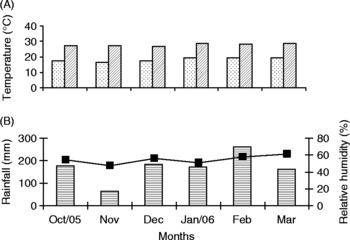
The present study was carried out in the city of Botucatu, São Paulo State, Brazil, from October 2005 to March 2006. During this period, mean monthly maximum and minimum temperatures ranged from 28.6°C to 19.5°C in January and from 27.1°C to 16.7°C in November, respectively (fig. 1).
(A) Mean minimum ( ) and maximum (
 ) temperatures, (B) total rainfall (
 ) and mean relative humidity (■) in Botucatu, São Paulo State, Brazil, from October 2005 to March 2006.

Twenty female Ile de France lambs, approximately 3 months old, were used. Before the beginning of the experiment, faecal samples were collected from each animal for faecal egg counts (FEC). The FEC values were determined using a modified McMaster technique, in which each nematode egg counted represented 100 eggs/g of faeces. Based on these values, animals were ranked in pairs. Then, a lamb of each pair was randomly allocated to one of the two experimental groups: one group was fed with the fungus (treated group) and the other group did not receive the fungus (control).
An area of 0.6 ha was divided into two paddocks (0.3 ha each) presenting similar pasture and topography. Control and treated groups were kept separately (each group in one of the paddocks) in continuous grazing from 7 October 2005 to 24 March 2006. Pastures consisted of Cynodon dactylon and Brachiaria decumbens and were kept without animals for 18 months prior to the experiment.
The nematode-trapping fungus D. flagrans (AC 001 isolate) was isolated from soil in Viçosa, Minas Gerais State, Brazil, according to the soil-spreading method of Duddington (Reference Duddington1955), modified by Santos et al. (Reference Santos, Ferraz and Muchovej1991). The isolate was replicated periodically and kept in test tubes containing 2% cornmeal agar (CMA 2%), at 4°C in the dark. Mycelia mass was obtained by inoculating culture discs (approximately 5 mm diameter) containing 2% CMA. The culture was incubated in 250 ml Erlenmeyer flasks containing 150 ml liquid GPY culture medium (glucose, sodium peptone and yeast extract, pH 6.5, kept under 120 rpm agitation), kept in the dark at 25°C, for 7 days.
Sodium alginate pellets were prepared with D. flagrans mycelia in liquid culture medium, as described by Walker & Connick (Reference Walker and Connick1983) and modified by Lackey et al. (Reference Lackey, Muldoon and Jaffe1993). Pellets contained 20% fungus and were administered to the animals of the treated group (1 g pellet for each 10 kg body weight) mixed in 1.2 kg of supplement feed (Ração Noel®, Cafenoel, São Manoẽl Brazil) twice a week (Tuesdays and Fridays). Animals of the control group received a similar amount of sodium alginate pellets prepared without fungus and mixed in the same quantity of supplement feed.
All animals were vaccinated against clostridial infections (Clostridium chauvoei, C. septicum, C. perfringens, C. novyi, C. sordellii and C. tetani, Sintoxan Polivalente® (Merial, Paulinia, Brazil) at the beginning of the experiment. Lambs had free access to mineralized salt (Nutrumin®, Nutrumin, Botucatu, Brazil) and drinking water throughout the experimental period.
High FEC were observed at the end of the first month (27 October 2005); thus, all animals were treated with anthelmintic 20 days after entering the experimental pasture. Therefore, animals with high FEC contaminated the pastures in the first weeks of the study, before of the treatment. To prevent deaths, salvage anthelmintic treatments were provided individually to animals presenting FEC higher than 4000 (Tembely et al., Reference Tembely, Lahlou-Kassi, Rege, Mukasa-Mugerwa, Anindo, Sovani and Baker1998; Amarante et al., Reference Amarante, Craig, Ramsey, El-Sayed, Desouki and Bazer1999) and/or packed cell volume lower than 21% (Amarante et al., Reference Amarante, Craig, Ramsey, El-Sayed, Desouki and Bazer1999). An association of levamisole phosphate at 10 mg/kg (Ripercol®, Fort Dodge, Campinas, Brazil) with albendazole at 10 mg/kg (Valbazen® 10 Cobalto, Pfizer, Guarulnos, Brazil) was used in the treatments. All animals were treated with trichlorfon (100 mg/kg; Neguvon®, Bayer, Belford Roxo, Brazil) on 1 February 2006, to control Oestrus ovis parasitism.
Measurements
All experimental samples were collected fortnightly. Faecal samples were collected directly from the rectum of animals for faecal cultures and FEC determination. Composite faecal cultures (Roberts & O'Sullivan, Reference Roberts and O'Sullivan1950) were prepared separately for each group, and the infective larvae obtained were identified according to Keith (Reference Keith1953). On the same day as faeces collection, blood samples were taken from the jugular vein using vacutainer tubes containing EDTA. Packed cell volume (PCV) was determined using micro-haematocrit centrifugation. Total serum protein concentration was estimated using an optical refractometer (Model SPR-N, Atago, Japan).
To determine the number of infective larvae (L3) per kilogram of dry matter (L3/kg DM), grass samples were collected manually from both paddocks, close to the soil, approximately every 3.5 m. The collector followed a W-track on the paddock (Taylor, Reference Taylor1939). Samples were processed in the laboratory according to Amarante & Barbosa (Reference Amarante and Barbosa1995), and the infective larvae obtained were identified according to Keith (Reference Keith1953).
Statistical analysis
The data obtained in the determination of FEC, PCV and total serum proteins were subjected to one-way analysis of variance using the Minitab program (release 11; State College, Pennsylvania, USA). Differences between means were considered significant when P < 0.05. The FEC values were analysed using logarithmic transformation − log10(x+1); however, in the figures, they were presented as arithmetic means. The numbers of larvae on herbage of both paddocks were classified as inferior or superior to 300 L3/kg DM and compared using the chi-square test.
Results
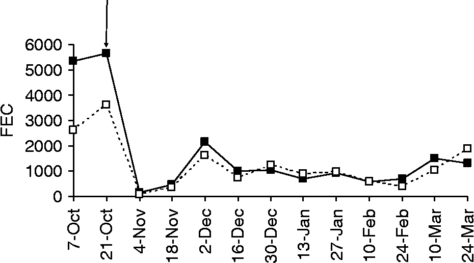
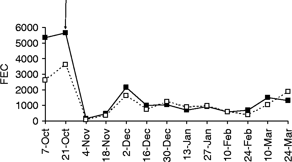
Groups showed similar mean FEC (fig. 2) throughout the experiment (P > 0.05), except on 10 February 2006, when mean FEC of the treated group was significantly lower than that of the control group (P < 0.05).
Arithmetic mean fecal egg counts (FEC) of lambs fed with the fungus Duddingtonia flagrans (□) or not fed (■). The arrow indicates the treatment with albendazole + levamisole.
To prevent deaths, individual anthelmintic treatments were administered to some animals. On 2 December 2005, three animals of the control group and two animals of the treated group required salvage anthelmintic treatment. On 16 December 2005 and on 27 January 2006, one lamb of each group was treated. Later, on 24 March 2006, two lambs of the treated group received anthelmintic treatment. Even after several anthelmintic treatments, one highly susceptible lamb of the control group died on 20 March 2006. On the other hand, six animals of the control group and eight of the treated group did not require individual treatments.
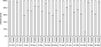
Only Haemonchus spp. and Trichostrongylus spp. infective larvae were identified in faecal cultures of both control and treated groups (fig. 3). On average, faecal cultures of the treated group showed 74.2% Haemonchus spp. and 25.9% Trichostrongylus spp.; faecal cultures of the control group presented 69.7% Haemonchus spp. and 24% Trichostrongylus spp.
Percentage of Haemonchus spp. () and Trichostrongylus spp. () infective larvae identified in faecal cultures of lambs fed with the fungus Duddingtonia flagrans (treated) or not fed this fungus (control).

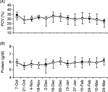
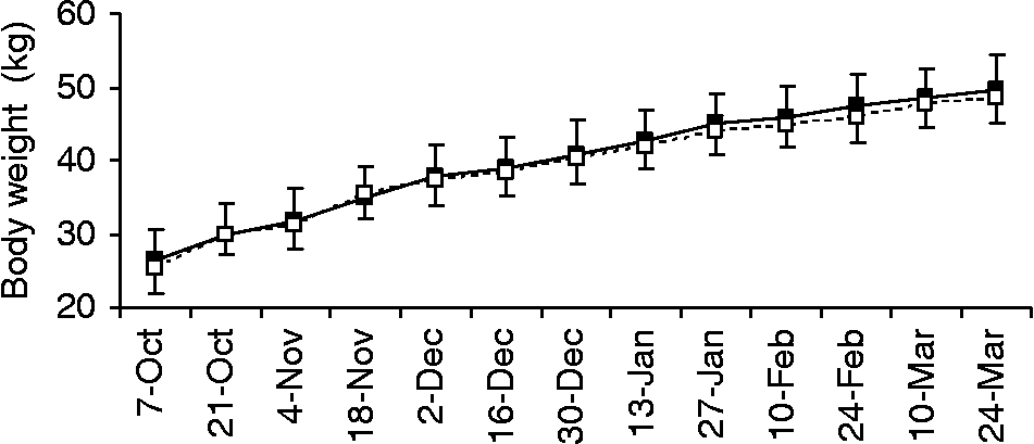
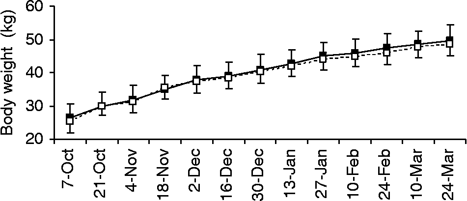
Comparison of mean PCV, total serum protein and weight gain showed no significant differences between groups (figs 4 and 5).
(A) Mean packed cell volume (PCV) and (B) total serum protein of lambs fed with the fungus Duddingtonia flagrans (□) or not fed this fungus (■). Bars: standard deviation.

Mean body weight (kg) of lambs fed with the fungus Duddingtonia flagrans (□) or not fed this fungus (■). Bars: standard deviation.
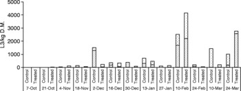
Similarly to faecal cultures, pastures presented only Haemonchus spp. and Trichostrongylus spp. infective larvae, generally at equivalent percentages on both paddocks (fig. 6). However, the paddock grazed by control animals presented higher numbers of Trichostrongylus spp. L3/kg DM on 4 November 2005, 16 December 2005, 13 January 2005 and 24 February 2006, and of Haemonchus spp. larvae on 18 November 2005 and 2 December 2005. The opposite occurred on 10 February 2006 and 24 March 2006, when a higher number of larvae was found in the paddock grazed by the treated group (fig. 6).
Number of Haemonchus spp. () and Trichostrongylus spp. () infective larvae per kilogram of dry matter (L3/kg DM) on herbage of the paddocks grazed by lambs fed with the fungus Duddingtonia flagrans (treated) or not fed this fungus (control).

Except for those obtained in the first grass collection, the total number of larvae was higher than 300 L3/kg DM on seven occasions in the paddock grazed by the control group and on four occasions in the paddock grazed by the treated group. Such occurrences were not statistically different (χ 2 = 1.51; P > 0.05).
Discussion
The treatment scheme using D. flagrans, tested in the present study, had no evident benefit to the prophylaxis of infections caused by Haemonchus spp. and Trichostrongylus spp. in sheep. Similar results were obtained in The Netherlands, where administration of D. flagrans had no effects on the weight gain, FEC or worm burden of lambs treated daily with 50,000 spores of the fungus (Eysker et al., Reference Eysker, Bakker, Kooyman, Olthuis and Ploeger2006). In Brazil, Graminha et al. (Reference Graminha, Monteiro, Silva, Oliveira and Costa2005) studied the efficacy of Arthrobotrys musiformis fungus in naturally infected sheep and did not detect differences between the FEC of the control group and that of treated group; however, they observed that A. musiformis reduced the number of Trichostrongylus colubriformis by more than 50% in tracer lambs.
Unlike the present study, Gómez-Rincón et al. (Reference Gómez-Rincón, Uriarte and Valderrábano2006), in Spain, reported that daily dosing grazing ewes with D. flagrans clearly reduced autumn and spring pasture contamination. Chandrawathani et al. (Reference Chandrawathani, Jamnah, Waller, Larsen, Gillespie and Zahari2003), in Malaysia, also observed efficacy in daily treatment of sheep with D. flagrans. Fontenot et al. (Reference Fontenot, Miller, Peña, Larsen and Gillespie2003), in the United States, reported L3 reduction on pasture and in faecal cultures after daily administration of D. flagrans; however, FEC, PCV or animal weight did not differ between fungus-fed and control groups. Terrill et al. (Reference Terrill, Larsen, Samples, Husted, Miller, Kaplan and Gelaye2004) also reported a reduction in larvae in the faeces of goats predominantly infected by Haemonchus contortus. These authors observed that daily supply of the fungus (D. flagrans) provided more consistent larval reduction than intermittent feeding (every 2 or 3 days).
In the studies cited above, good results were obtained when animals had daily access to the fungus. The frequency of the treatments used in the present study (only twice a week) was probably insufficient to reduce pasture contamination. However, promising results have been obtained in Brazil with the administration of Monacrosporium thaumasium fungus to cattle twice a week; treated animals presented a lower degree of infection than those of the control group (Alves et al., Reference Alves, Araújo, Guimarães, Assis, Sarti and Campos2003; Araújo et al., Reference Araújo, Guimarães, Campos, Sá, Sarti and Assis2004).
The dosage used (1 g/10 kg live weight) may also have been insufficient. Araújo et al. (Reference Araújo, Freitas, Vieira and Campos2006) treated goats with a higher dose (20 g/animal) of the same fungus and observed a 59.3% reduction in the production of H. contortus larvae in faecal cultures.
Replicates of the experimental paddocks were not used in the present study, which may have influenced the results, although the two paddocks used were contiguous and pastures were alike. This was only an initial study carried out with sheep in Brazil; further studies aimed at increasing the efficacy of nematophagous fungi in the prophylaxis of sheep parasitic gastroenteritis are needed. Other fungal species may be more adapted to the Brazilian environmental conditions and should be tested for the biological control of gastrointestinal nematodes.
The results obtained also reinforced the importance of identifying animals in a flock that are highly susceptible to parasites. This problem was observed in the present study: some highly susceptible individuals were present in both groups. Susceptible animals are responsible for a great part of pasture contamination and, once identified, they must be removed from the flock.
Acknowledgements
The authors thank CAPES for the grant to R.A.R. and the National Council for Scientific and Technological Development (CNPq) for the grants to A.F.T.A and J.V.A.