Introduction
Massive bone defects caused by infection, tumor, abnormalities, and trauma need treatments to facilitate their repair, replacement, or regeneration. The developing field of tissue engineering (TE) aims to regenerate damaged tissues by combining cells from the body with highly porous scaffold biomaterials, which act as templates for tissue regeneration, to guide the growth of new tissue [Reference Kneser, Schaefer, Munder, Klemt, Andree and Stark1]. The chance to design a patient-specific implant also opens the possibility of designs that improve biomechanical outcomes and promote the standard bony healing response in tissue-engineered grafts [Reference Dean, Min and Bond2, Reference Siu, Rogers, Lin, Thompson and Owbridge3], which should encompass some vital properties, like biocompatibility, osteoproductive potential, degradability, compatible mechanical strength, and appropriate spatial configuration [Reference Hutmacher4, Reference Hollister5].
Composites including polymer and ceramic phases are regarded as particularly effective in obtaining scaffolds appropriate to hard-tissue regeneration by summing up the favorable mechanical strength and bioactivity given by the ceramic phase, and biodegradability, toughness, and flexibility provided by the polymer phase. In this context, hybrid scaffolds comprising biodegradable polymers as poly(lactic acid) (PLA), poly(glycolic acid) (PGA), poly(ɛ-caprolactone) (PCL), and inorganic bioactive phase [hydroxyapatite (HA), bioactive glasses, and tricalcium phosphate (TCP)] as fillers which yielded better mechanical properties and improved biological activity have been successfully manufactured to satisfy the requirements of scaffolds in different areas [Reference Armentano, Dottori, Fortunati, Mattioli and Kenny6]. However, there are still drawbacks in the inherent brittleness of bioceramics and phases separation during processing, both limiting their use in load-bearing area [Reference Abu Bakar, Cheang and Khor7, Reference Li and Tjong8].
Carbon nanotube (CNT) with extraordinary mechanical strength, excellent electrical conductivity, superior flexibility, nanodimensionality, and large aspect ratio was proved to be an outstanding candidate nanofiller to improve the functional properties of the polymer composite [Reference Tasis, Tagmatarchis, Bianco and Prato9]. These characteristics have motivated many investigators to address their applicability in bone TE. Coincidentally, researchers reported that polymeric biocomposites reinforced with multiwalled carbon nanotube (mwCNT) could be used as a bone replacement through enhancing the stiffness, tensile strength, and impacting toughness of the nanocomposite, apart from increasing the number of osteoblasts [Reference Sahithi, Swetha, Ramasamy, Srinivasan and Selvamurugan10, Reference Cheng, Rutledge and Jabbarzadeh11, Reference Liao, Li, Wong, Tong, Yeung and Tjong12, Reference Shokuhfar, Makradi, Titus, Cabral, Ahzi, Sousa, Belouettar and Gracio13]. In addition, the functionalization process of CNTs was at first adopted to improve their solubility, processability, and biocompatibility; however, the functionalized CNTs were proved to further improve the compatibility between non-intermingle components, and the functional groups on their surfaces could serve as nucleation sites for the deposition of hydroxyapatite as a bone matrix in vivo [Reference Zhang, Wen, Zhao and Dai14, Reference Liao, Xu, Wang, Watari, Cui, Ramakrishna and Chan15].
In the present study, we propose the development of nanocomposite scaffolds for bone regeneration using a solvent evaporation method comprising of nanocrystalline hydroxyapatite (HA) and polycaprolactone, enhanced by CNTs. Polycaprolactone is an FDA-approved bioresorbable polyester with slower degradation rate, good thermoplastic processability, and great elastic modulus [Reference Woodruff and Hutmacher16]. Nanocrystalline HA is a suitable ceramic material to manufacture scaffolds for bone TE because the chemical composition is comparable to the inorganic part of the bone and because of excellent biological responses to the physiological environment [Reference Huang, Best, Bonfield, Brooks, Rushton, Jayasinghe and Edirisinghe17]. Previous investigators have confirmed the positive effect of mwCNT as a functional nanofiller for PCL/nHA nanocomposite with 3D porous model. However, the mechanical strength indicated by compressive yield strength is barely satisfactory for the high-weight-bearing areas, probably attributing to the higher nHA content (nearly 50%) chosen for the technological insufficiency dealing with viscous fluid during the extrusion process [Reference Dorj, Won, Kim, Choi, Shin and Kim18, Reference Goncalves, Oliveira, Silva, Neto, Fernandes, Amaral, Regí and Vila19], while high quantity of nHA could blemish the elastic property of the composite by agglomeration within composites [Reference Baji, Wong, Liu, Li and Srivatsan20]. Also, the vital degradation properties which should be comparative with the new bone formation need further discussion, since both the alkaline degradation product of nHA and mwCNTs loading influence degradation properties of PCL in a blend ratio dependent manner [Reference Kim, Knowles and Kim21, Reference Cadek, Coleman, Barron, Hedicke and Blau22]. Taking all these into account, porous architecture, mechanical strength, biological properties, and degradation rate of the raw composites will influence the performance of the advanced engineered template, and should be optimized as far as possible.
In this study, the mwCNT, nHA, and PCL were used as the parent material, and PCL, nHA/PCL, mwCNT/PCL, and mwCNT/nHA/PCL scaffolds were prepared at different blend ratios by a solvent evaporation method. The structures of these composites were characterized by scanning electron microscope and X-ray diffraction, and their mechanical features were compared using the universal material testing system. To learn about the degradation properties in vitro, we soaked the PCL, nHA/PCL, mwCNT/PCL, and mwCNT/nHA/PCL materials in SBF liquid and observed the mass loss, pH value of leaching liquors, and microstructure subsequently. Furthermore, we estimated the bioactivities about MG63 attachment, proliferation, and differentiation on these scaffolds. The results from our present work may contribute to further development of more suitable composites in tissue regeneration application.
Results and discussion
Structure characterization of the composite scaffolds
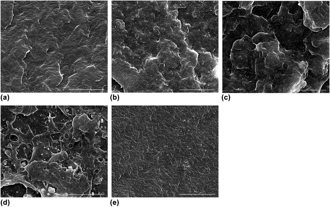
In this study, composite scaffolds were fabricated by a solution evaporation method, and the sonication technique was used to break the agglomeration of carboxyl mwCNT and nHA, which was vital for the dispersion of nanofiller in a polymeric matrix. Figure 1 represents the SEM images of different composite materials. According to Fig. 1(b), after the ultrasonic process, the functionalized mwCNTs of 1 wt% were distributed uniformly throughout the scaffold and aligned with certain direction into the matrix, which was attributed to the enhanced interaction between carboxyl groups and polymer. All the mwCNTs were surrounded by PCL, which indicated good compatibility between mwCNTs and PCL. However, increasing concentration of mwCNTs agglomerated together and the roughness of the scaffold surface increased [Fig. 1(e)]. Previous researchers have described the reason for this phenomenon as Van der Waals force between mwCNTs [Reference Li and Chou32]. The morphology of nHA in PCL was also shown for comparison, granular nHA dispersed well with a diameter of 10–20 nm and was tightly wrapped by PCL matrix with a concentration of 15%, and the composite surface looked smooth [Fig. 1(c)], but when its weight ratio reached 30%, block mass of nHAs and obvious phase separation between nHA and PCL could be observed [Fig. 1(d)]. A similar clustering behavior of nHA due to the large surface areas was found in the polymer matrix of binary PCL/nHA nanocomposites with high filler loadings, which may become the point where the stress was concentrated and contributed to crack formation [Reference Zhang, Venugopal, El-Turki, Ramakrishna, Su and Lim33, Reference Fu, Ni, Wang, Chu, Peng, Zheng, Zhao, Luo, Wei and Qian34].
SEM images of composite materials. (a) PCL only. (b) mwCNT/nHA/PCL. The mass fraction of mwCNT and nHA is 1% and 15%, respectively. (c) nHA/PCL with 15 wt% nHA. (d) nHA/PCL with 30 wt% nHA (e) mwCNT/PCL with 2 wt% mwCNT (black part is PCL matrix, lighter part is mwCNT or nHA).

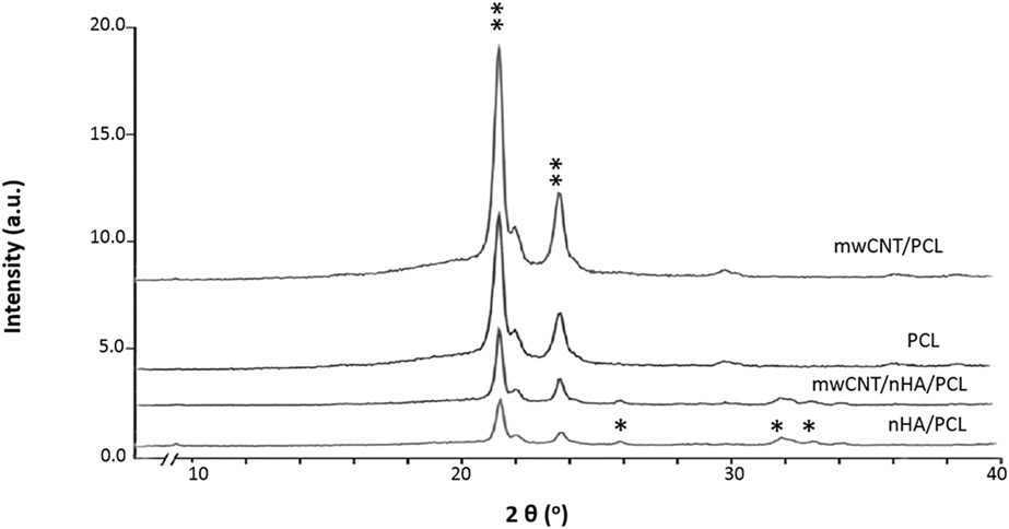
Figure 2 shows typical XRD patterns of pure PCL, mwCNTs/PCL, nHA/PCL, and mwCNT/nHA/PCL scaffolds. In all composites, two peaks at Bragg angles 2θ = 21.4° and 23.8° were observed, which were assigned to the diffraction of the (110) lattice plane and the (200) lattice plane of the semi-crystalline PCL, respectively [Reference Zhang, Leblanc-Boily, Zhao and Prud’homme35]. Also, it was observed that the XRD patterns of the nHA/PCL and mwCNT/nHA/PCL composites still had the characteristic peaks of nHA at 25.8°, 32°, and 32.9°. These were consistent with the results of the previous studies that incorporation of mwCNTs did not significantly change the chemical structure of PCL and nHA components [Reference Dorj, Won, Kim, Choi, Shin and Kim18]. It is well known that the peak intensity in XRD patterns can intuitively represent the maximum extent of crystallization [Reference Cho, Saheb, Yang, Kang, Kim and Lee36]. In our case, the intensities of the diffraction peak in PCL and mwCNT/PCL composites were visibly higher than in nHA/PCL and mwCNT/nHA/PCL materials, which may be attributed to the addition of 15% lower nHA in the PCL content consequently. Moreover, after blending with 1% of mwCNT the crystalline peak intensity for PCL at 21.4 and 23.8° 2θ in both mwCNT/PCL and mwCNT/nHA/PCL composites increased compared with the corresponding PCL and nHA/PCL samples, which may be explained by the good disparity of mwCNT facilitating crystallization and nucleation of PCL to some extent [Reference McCarthy, Coleman, Czerw, Dalton, Panhuis, Maiti, Drury, Bernier, Nagy, Lahr, Byrne, Carroll and Blau37].
XRD patterns of the composites (** represents the characteristic peaks of PCL; * indicates the peaks of nHA).

Mechanical properties
Mechanical test results showed that the incorporation of mwCNT and nHA enhanced the compressive and tensile strength of the composites. As the weight ratio of nHA in PCL increased, the compressive modulus of the scaffolds increased accordingly, which implied that nHA helped to enhance the rigidity of nHA/PCL composite scaffolds. By adding 30 wt% nHA, the modulus enhancement was nearly 95% (39.2 ± 2.0 versus 22.6 ± 1.4, n = 6, Table I). The stiffness of the nHA/PCL composites could be further increased by incorporating low loading levels of mwCNTs (≤1 wt%) into the matrices. Take the mwCNT/nHA/PCL composite with 5 wt% nHA and 0, 0.5, 1 wt% mwCNTs as an example; the compressive strength turned out to be 25.0 ± 1.7, 28.3 ± 0.8, and 32.4 ± 1.6 MPa. Considering the tensile strength of PCL/nHA composites, it increased and saturated at 5–15 wt% nHA content and then decreased slightly with further nHA addition. The strength reduction was related to the agglomeration of nHA fillers at high concentrations and may spoil the force transduction property of the material. Similarly, the tensile strength of PCL/nHA composites is also improved considerably by adding mwCNTs, which increased and saturated at 0.5–1 wt% content. From Table I, it is clearly seen that when the weight ratio of mwCNT is 1 wt% and nHA is 15 wt%, the composite had maximal mechanical strength with compressive and tensile modulus increasing by 91% and above 2-folds, respectively, compared with pure PCL bulk, and both were significantly enhanced compared with groups with 2 wt% mwCNTs added (42.4 ± 1.3 versus 34.7 ± 0.7; 35.3 ± 0.3 versus 23.4 ± 1.4. P < 0.01). This result was contrary to the conclusion of previous studies by Pan et al. and Luo et al. [Reference Pan, Pei, He, Wan and Wang38, Reference Luo, Pan, Hong, Wang, Pei, Wang and Wan39], who reported that 2 wt% might be the threshold concentration of mwCNTs in the matrix for enhancement, while 0.5 wt% mwCNTs/PCL had better induction effects on growth and proliferation of BMSCs. It is worth noting that those studies were binary without the participation of nHA. Considering that bioactive property was as important as mechanical strength for ideal bone-repair materials, we choose mwCNT/nHA/PCL (1/15/84 wt%), mwCNT/PCL (0.5/99.5 wt%), and nHA/PCL (15/85 wt%) scaffolds for the following experiments.
Compressive strength and tensile strength of composites.

In vitro immersion test in SBF
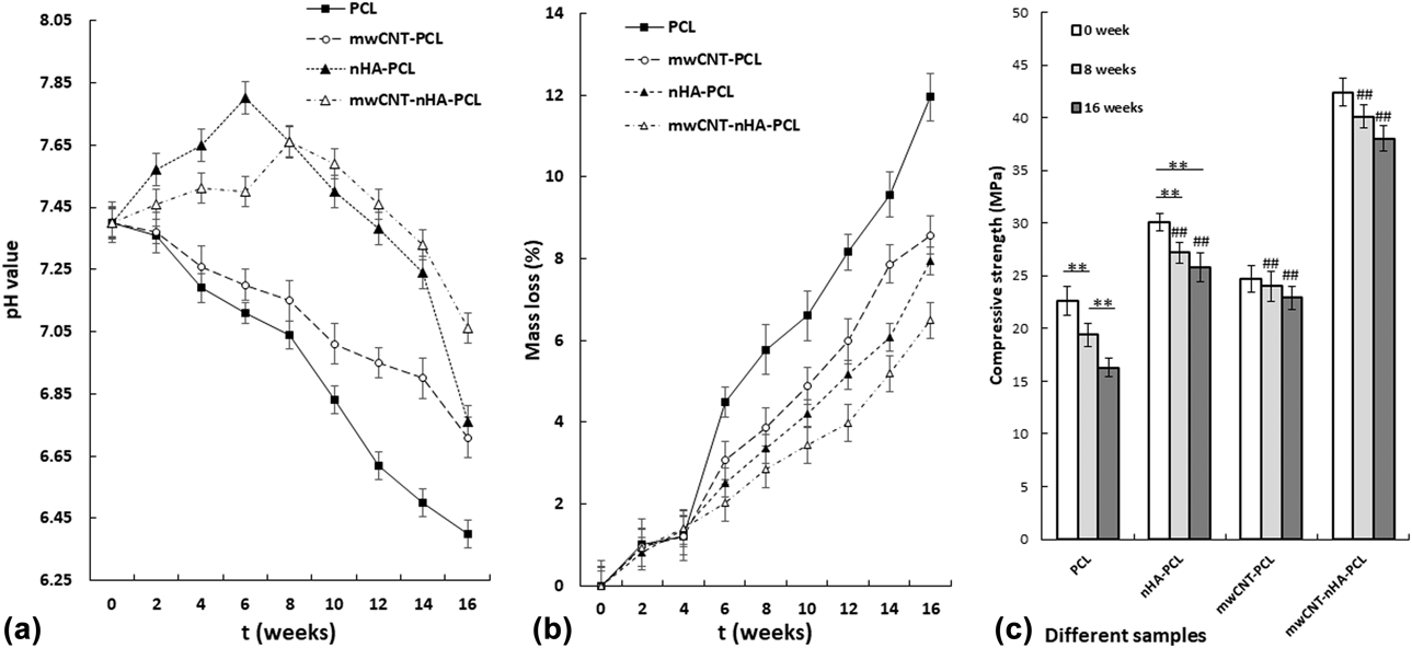
The ideal degradation rate of material is supposed to coordinate with the rate of new bone formation, initially slowly to maintain mechanical strength, and subsequently accelerated to provide sufficient space for new bone formation. Also, a proper degradation rate always companied better biocompatibility due to less immunological response [Reference Babensee, Anderson, McIntire and Mikos40]. According to Fig. 3(a), the pH values of the pure PCL experienced a significant decrease from 7.4 to 6.25–6.5 after being soaked in the SBF solution for 4 months. The decrease in pH values was attributed to the continuous generation of carboxylic groups during the PCL degradation process [Reference Persenaire, Alexandre, Degee and Dubois41]. The mwCNT/PCL group shared similar pH values and a similar degradation trend with PCL groups, which indicated that the addition of mwCNTs had little effect on the degradation of the PCL. However, adding of hydrophilic nHA leads to an increase in pH value at sixth week because of the OH− product by nHA dissolution [Reference Kim, Knowles and Kim21]. Lately, pH values of both nHA/PCL and mwCNT/nHA/PCL group dropped as a result of accumulated acidic products, implicating the scaffolds were gradually degraded in SBF and could leave sufficient space for cell proliferation and new tissue growth. To our surprise, the pH of mwCNT/nHA/PCL group was kept near the physiological range (7.2–7.6), which implicated better biocompatibility than other composites.
(a) PH values, (b) weight loss, (c) the compressive strength of the composites immersed in SBF for 4 months. (Mean ± SD, n = 6). **indicates significant difference (P < 0.01) between groups connected by the underline, and ## means the significant difference (P < 0.01) between the group and the corresponding PCL group at each time point.

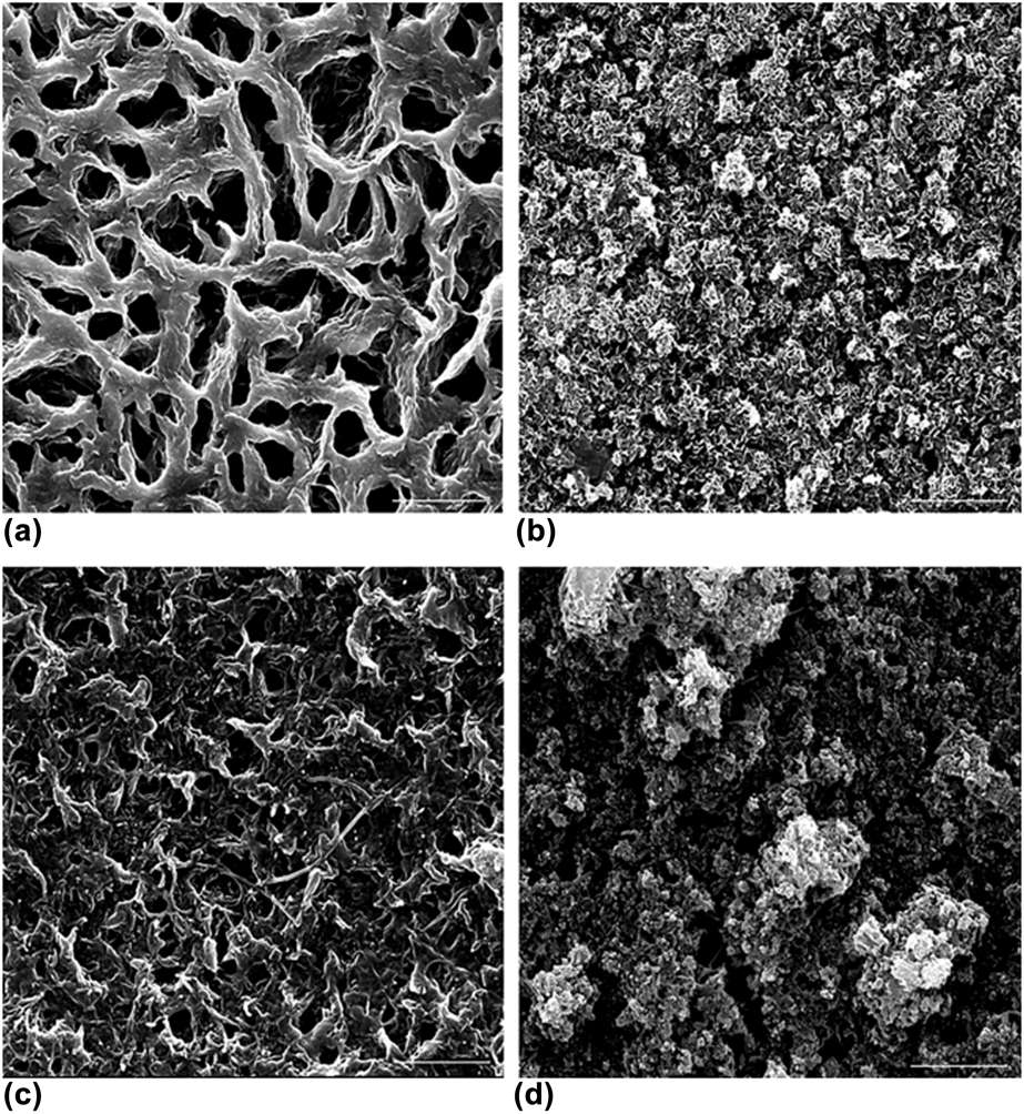
Mass loss would indicate the onset of degradation, which is deduced via a surface degradation pathway for PCL, and the surface erosion is in agreement with consistent mass loss as the material is removed from the surface inwards towards the core, throughout the degradation period [Reference Lam, Savalani, Teoh and Hutmacher42]. Weight loss of the four scaffolds synchronized with the pH change in the SBF [Fig. 3(b)]. The PCL group showed the fastest decline, which was due to autocatalytic hydrolysis of PCL due to acidic by-product retained within the pits and caverns as surface erosion sites [Fig. 4(a)] [Reference Meseguer-Duenas, Mas-Estelles, Castilla-Cortazar, Escobar Ivirico and Vidaurre43]. Noticeably, although the initial alkaline environment could retard the degradation rate of the PCL, the nHA/PCL and mwCNT/nHA/PCL group displayed more weight loss than the PCL and mwCNT/PCL groups in the first four weeks because of exceptional nHA dissolution, which act as ‘defects’ for water ingression in the polymer matrix tipping the PCL degradation. With increasing time, OH− began to enrich on the composite surface and attracted Ca2+, CO32−, and PO43− in SBF solution, which promoted the CHA nucleation and growth compensating the degradation mass [Reference Lei, Rai, Ho and Teoh44]. Moreover, mwCNT/PCL group showed less weight loss compared with PCL, and mwCNT/nHA/PCL group displayed the least weight loss among the four scaffolds, both indicating that mwCNTs could decrease the degradation rate of composites. The mwCNTs might be attributed to the enhanced crystallization of PCL, and the well-dispersed mwCNTs could interweave into a network preventing the dissociation of composite material into SBF solution and incursion of water into the material [Fig. 4(c)] [Reference Cadek, Coleman, Barron, Hedicke and Blau45].
SEM images of the four scaffolds after being soaked in SBF solution for 16 weeks (a) PCL, (b) nHA/PCL, (c) mwCNT/PCL, and (d) mwCNT/nHA/PCL. Scale bar = 5 μm.

The compressive property of the composites after degradation is illustrated in Fig. 3(c). The PCL and nHA/PCL groups experienced dramatic weight loss after degradation for four months (P < 0.01) and the compressive modulus of the PCL group decreased to 70.2% compared to its original value. In contrast, the mwCNT/PCL and mwCNT/nHA/PCL composites behaved differently within the same observation period. They held their mechanical properties and degraded at a similar ratio. After degradation, the compressive strength of the nHA/PCL, mwCNT/PCL, and mwCNT/nHA/PCL scaffolds were significantly higher than pure PCL (P < 0.01), and the mwCNT/nHA/PCL group possessed the best compressive property, which enabled the scaffolds to provide sufficient mechanical support and to ensure the structural stability and the integrity, particularly, at the early stage of implantation.
The presence of bone-like apatite on the scaffolds was confirmed by SEM analyses [Figs. 4(b) and 4(d)]. The surface of the nHA/PCL group and mwCNT/nHA/PCL group appeared rough, coated with spherical globes, and formed a white flocculent sediment layer. The amount and size of the spherical globules of mwCNT/nHA/PCL group were more significant than the nHA/PCL group and clusters of globules obscured the micropores on the surface of the material, while PCL and mwCNT/PCL groups appeared smooth with no precipitates. As reported previously, bone-like hydroxyapatite formed on the scaffold surface in SBF was useful for predicting in vivo bioactivity of the material not only qualitatively but also quantitatively [Reference Kokubo and Takadama46]. The “microrough” implants were able to enhance the rate and quality of osseointegration, with greater bone-to-implant contact and greater resistance to torque removal [Reference Thalji, Gretzer and Cooper47, Reference Costa, Prowse, Chrones, Sims, Hamilton, Rizkalla and Jeffrey Dixon48]. In summary, the mwCNT/nHA/PCL composite seemed to have better mechanical property and bioactivities for bone TE application.
Effects on attachment, proliferation, and differentiation of MG63
Cell adhesion assay is one of the most important indexes for evaluating the biocompatibility of bioactive materials, for bone repair material; it promotes osteoblasts to adhere and proliferate on the surface of the material which may facilitate the osseointegration of implants [Reference Freed, Vunjak-Novakovic, Biron, Eagles, Lesnoy, Barlow and Langer49]. After incubation for 4 h, the vast majority of MG63s attached to the nHA/PCL and mwCNT/nHA/PCL scaffolds with an adhesion rate of 92.5% and 95.7%, respectively. The cellular adhesion of PCL and mwCNT/PCL scaffolds was slightly inferior to the former groups, being 76.4% and 84.1%, respectively. It is well known that surficial physicochemical characteristics of the implanted materials would affect their interaction with cells, including the surface composition, morphology, energy, charge, and even degradation activity [Reference Yamashita, Machigashira, Miyamoto, Takeuchi, Noguchi, Izumi and Ban50]. In our study, both mwCNTs and nHA have large aspect ratios and exceptionally high surface energy, and the ability to absorb proteins onto the material surface enhanced the cell affinity of materials [Reference Huang, Best, Bonfield, Brooks, Rushton, Jayasinghe and Edirisinghe17, Reference Mata, Oliveira, Ferro, Gomes, Fernandes, Lopes and Silva51], while nHA prompted MG63s adhesion to a more considerable extent which may be attributed to its high content in the composite.
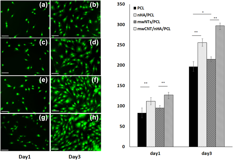
The morphology of MG63s cultured on the surface of composite scaffolds was observed using a fluorescence microscope at 24 and 72 h. As is known universally, the interaction between cells and the physical surface plays an essential role in the outcome of the cells, influencing biological processes such as cell proliferation and differentiation [Reference Behring, Junker, Walboomers, Chessnut and Jansen52]. On the first day, cells anchored tightly on the surface of all scaffolds and exhibited a spindle shape morphology, indicating that cells had finished attachment and were in the progress of spreading. MG63 cells cultured on the nHA/PCL and mwCNT/nHA/PCL scaffolds demonstrated a higher occurrence of the protrusive subcellular features such as filopodia (microspikes), indicating that the cells preferred the surface of nHA-composited scaffolds over that of mwCNTs/PCL and pure PCL scaffolds, while the morphology of MG63s was inferior on pure PCL scaffolds. Moreover, the denser and divided nucleus of nHA/PCL and mwCNT/nHA/PCL groups represented flourishing mitosis activity on these two scaffolds [Figs. 5(a)–5(d)]. On the third day, the morphological differences of MG63 cells cultured on the nHA/PCL or mwCNT/nHA/PCL scaffolds compared with PCL scaffold were more significant, and the cells demonstrated tighter packed manner [Figs. 5(e)–5(h)]. It is worth noting that the amount and morphology of MG63 on mwCNTs/PCL scaffolds were better than on pure PCL scaffold [Figs. 5(e) and 5(f)] that might be associated with the rough surface topography caused by the addition of mwCNTs [Reference Raffaini and Ganazzoli53]. After quantifying the immunofluorescence data, we find that the cell numbers on the mwCNTs/PCL, nHA/PCL, and mwCNT/nHA/PCL scaffolds were all significantly higher than in the pure PCL scaffolds after 72-h culture.
Images and quantification results of MG63 cultured for 1 day and 3 days under fluorescence microscope. (a, e) MG63s cultured on pure PCL. (b, f) MG63s cultured on mwCNTs/PCL scaffold. (c, g) MG63s cultured on nHA/PCL scaffold. (d, h) MG63s cultured on mwCNT/nHA/PCL scaffold. Scale bar is 100 µm.

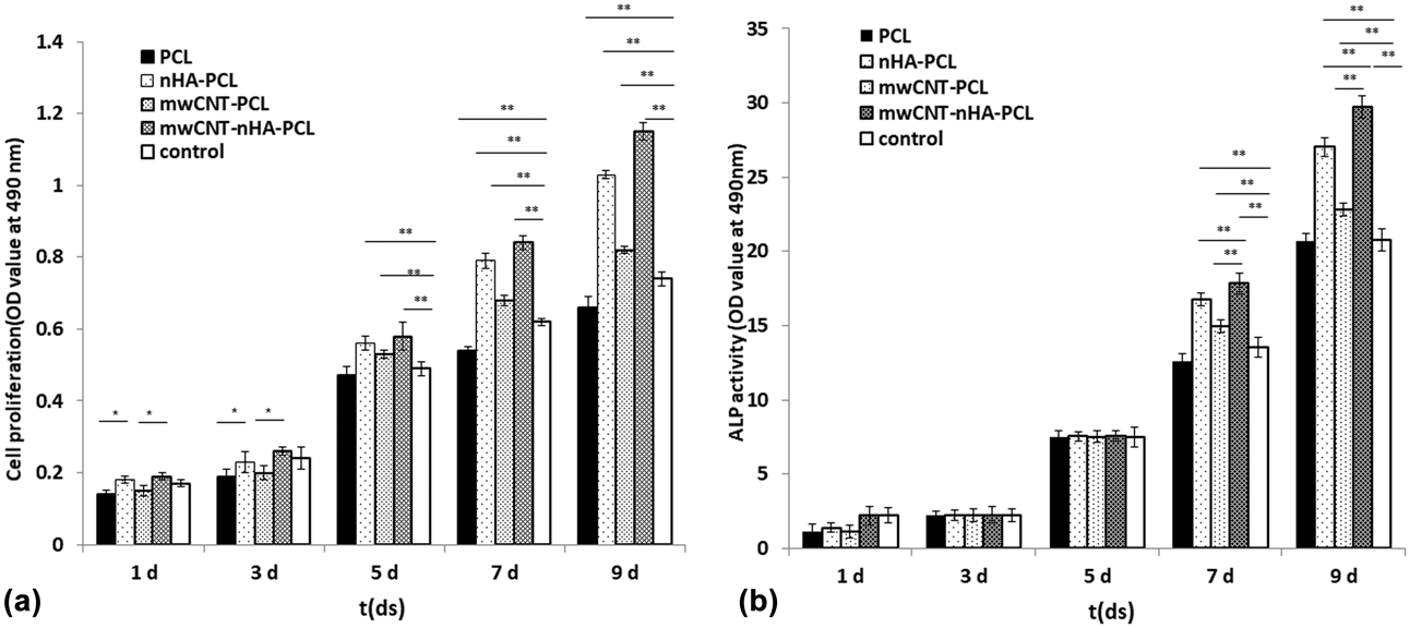
The proliferation of MG63s cultured on each scaffold was also quantitatively assessed by an MTT assay. As shown in Fig. 6, there was no significant difference in cell proliferation between nHA/PCL and mwCNT/nHA/PCL groups with the control group on the first and third days, but the OD values of MG63s in nHA/PCL and mwCNT/nHA/PCL composite groups remarkably overmatched pure PCL and mwCNT/PCL groups (P < 0.05). From the fifth day on, cell proliferation of mwCNT/PCL, nHA/PCL, and mwCNT/nHA/PCL groups were significantly higher than those of control groups (P < 0.01), indicating a promotion effect of mwCNTs and nHA on cell proliferation. On the contrary, OD values of PCL group were always significantly lower than the control group (P < 0.01), which may be attributed to the poor adhesion affinity of PCL [Reference Lee, Jeong, Jeong, Park, Bae, Kim and Cho54]. In addition, the OD values of all groups increased with the culture time, which proved their good biocompatibility.
(a) Cell proliferation ability and (b) ALP values of MG63 cells cultivated for 1, 3, 5, 7, and 9 days on four scaffolds (OD, Mean ± SD, n = 3).

ALP activity was usually used as a biochemical marker for determining osteoblast phenotype and was considered as an important factor in determining bone differentiation and mineralization [Reference Marom, Shur, Solomon and Benayahu55]. The results of ALP activity assay of MG63s are shown in Fig. 6(b). During the first 5 days of incubation, low ALP activity was detected in all scaffolds with the pure PCL group demonstrating the least activity, and there was no significant difference between the four scaffolds and the control group (P > 0.05), which could be explained as follows: cell mineralization activity had not yet started in the early period of cultivation and the ALP value was mainly connected with the number of attached cells on the scaffold. On the 7th day, cells cultured on the surface of composite scaffolds presented extremely high activity than that on cell culture plates or pure PCL scaffolds (P < 0.01), confirming the promotion effects of nHA and functional mwCNTs on cellular differentiation as previously reported [Reference Morelli, Salerno, Holopainen, Ritala and Bartolo56, Reference Abarrategi, Gutierrez, Moreno-Vicente, Hortiguela, Ramos, Lopez-Lacomba, Ferrer and Monte57, Reference Im, Li, Wang, Zhang and Keidar58]. In addition, cells cultured on the mwCNT/nHA/PCL scaffolds showed higher ALP activity than nHA/PCL and mwCNTs/PCL group (P < 0.01), and the mwCNT/nHA/PCL scaffold group presented a peak of ALP secretion after 9 days of culture, suggesting that this scaffold provided a more favorable microenvironment for cell differentiation into bone.
Conclusions
The perfect graft material would, for example, be readily available, require a minimal donor site, be an ideal match for the defect, be highly biocompatible, and allow the host bone to remodel. Within the limitations of this study, mwCNT/nHA/PCL composites with a weight content of 1/15/84 wt% possessed superior mechanical properties, modest degradation rate, and better inducing effects on the proliferation and differentiation of MG63s than nHA/PCL, mwCNT/PCL, and pure PCL materials. It was concluded that it has the potential to be used as a raw material for bone TE scaffold. Further investigations may be aimed at facilitating the application of this promising raw material in 3D porous structure, even in further individualized medicine through reasonable processing.
Materials and method
Preparation of composite materials and molding
Functionalized multiwalled carbon nanotubes (mwCNTs-COOH) were supplied by Chengdu Organic Chemicals Co., Ltd. (Chengdu, Sichuan, China), with the diameter being less than 8 nm and the length ranging from 10 to 30 μm. The nHA powder with particle size of 160–200 mesh was provided by Bioengineering Materials Institute of Sichuan University (Chengdu, Sichuan, China). The PCL pellets (Mw = 50,000 g/mol, density = 1.1 g/cm3) were supplied by Shenzhen Brightchina Industrial Ltd. (Shenzhen, Guangzhou, China). Weight content of 0, 0.5, 1, and 2 wt% for mwCNTs was adopted, with 0, 5, 15, and 30 wt% for nHA, and PCL was added as a balance. Sixteen kinds of experimental specimens were prepared and divided into pure PCL, mwCNTs/PCL, nHA/PCL, and mwCNTs/nHA/PCL groups according to their different constituents.
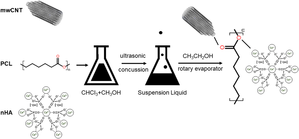
For composite fabrication, the ingredients were added to a ψ solution (trichloromethane:methanol = 1:3) simultaneously and went through ultrasonic concussion for 2 h, then were stirred for 12 h on a magnetic stirrer followed by sonication for 2 h on an ultrasonic cell crusher (JY92-II, Ningbo Scientz Biotechnology Co., Ltd., Ningbo, China) to achieve a homogeneous solution; the scheme is represented in Fig. 7. The ψ solvent was removed in a rotary evaporator (RE III, BÜCHI Labortechnik AG, 9230 Flawil, Switzerland) after adding high quantity of absolute ethyl alcohol. Each mixture was dried at 40 °C in vacuum overnight and continually placed in ventilation environment for one week to volatilize residual solvent. HAAKE MiniJet (Thermo Electron (Karlsruhe) GmbH Dieselstr, 76227 Karlsruhe, Germany) was adopted to cast them into a substantial cylinder or dumbbell shape slowly after preheating for 3 min at 80 °C, and the mold was kept at 35 °C to avoid any air residue.
Scheme for preparation of mwCNT/nHA/PCL composites.

Structure characterization of composite scaffolds
The morphology of the composites was observed under scanning electron microscopy (SEM). Before observation, samples were cryofractured by hand after being immersed in liquid nitrogen.
Micrographs were taken with an accelerating voltage of 20 kV. The constituents were analyzed with an X-ray diffractometer (Rigaku D/max-rA) equipped with Cu Kα radiation source (35 kV, 20 mA); all the composite scaffolds were cut into thin pieces with 1 mm thickness and the surface sections of these pieces were scanned at the rate of 5°·min−1.
Mechanical properties
The tensile and compressive strengths were obtained after the composite specimens were immersed for 24 h in simulated body fluid (SBF) and excess moisture was absorbed by filter paper. A universal material testing system (Instron 5567, Canton, Massachusetts) was used for all mechanical tests at room temperature. Specimens for the tensile test were fabricated to dumbbell shape, and tensile tests were performed by ASTM D638 with a crosshead speed of 5.0 mm/min and a gauge length of 10 mm. The compressive tests were conducted following ASTM standards D695, a loading rate at 5 mm/min was applied to the compressive specimens till half of its original length was reached. At least six specimens were used in both mechanical tests for each group.
In vitro immersion test in SBF
SBF solution was an acellular aqueous solution with inorganic ion composition nearly equal to the composition of human plasma as proposed by Kokubo et al. [Reference Kokubo, Kim and Kawashita23], and it was made following the same protocol. The solution was previously filtered with a 0.23-mm Millipore filter to avoid any microorganism contamination. Scaffolds with specified weight content of mwCNTs and nHA in PCL, namely mwCNTs/PCL scaffold (0.5/99.5 wt%), nHA/PCL scaffold (15/85 wt%), and mwCNTs/nHA/PCL (1/15/84 wt%) scaffold, together with the pure PCL scaffold, were precisely weighed and placed in a plastic tube containing quantitative SBF (pH 7.4) at 37 °C for up to 16 weeks. The volume of SBF was determined by the equation VS = Sa/10 (mL) (Sa represents the apparent surface area of the specimen; VS is the volume of SBF) [Reference Boyd, Towler, Wren, Clarkin and Tanner24]. The SBF was replaced every week. At set time-points (8 and 16 weeks), three specimens were taken off from each group, washed by deionized water for three times and cryodesiccated at −75 °C in vacuum to constant weight. The weight loss was determined gravimetrically by comparing the dry weight at specific time points with the initial weight using an electronic balance (Sartorius BP 210-S, ±0.0001 g, Sartorius AG, 37079 Goettingen, Germany), and pH value of the harvested SBF was measured at 37 °C with an Oakton 1100 pH meter. The dried specimens were further measured on Model 5567 Instron Universal Testing Machine (ADMET, Norwood, Massachusetts) to study the change in the compressive modulus. At the 16th week, coated samples were thoroughly washed with distilled water and air-dried. The surface morphology of the composites was assessed by SEM (Fig. 1).
Effects of the composite scaffolds on attachment, proliferation and differentiation of MG63
The biological properties of the four special composite scaffolds were investigated with human osteoblast-like cell line MG63. Prior to cell seeding, all the samples were cut into cylinders with a diameter of 10 mm and thickness of 2 mm, sterilized in 75% ethanol for 24 h, washed with fresh PBS solution for 2 h, and then immersed in the culture medium with 10% fetal bovine serum (FBS) (Hyclone Laboratories, Inc., Logan, Utah) and 1% penicillin–streptomycin (Beijing Solarbio Science & Technology Co., Ltd., Beijing, China) overnight for use [Reference Zhong, Lu, Valtchev, Wei, Zreiqat and Dehghani25, Reference Hwang, Sangaj and Varghese26]. Human osteoblast-like cell line MG63 was provided by the State Key Laboratory of Oral Disease Research of Sichuan University and cultured in 75-cm2 cell culture flasks with Dulbecco’s modified eagle medium (DMEM) (Gibco, Waltham, Massachusetts), supplemented by 10% FBS (Hyclone Laboratories, Inc., Logan, Utah) and 1% penicillin–streptomycin (Beijing Solarbio Science & Technology Co., Ltd., Beijing, China) at 37 °C with 5% humid CO2. After passaging twice, 500 μL MG63s was seeded at a density of 5 × 10³ cells/mL in a 24-well plate for each scaffold. Control group with the absence of the scaffolds was also prepared. All the following tests were performed in triplicate.
Cell adhesion rate
After incubating for 4 h, the scaffolds were removed, and the old cell culture medium was collected. Then, MG63s adhered to the 24-well plate were detached with 200 μL of trypsin–EDTA solution (Cat. No. T4174, Sigma-Aldrich, Inc., St. Louis, Missouri) for 3 min at 37 °C, and 400 μL of DMEM (Gibco, Waltham, Massachusetts), supplemented with 10% fetal bovine serum (FBS) (Hyclone Laboratories, Inc., Logan, Utah), and 1% penicillin–streptomycin (Beijing Solarbio Science & Technology Co., Ltd., Beijing, China) was added [Reference Anselme, Linez, Bigerelle, Le Maguer, Hardouin, Hildebrand, Iost and Leroy27]. The cell density per mL in each well was analyzed with a TC20™ Automated Cell Counter (Cat. No. 145-0102EDU, Bio-Rad, Hercules, California), and cell population was calculated as A 1. The adhesion rate was calculated using the following equation: Adhesion rate = [(A 0 − A 1)/A 0] × 100% (where A 0 is the number of seeded cells).
Acridine orange staining
Acridine orange (AO) is a kind of fluorescent pigment, which can penetrate the cell membrane nondestructively and stain the nuclear acid, and considered a useful tool for assessing the physical activity of cells [Reference Tucker and Lardelli28]. On the first and third days, AO staining was performed to show cell morphology and population on the surface of scaffolds. Briefly, the scaffolds were taken out from the cell culture and rinsed with PBS twice; then 0.01% acridine orange solution (Sigma-Aldrich, Inc., St. Louis, Missouri) was added at room temperature for 15 min; after washing with PBS for two more times to remove the dye absorbed on the scaffolds, the cells were observed by fluorescence microscope (Olympus BX51, Olympus Corporation, Tokyo, Japan). Three randomly chosen fields of view (2 mm2) were photographed from each well, and the number of attached cells was counted.
MTT detection
MTT assay was used to quantitatively evaluate the viability of cells cultured onto the scaffolds. This assay usually shows high correlation with the number of living cells, cell proliferation, and release of mitochondrial matrix enzymes [Reference Vega-Avila and Pugsley29]. After incubation for 1, 3, 5, 7, and 9 days, the MTT assays for each sample were performed. Briefly, 40 μL of 2 mg/mL MTT solution was added to each well and incubated at 37 °C for 4 h; then 420 μL of dimethyl sulfoxide (DMSO) was used for dissolving the produced formazan crystals. From each sample, 200 μL of the dissolved formazan solution was subsequently added into a 96-well plate, and the absorbance at 490 nm was measured using a microplate reader (Thermo Scientific Multiskan MK3, Thermo Fisher Scientific Inc., Waltham, Massachusetts).
Alkaline phosphatase (ALP) activity
Alkaline phosphatase (ALP) is one of the important markers of osteoblast differentiation and maturation, and its activity can reflect the ability of differentiation and calcification of osteoblasts [Reference Anselme30]. ALP activity of MG63s cultured on the composite scaffolds was assayed at the first, third, fifth, seventh and ninth day, respectively. The scaffolds were removed and rinsed with PBS before 300 μL Triton X-100 lysate was added, then submitted to three freeze-thaw cycles, and centrifuged at 14,000 rpm for 10 min at 4 °C. Alkaline phosphatase activity was examined using an ALP assay kit (Nanjing Jiancheng Technology Inc., Nanjing, China), and the optical density (OD) values were read at 405 nm with a Hitachi U-3010 spectrophotometer (Hitachi, Ltd., Tokyo, Japan). Total protein content was determined using Coomassie brilliant blue method as described elsewhere [Reference Neuhoff, Stamm and Eibl31]. The ALP activity was expressed as unit per mg protein.
Statistical method
SPSS version 17.0 software (IBM, Chicago, Illinois) was used for statistical analysis. The data were all expressed as means ± standard deviation. For comparisons between pure PCL, mwCNTs/PCL, nHA/PCL and mwCNTs/nHA/PCL group, one-way factorial analysis of variance (ANOVA) was performed, and differences were considered significant when *P < 0.05 and very significant when **P < 0.01, respectively.