INTRODUCTION
Babesiosis is a tick-transmitted haemoprotazoan disease of domestic animals causing fever, anaemia, haemoglobinuria and even death. To date, 3 Babesia species infective for small ruminants, B. ovis, B. motasi and B. crassa, have been recognized as valid species. B. ovis and B. motasi are two old-line parasites and the vector ticks are Rhipicephalus bursa and Haemaphysalis punctata, respectively (Alani and Herbert, Reference Alani and Herbert1980; Uilenberg et al. Reference Uilenberg, Rombach, Perie and Zwart1980; Levine, Reference Levine1985; Friedhoff, Reference Friedhoff and Ristic1988, Reference Friedhoff1997). Nevertheless, Uilenberg (Reference Uilenberg2006) recently considered that B. motasi may well consist of at least 2 species or subspecies, namely a low pathogenicity group and a high pathogenicity group. B. crassa is a large Babesia species isolated from Iran. It is generally recognized as a valid Babesia species infective for small ruminants, based on morphology, pathogenicity, serology and phylogeny data (Hashemi-Fesharki and Uilenberg, Reference Hashemi-Fesharki and Uilenberg1981; Schnittger et al. Reference Schnittger, Yin, Gubbels, Beyer, Nieman, Jongejan and Ahmed2003) although its vector tick is still unknown.
In China, Chen (Reference Chen1982) and Zhao et al. (Reference Zhao, Li and Min1986) published the first reports of ovine babesiosis in Sichuan and Heilongjiang province, respectively, and identified the pathogens as B. ovis. After that, several strains were isolated in China. These strains can be divided into 2 groups based on the 18S rRNA gene sequences. One group includes Babesia sp. Liaoning, Babesia sp. BQ1 (Lintan), Babesia sp. BQ1 (Ningxian), Babesia sp. Tianzhu, Babesia sp. Madang and Babesia sp. Hebei, and the other just contains Babesia sp. Xinjiang (Liu et al. Reference Liu, Yin, Guan, Schnittger, Liu, Ma, Dang, Liu, Ren, Bai, Ahmed and Luo2007). Recent analysis based on the ribosomal DNA internal transcribed spacer (ITS) sequences suggested that, except for Babesia sp. Xinjiang, all these strains should be considered as B. motasi (=‘B. motasi group in China’) (Niu et al. Reference Niu, Luo, Guan, Liu, Ma, Liu, Gao, Ren, Li, Qiu and Yin2009).
To date, the main detection method for Babesia sp. BQ1 (Lintan) is still the microscopic examination of blood smears, because of its ease and rapid application. However, in subclinical or latent infections, parasites may not be observed because of the low parasitaemia. Guan et al. (Reference Guan, Chauvin, Luo, Inoue, Moreau, Liu, Gao, Oriel, Ma, Liu, Dang, Liu, Ren, Jin, Sugimoto and Yin2008) developed a loop-mediated isothermal amplification (LAMP) method for the diagnosis of Babesia sp. BQ1 (Lintan) infection. This simple and rapid technique can detect parasites only up to 63 days post-infection (p.i.). An enzyme-linked immunosorbent assay (ELISA) has proved useful for the detection of subclinical cases and large-scale field surveys (Birkenheuer et al. Reference Birkenheuer, Levy, Stebbins, Poore and Breitschwerdt2003) because it is sensitive, can be automated for high throughput and is easily standardized. Our aim in this study was to develop an indirect ELISA specific for infection of Babesia sp. BQ1 (Lintan), using merozoite antigen (BQMA) derived from in vitro culture, which would be suitable for sero-epidemiological survey of the disease in China. This ELISA was used to assess both the humoral immune response in experimentally infected sheep and the prevalent situation of Babesia sp. BQ1 (Lintan) on a small scale. The serological relationships between the Chinese Babesia strains were also explored. Finally, the main proteins from BQMA, potentially suitable for use in serodiagnosis, were also identified by Western blot and mass spectrometry.
MATERIALS AND METHODS
Parasites
A monoclonal line (G7) of Babesia sp. BQ1 (Lintan) derived from in vitro culture by limiting dilution, as previously described (Malandrin et al. Reference Malandrin, L'Hostis and Chauvin2004) was maintained and cryopreserved in liquid nitrogen at the Veterinary School of Nantes (Ecole Nationale Vétérinaire de Nantes (ENVN), France) and at the Lanzhou Veterinary Research Institute (LVRI), China (Unpublished data). Babesia sp. BQ1 (Ningxian), Babesia sp. Tianzhu, Babesia sp. Hebei, Babesia sp. Xinjiang and the original strain of Babesia sp. BQ1 (Lintan) from infected sheep were cryopreserved in liquid nitrogen and provided by LVRI (Guan et al. Reference Guan, Yin, Luo, Lu, Zhang, Ma, Yuan, Lu, Wang and Muhe2001, Reference Guan, Yin, Luo, Lu, Zhang, Gao and Lu2002, Reference Guan, Ma, Moreau, Liu, Lu, Bai, Luo, Jorgensen, Chauvin and Yin2009; Bai et al. Reference Bai, Liu, Liu, Ren and Li2002; Liu et al. Reference Liu, Yin, Guan, Schnittger, Liu, Ma, Dang, Liu, Ren, Bai, Ahmed and Luo2007).
Sheep and immune sera
Six 1 to 1·5-year-old spleen-intact Chinese sheep (Tan mutton) purchased from a Babesia-free region in Jingtai county, Gansu province of China and three 1·5-year-old French spleen-intact sheep (‘Vendéen’ breed), born and kept in a sheep-fold without contact with ticks, were used in the Babesia infection experiment.
Spleen-intact sheep No. 3201 was infected with cryopreserved stabilate of Babesia sp. Xinjiang and parasites in the blood stream were monitored with loop-mediated isothermal amplification (LAMP) (Guan et al. Reference Guan, Chauvin, Luo, Inoue, Moreau, Liu, Gao, Oriel, Ma, Liu, Dang, Liu, Ren, Jin, Sugimoto and Yin2008). Spleen-intact sheep Nos 08040, 08026 and 08020 were infected subcutaneously with 15 ml of cryopreserved stabilate (10% parasitaemia) of Babesia sp. Hebei, Babesia sp. BQ1 (Ningxian) and Babesia sp. Tianzhu, respectively, and the presence of the parasites was detected with microscopic examination of Giemsa-stained smears made with ear venous blood. Parasites were found in all infected sheep during the first 15 days after inoculation. Sera collected from these infected sheep on day 30 p.i. were used as immune positive sera of Babesia sp. Hebei, Babesia sp. BQ1 (Ningxian) and Babesia sp. Tianzhu in this study. Two Tan mutton sheep (Nos 3216 and 2007) (Guan et al. Reference Guan, Chauvin, Luo, Inoue, Moreau, Liu, Gao, Oriel, Ma, Liu, Dang, Liu, Ren, Jin, Sugimoto and Yin2008) were infected with the original strain. Three French sheep No. 3390, No. 3533 and No. 3446 were inoculated with 107, 108 and 109 red blood cells (iRBC) infected with the monoclonal line (G7) of Babesia sp. BQ1 (Lintan) by in vitro culture. Sera collected from these 5 sheep during 8 months p.i. were used to evaluate antibody kinetics. Sera collected from the 2 Chinese sheep on day 30 were considered as positive control sera. All animals were kept in a sheep-fold with tick-free food and water available ad libitum in ENVN and LVRI. Sera positive for Theileria luwenshuni, T. uilenbergi and Anaplasma ovis were provided by the Vector and Vector-borne Disease (VVBD) laboratory of LVIR (Liu et al. Reference Liu, Dang, Luo, Yin, Ahmed and Seitzer2008; Li et al. Reference Li, Luo, Gao, Guan, Ma, Liu, Liu, Liu, Ren, Dang, Lu, Liu, Bai and Yin2009).
Twelve 1·5 to 2-year-old sheep that were positive for Babesia sp. BQ1 (Lintan) by identification with LAMP (Guan et al. Reference Guan, Chauvin, Luo, Inoue, Moreau, Liu, Gao, Oriel, Ma, Liu, Dang, Liu, Ren, Jin, Sugimoto and Yin2008), grazed on pasture in Lintan county, were bled every month for 1 year and the sera were used to evaluate annual kinetics of anti-BQMA antibodies under natural infection.
Sera collected from 198 sheep, 4 to 6-month-old, purchased from a Babesia-free region in Jingtai county, Gansu province of China from 2005 to 2007 before experiment were involved to evaluate specificity of the ELISA and the mixture of these sera was used as the negative control sera.
Sera were randomly collected from 974 field-grazing sheep in Lintan, Zhuoni and Xiahe county of Gannan Tibet Autonomous Region, Gansu Province where Haemaphysalis spp. (the vector tick of Babesia sp. BQ1 (Lintan) (Guan et al. Reference Guan, Yin, Luo, Lu, Zhang, Gao and Lu2002; Sun et al. Reference Sun, Liu, Gao, Guan, Ma, Luo and Yin2008 and unpublished data)) are distributed, during May to June 2004.
Preparation of the soluble antigens from Babesia sp. BQ1 (Lintan) merozoites
An in vitro culture of Babesia sp. BQ1 (Lintan) in sheep erythrocytes was maintained in RPMI-1640 medium supplemented with 20% FBS in 75 cm2 flasks. When parasitaemia reached 8–10%, the cultures were harvested into 50 ml tubes and centrifuged at 900 g for 10 min. The pellets of erythrocytes were transferred into flasks and supplemented with fresh erythrocytes up to 1 ml to continue cultivation. The supernatants were collected into 50 ml tubes and the free merozoites were harvested by centrifugation at 3000 g for 15 min. The pellets of merozoites were washed 3 times with phoshphate-buffered saline, pH 7·2 (PBS). The collected merozoites were re-suspended with PBS and subjected to 5 freeze-thawing cycles, sonicated (3×30 sec with 30 sec interval), centrifuged (14 000 g, 15 min) and the supernatant, containing soluble protein, was used as antigen (BQMA). The protein concentration was measured with the kit of BC Assay (UP40840A, Interchim).
Western blot procedure
BQMA were separated using SDS-PAGE. Western blot analysis was performed using the procedure previously described by Chauvin et al. (Reference Chauvin, Bouvet and Boulard1995a) with some modifications. Briefly, the protein bands were transferred to nitrocellulose (NC) membranes of 0·45 μm pore size (RPN303E, Amersham). The NC sheet was then cut into strips 0·5 cm or 0·25 cm in width and blocked with 10% skimmed milk powder in 0·1 m Tris-buffered saline (pH 7·6) with 0·1% Tween (TBST), overnight at +4 °C. The NC strips were incubated for 1 h with each test serum diluted at 1/100 in TBST. Following 4 washes with TBST, the strips were incubated with monoclonal anti-goat/sheep IgG-alkaline phosphatase conjugate, antibody produced in mouse (A8062-5 ml, Sigma) diluted at 1/2000 in TBST for 1 h. After 5 washes with TBST, positive signals were revealed using 5-bromo-4-chloro-3-indolyl phosphate/nitro blue tetrazolium (BCIP/NBT) liquid substrate system (B1911, Sigma). The approximate molecular weights were calculated by comparing their migrations with the standard molecular weight markers (TIANGEN, Beijing, China or ProSieve Color protein markers, Lonza, Levallois-Perret, France) using polynomial regression.
ELISA procedure
An ELISA procedure previously described by Chauvin et al. (Reference Chauvin, L'hostis, Valentin, Precigout, Cesbron-Zeggane and Gorenflot1995b) was employed in this study with some modifications. The optimum concentration of BQMA, serum and conjugate were determined using different concentrations for each reagent. The dilutions with the highest difference between positive and negative sera were then selected for use in the assay. Briefly, microplates (Nunc) were coated with 5 μg/ml of BQMA in 0·1 m carbonate buffer, pH 9·6, at 37°C for 1 h and then at 4°C overnight. After washing 3 times with PBS containing 0·1% Tween 20 (PBST), the plates were blocked with 150 μl of 2% gelatin in carbonate buffer at 37°C for 30 min. After drying the plate, the samples, blank (PBST), standard positive and negative control (dilution of 1:200) were distributed in duplicate and the plates incubated at 37°C for 1 h. After washing as above, a peroxidase conjugate of monoclonal anti-goat/sheep IgG clone GT-34 (A-9452, Sigma) diluted at 1:1000 was added to each well and the plates were again incubated at 37°C for 1 h. After washing, 50 μl of TMB (UP664781, Interchim, France) were added to each well and incubated at room temperature for 15 min. The reaction was stopped by adding 50 μl of 0·1M H2SO4 and the plates were read at 450 nm with an ELISA automat (MRX microplate reader, USA). The results are expressed as percentage of the specific antibody mean rate (AbR %) determined with the following formula:

Mass spectrometric analysis
Mass spectrometry analyses were conducted by the platform ‘Biopolymers-Interaction-Structural Biology’ located at the INRA Center of Angers-Nantes (http://www.angers-nantes.inra.fr/plateformes_et_plateaux_techniques/plateforme_bibs).
The protein bands to be identified were manually excised from Coomassie-stained SDS-PAGE gels and in-gel trypsin digestions were applied to each band.
LC-MS/MS analysis
Nanoscale capillary liquid chromatography-tandem mass spectrometry (LC-MS/MS) analyses of the digested proteins were performed using a Switchos-Ultimate II capillary LC system (LC Packings/Dionex, Amsterdam, the Netherlands) coupled to a hybrid quadrupole orthogonal acceleration time-of-flight mass spectrometer (Q-TOF Global, Micromass/Waters, Manchester, UK). Chromatographic separation was conducted on a reverse-phase capillary column (Pepmap C18, 75 μm i.d., 15 cm length, LC Packings) at a flow rate of 200 nl/min. The gradient consisted of a linear increase from 2% to 40% of acetonitrile in 50 min, followed by a rapid increase to 50% of acetonitrile within 10 min. Mass data acquisitions were piloted by the Mass Lynx software (Micromass/Waters) using the so-called ‘data-dependent acquisition’ mode: MS data were recorded for 1 sec on the mass-to-charge (m/z) range 400–1500, after which the 3 most intense ions (doubly, triply or quadruply charged ions) were selected and fragmented in the collision cell (MS/MS measurements).
Protein identification – Databank searching
Raw data were processed using the Protein Lynx Global Server software (version 2.1, Micromass/Waters). Proteins were identified by comparing the collected LC-MS/MS data with the Uniprot sequence databank (http://www.uniprot.org/) (version 14.7; 7409116 sequences). Databank searches were performed using the Mascot server 2.2 program (Matrix Science). The mass tolerance was set at 250 ppm for parent ions (MS mode) and 0·3 Da for fragment ions (MS/MS mode); one missed cut per peptide was allowed, and the oxidation of methionines was set as a variable modification.
Validation of protein identifications
For MS/MS experiments, protein identification was based on a minimum of 2 MS/MS spectra matching the Databank sequence, with individual MASCOT ion scores above the significance threshold (P<0·05), i.e. individual peptide scores above 52 in the present case. Results were validated and exported in an analytical report using the OVNIp software developed by INRA Nantes (http://wwwappli.nantes.inra.fr:8180/OVNIp/). Protein identifications generated by single MS/MS spectra were validated only if the expected value was below 0·005 and/or when the MS/MS spectra displayed a wide series of intense fragments that could be assigned, in turn, to major predicted fragments (i. e. b or y ions) of the proposed peptide sequence. These single hits were carefully inspected to validate protein identification.
RESULTS
Evaluation of positive threshold value and specificity of ELISA
The positive threshold value and specificity were determined with 198 negative sera collected from 4 to 6-month-old lambs purchased from a Babesia-free region in Jingtai County, Gansu Province of China, according to Chauvin's (Reference Chauvin, Bouvet and Boulard1995b) method as described previously. The AbRs of each serum were calculated and divided into 14 classes based on the number of AbRs, i.e. first class: less than −10%; second: from −10% to −5%; third: from −5% to 0%, and so on, the last class comprising more than 50%. For calculated positive threshold values of 20%, 25%, 30%, 35%, and 40% respectively, the numbers of false positive sera were 15, 11, 9, 6, and 3, and the corresponding specificities were 92·4±0·8%, 94·4±0·9%, 95·5±1·0%, 97·0±1·1%, and 98·5±1·3% (Fig. 1). We chose 30% as the positive threshold value of the ELISA in this experiment.
Distributions of mean antibody rates (AbR) of negative sera for evaluation of the positive threshold value. Animal number: the number of sheep corresponding sera that have an AbR value in the AbR class; the number on top of each column represents the animal number in this AbR class.

Cross-reactivity with other haemoparasites
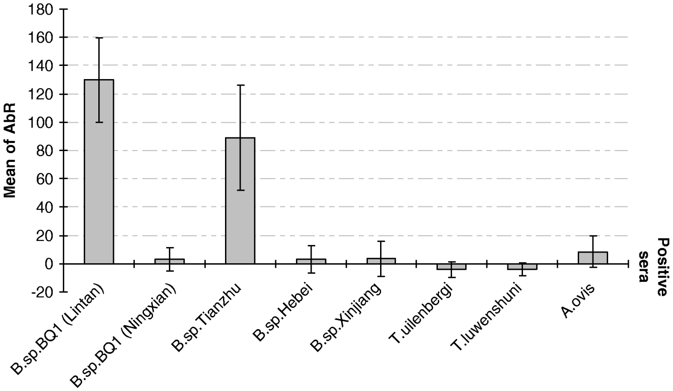
Cross-reactivities between BQMA and positive sera against Babesia sp. BQ1 (Ningxian), Babesia sp. Tianzhu, Babesia sp. Hebei, Babesia sp. Xinjiang, Theileria lunwenshuni, T. uilenbergi, and Anaplasma ovis were evaluated with ELISA and Western blot techniques. The tests were repeated twice and the mean OD and standard diversity were calculated with Excel 2000. No cross-reactions were observed between BQMA and anti-Babesia sp. BQ1 (Ningxian), -Babesia sp. Hebei, -Babesia sp. Xinjiang, -T. lunwenshuni, -T. uilenbergi, and -A. ovis antibodies in the sera by ELISA. In contrast, anti-Babesia sp. Tianzhu antibodies cross-react with BQMA (Fig. 2).
Cross-reaction of merozoite antigen of Babesia sp. BQ1 (Lintan) with positive sera of Babesia, Theileria and Anaplasma infective to sheep in China, as determined by ELISA (AbR: antibodies rate).

Western blot analysis confirmed a strong reaction with positive sera from Babesia sp. BQ1 (Lintan) or Babesia sp. Tianzhu-infected sheep. Antibodies specifically recognized at least 9 common proteins, with molecular weights of 171, 140, 92, 74, 48, 45, 35, 33 and 32 kDa. Anti-Babesia sp. BQ1 (Ningxian) antibodies could recognize 3 proteins (140, 48 and 33 kDa) that co-migrated with those of Babesia sp. BQ1 (Lintan) and Babesia sp. Tianzhu. No specifically recognized antigens, compared with that of negative sera, were obtained for the reactions with positive sera of Babesia sp. Hebei, Babesia sp. Xinjiang, T. lunwenshuni, T. uilenbergi, and A. ovis, (Fig. 3).
Western blot of merozoite antigens derived from in vitro culture of Babesia sp. BQ1 (Lintan). Lanes: M, the molecular weight markers (kDa); 1, a pool of negative sera, 2–9, positive sera of A. ovis, T. uilenbergi, T. lunwenshuni, Babesia sp. Xinjiang, Babesia sp. Hebei, Babesia sp. Tianzhu, Babesia sp. BQ1 (Ningxian), Babesia sp. BQ1 (Lintan). Standard molecular weight markers are indicated on the left (kDa). Arrows and numbers on the right show the antigens recognized by sera and their molecular weights (kDa).

Kinetics of anti-Babesia sp. BQ1 (Lintan) antibodies in experimentally and naturally infected sheep
Two Tan mutton sheep (Chinese sheep) (Nos 3216 and 2007) and 3 Vendean sheep (French sheep) (Nos 3390, 3533 and 3446) were experimentally inoculated with Babesia sp. BQ1 (Lintan) infected erythrocytes, and the sera collected during infection were assessed by ELISA (Fig. 4). Animals experimentally infected with Babesia sp. BQ1 (Lintan) had seroconverted by 1 week post-inoculation and the highest level of antibodies was attained after 2 or 3 weeks. The high level of antibodies usually lasted 8 months (240 days p.i.) although it decreased after 3 months (84 days p.i.) in sheep 3446 (the OD values at 0, 84 and 240 days p.i. were 0·19, 2·96 and 2·13, respectively).
Kinetics of anti-Babesia sp. BQ1 antibody in 3 French sheep experimentally infected with culture (Nos 3390, 3533 and 3446) and 2 Chinese sheep infected with blood (Nos 3216 and 2007). The bold curve shows the mean OD value corresponding to each time point and the error bars indicate the standard deviation.

Twelve 1·5 to 2-year-old sheep grazed on pasture in Lintan county were bled every month for 1 year to evaluate the kinetics of anti-BQMA antibody production during natural infection with Babesia sp. BQ1 (Lintan) by the ELISA (Fig. 5). No fluctuation of antibody titre was apparent in naturally infected sheep.
Kinetics of anti-Babesia sp. BQ1 (Lintan) antibodies production in 12 naturally-infected Chinese sheep studied by ELISA. The bold curve shows the mean OD value corresponding to each month and the error bars indicate the standard deviation.

Estimation of serological prevalence of Babesia sp. BQ1 (Lintan) in different counties of Gansu province, China with ELISA
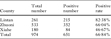
The ELISA was applied to 974 sera collected from field-grazing sheep in Lintan, Zhuoni and Xiahe county of Gannan Tibet Autonomous Region, Gansu Province, northwestern China during May to June 2004, to evaluate the infection situation in these areas. When the positive threshold value was taken as 30%, the results showed that the positive rates for Lintan, Zhuoni and Xiahe County were 82·38%, 66·04% and 46·67%, respectively, and the average positive rate in these regions was 66·84% (Table 1).
Examination of anti-Babesia sp. BQ1 (Lintan) antibodies from field samples with ELISA

Identification of potential antigens for serological diagnosis
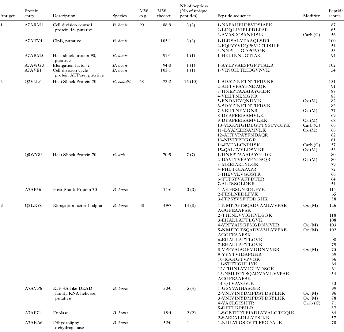
Western blot was performed with sera from 3 French sheep infected with Babesia sp. BQ1 (Lintan) to study the kinetics of the recognition of antigens during infection. Three antigens, out of approximately 20 molecules, were selected because they were recognized early during infection (between 6 and 9 days p.i.), with high intensity, by all Babesia sp. BQ1 (Lintan)-infected sheep (Fig. 6). All of them seemed to cross-react only with sera from Babesia sp. Tianzhu-infected sheep (Fig. 3). The 3 corresponding proteins were identified by comparing the collected LC-MS/MS data with the Uniprot sequence databank. Table 2 indicates only the proteins identified as proteins of Babesia spp. Host proteins were also identified but they were probably contaminants because they were not recognized by antibodies from sheep sera taken before infection.
Western blot analysis of IgG response to BQMA of a representative sheep infected with Babesia sp. BQ1 (Lintan). Standard molecular weight markers are indicated on the left (M, kDa). Arrows and numbers on the right show the 4 antigens which could be potentially used for the serodiagnosis of Babesia sp. BQ1 (Lintan) infection.

Identification of proteins by mass spectrometry

Note: ox (M): oxidation of methionine; Carb (C): carbamidomethylation of cysteine, Nb: number, MW Exp: experimental molecular weight, MW Theorit: theoretical MW, unique peptide: in terms of amino acid sequence.
DISCUSSION
Sero-diagnostic techniques of babesiosis are essential tools for epidemiological studies and for the evaluation of preventive measures, e.g. vaccination, premunition and tick control. The enzyme-linked immunosorbent assay (ELISA) provides a quantitative result, is more sensitive and non-subjective, can be automated for high throughput and is easily standardized, compared with the complement fixation test (CFT) and indirect fluorescent antibody test (IFAT) (Tenter and Friedhoff, Reference Tenter and Friedhoff1986; Brüning, Reference Brüning1996). Thus, a number of ELISAs have been designed recently for the diagnosis of Babesia spp. infection (Molloy et al. Reference Molloy, Bowles, Jeston, Bruyeres, Bowden, Bock, Jorgensen, Blight and Dalgliesh1998; Ikadai et al. Reference Ikadai, Osorio, Xuan, Igarashi, Kanemaru, Nagasawa, Fujisaki, Suzuki and Mikami2000; Goff et al. Reference Goff, McElwain, Suarez, Johnson, Brown, Norimine and Knowles2003). Few studies, however, have focused on the development of simple, rapid and stable routine serological techniques to detect ovine babesiosis. Current detection in both domestic and wild animals is mainly based on determination of the sero-prevalence by IFAT, and a cross-reaction between B. motasi and B. crassa has been demonstrated (Lewis et al. Reference Lewis, Holman, Purnell and Young1981; Habela et al. Reference Habela, Reina, Nieto and Navarrete1990; Kjemtrup et al. Reference Kjemtrup, Thomford, Gardner, Conrad, Jessup and Boyce1995; Papadopoulos et al. Reference Papadopoulos, Perié and Uilenberg1996; Ferrer et al. Reference Ferrer, Castellà, Gutiérrez, Lavín and Marco1998). Duzgun et al. (Reference Duzgun, Wright, Waltisbuhl, Gale, Goodger, Dargie, Alabay and Cerci1991) developed an ELISA for B. ovis using a recombinant B. bovis-derived antigen with cross-reactivity between B. ovis and B. bovis. In the present study, an ELISA was developed to detect infection of Babesia sp. BQ1 (Lintan) using soluble antigens of merozoite (BQMA) derived from in vitro culture. The calculated specificity of this ELISA was 95·5±0·97% with a positive threshold value of 30%.
The ELISA described in this manuscript was then used to investigate the seroconversion and persistence of specific antibodies during experimental and natural infection of sheep with Babesia sp. BQ1 (Lintan). Comparison of the kinetics of antibody levels in French sheep and Chinese sheep experimentally infected by Babesia sp. BQ1 (Lintan) revealed that the humoral immune response of different sheep breeds to this parasite was identical. The production of specific antibodies against Babesia sp. BQ1 (Lintan) was similar to those of other ovine Babesia, B. motasi, B. crassa and B. ovis, i.e. first produced 5–7 days p.i. then peaked between 14 and 28 days p.i. and persisted for at least 330 days p.i. (12–13 months) (Uilenberg et al. Reference Uilenberg, Rombach, Perie and Zwart1980; Christensson and Thunegard, Reference Christensson and Thunegard1981; Hashemi-Fesharki and Uilenberg, Reference Hashemi-Fesharki and Uilenberg1981; Habela et al. Reference Habela, Reina, Nieto and Navarrete1990).
The antibody levels in experimentally infected sheep (No. 3446) decreased during infection whereas those of 12 naturally infected grazing sheep showed no significant fluctuations over 1 year in prevalent areas. This could be explained by the presence of 2 activity peaks per year in H. qinghaiensis, the vector of Babesia sp. BQ1 (Lintan), one in April–July and the other in September (Teng, Reference Teng1991). In any one year, grazing animals may therefore be infected several times by Babesia sp. BQ1 (Lintan), which each time may stimulate the immune response and thus explain the maintenance of a high level of specific antibodies. The decrease in the level of antibodies in experimentally infected sheep could be explained by the absence of periodic antigen stimulation and by the decrease of parasitaemia, as observed in Babesia canis-infected dogs (Brandao et al. Reference Brandao, Hagiwara and Myiashiro2003).
Guan et al. (Reference Guan, Chauvin, Luo, Inoue, Moreau, Liu, Gao, Oriel, Ma, Liu, Dang, Liu, Ren, Jin, Sugimoto and Yin2008) recently showed that the positive rate determined by LAMP for Babesia sp. BQ1 (Lintan) was 14·3% (52/365) in Gannan Tibet Autonomous Region, the most important stock-raising region of Gansu Province, People's Republic of China. The sero-epidemiological analysis established with ELISA indicates that infection with Babesia sp. BQ1 (Lintan) poses a very serious problem, the positive rate attaining 66·84% in Gannan Tibet Autonomous Region. The difference in prevalence measurements obtained with the 2 methods is due to the different parameters detected by these methods. The LAMP technique, developed by Guan et al. (Reference Guan, Chauvin, Luo, Inoue, Moreau, Liu, Gao, Oriel, Ma, Liu, Dang, Liu, Ren, Jin, Sugimoto and Yin2008), is able to detect the parasite for 2 months after infection in experimentally infected sheep. In contrast, high levels of antibodies persist for at least 8 months and can be detected by BQMA-based ELISA (this study). Therefore, LAMP should be used to identify sheep recently infected with Babesia sp. BQ1 (Lintan) whereas ELISA can detect both early and latent infection.
Merozoite antigens from Babesia sp. BQ1 (Lintan) do not show cross-reactivity with sera from sheep infected with Babesia sp. Hebei, Babesia sp. Xinjiang, T. luwenshuni, T. uilenbergi or A. ovis, which are the dominant haemoparasites of small ruminants in China. Otherwise, the positive serum of Babesia sp. Tianzhu gives a strong cross-reaction with BQMA in both ELISA and Western blot and a weak cross-reaction between Babesia sp. BQ1 (Ningxian) and Babesia sp. BQ1 (Lintan) was detected only by Western blot. Based on internal transcribed spacer (ITS) gene sequences, Niu et al. (Reference Niu, Luo, Guan, Liu, Ma, Liu, Gao, Ren, Li, Qiu and Yin2009) demonstrated that these parasites should be included in 2 Babesia species, B. motasi and Babesia sp. Xinjiang. In B. motasi, the molecular phylogeny (Niu et al. Reference Niu, Luo, Guan, Liu, Ma, Liu, Gao, Ren, Li, Qiu and Yin2009) and serological evidence (this study) suggest that the ‘B. motasi in China’ group contains 2 species or subspecies, one including Babesia sp. BQ1 (Lintan) and Babesia sp. Tianzhu, explaining the strong cross-reactivity between these 2 strains, and the other with Babesia sp. BQ1 (Ningxian) and Babesia sp. Hebei, explaining the absence or the weak cross-reactivity between these 2 strains and Babesia sp. BQ1 (Lintan).
We have preliminarily studied 3 potential serodiagnosis antigens in order to increase the specificity of the BQMA-based ELISA and limit cross-reaction with other Babesia sp. in China. Some of them, such as heat shock protein 70 or enolase (antigen 2 and 3 respectively), have been used to detect other Apicomplexa such as B. orientalis (He et al. Reference He, Liu, Quan, Zhou, Zhou and Zhao2009), Theileria sp. (China) (Miranda et al. Reference Miranda, Bakheit, Liu, Yin, Mu, Guo, Beyer, Oliva, Ahmed and Seitzer2006), Plasmodium vivax (Na et al. Reference Na, Park, Lee, Lin, Kim, Bae, Sohn, Kim and Kong2007), and Neospora caninum (Shin et al. Reference Shin, Lee, Shin, Kim, Lee, Kim, Jang, Gershwin, Kim, Kim, Kim, Suh and Jung2004). This preliminary study now needs to be confirmed by testing the corresponding recombinant proteins and developing an ELISA able to detect only Babesia sp. BQ1 (Lintan) and Babesia sp. Tianzhu-infected sheep and thereby distinguish the 2 species or subspecies of the group of ‘B. motasi in China’.
In conclusion, an ELISA has been developed for the diagnosis and epidemiological study of Babesia sp. BQ1 (Lintan). This ELISA is a rapid, cheap and robust technique for detecting sheep infected with Babesia sp. BQ1 (Lintan) early after infection and during the persistent phase of the parasite. This ELISA provides an important tool for further investigating the prevalence of this parasite in pasture-grazing small ruminants in Inner Mongolia, Xinjiang, Qinhai, Gansu and Sichuan provinces of China. Further studies on antigens are needed to distinguish Babesia sp. BQ1 (Lintan) by ELISA from some other Babesia spp. in China.
ACKNOWLEDGEMENTS
We are grateful to Stéphanie Deligny-Penninck for excellent technical assistance in the mass spectrometry analyses.
FINANCIAL SUPPORT
This study was financially supported by the Key Project of Gansu Province (0801NKDA033); ‘863’ Project (2006AA10A207), Supporting Plan (2007BAD40B00), National Natural Sciences Foundation of China (30800820; 30571397), the National Natural Resource Platform Project (2005DKA21100), Specific Fund for Sino-Europe Cooperation, MOST, China; State Key Laboratory of Veterinary Etiological Biology Project-(SKLVEB 2008ZZKT019) and National Public Interests Research Institute Basic Scientific Research Expenses Special Fund Project. The research was also facilitated by SSA-income Projects, ICTTD 3 and EPIZONE of the European Commission, Brussels, Belgium.