Takayasu’s arteritis is a rare inflammatory, granulomatous, fibrosing arteritis of the aorta, its branches, and the pulmonary arteries, resulting in stenosis (98%), occlusion, dilatation, or aneurysm formation (27%).Reference Serra, Butrico and Fugetto 1 , Reference Kerr, Hallahan and Giordano 2
Although the aetiology is not known, various stimuli to the development of Takayasu’s arteritis have been proposed. These include tuberculosis,Reference Kinare 3 – Reference Kothari 6 an autoimmune trigger,Reference Scott, Salmon and Scott 7 – Reference Seko, Minota and Kawasaki 10 and a genetic link with human leucocyte antigen associations.Reference Kimura, Ota and Katsuyama 11 , Reference Yajima, Numano, Park and Sagar 12 It has been diagnosed world-wide across both low- and high-income countries.Reference Lande, Bard, Rossi, Passariello and Castrucci 13 – Reference Waern, Andersson and Hemmingsson 15 The extent and patterns of vascular involvement of the aorta and its branches vary among different populations and age groups.Reference Yajima, Numano, Park and Sagar 12 It is mainly a disease of young women in the second and third decades of life, but has been described in children as young as 2 years of age.Reference Kerr, Hallahan and Giordano 2 , Reference Ladapo, Gajjar, McCulloch, Scott, Numanoglu and Nourse 5 , Reference Ladhani, Tulloh and Anderson 16 – Reference Shrivastava, Srivastava and Tandon 18 The diagnosis is challenging particularly in the early stages of the disease when symptoms are often vague and include fever, abdominal pain, headaches, and rashes. Nonspecific inflammatory markers are frequently raised during the initial active phase of the illness, but the disease cannot be confirmed because there is no specific diagnostic test.Reference Brunner, Feldman and Tyrrell 19
Consequently, the diagnosis is often made clinically very late in the vascular phase when arterial stenoses have occurred, resulting in hypertension, absent pulses, stroke, and claudication.Reference Brunner, Feldman and Tyrrell 19 Renal artery stenosis is common in childhood Takayasu’s arteritis, resulting in malignant hypertension and chronic renal disease.Reference Hahn, Thomson, Kala, Beale and Levin 20 , Reference Wiggelinkhuizen and Cremin 21 Children also manifest more frequently with heart failure. The reason for this is not known.Reference Lupi-Herrera, Sanchez-Torres, Marcushamer, Misipreta, Horwitz and Vela 22 Left ventricular dysfunction as the underlying mechanism of heart failure is often attributed to hypertension, heart valve lesions such as aortic or mitral valve regurgitation, coronary artery stenosis, and myocardial involvement itself.Reference Talwar, Kumar and Chopra 23 , Reference Panja, Kar, Dutta, Chhetri, Kumar and Panja 24
The management of advanced Takayasu’s arteritis is very challenging and includes arresting progression of existing lesions using anti-inflammatory and chemotherapeutic agents, and revascularisation of stenotic lesions.Reference Liang, Tan-Ong and Hoffman 25 Established lesions are not usually reversed by immunosuppressive therapy.Reference Kerr, Hallahan and Giordano 2 , Reference Weaver, Yellin and Campen 26 Treatment of renal artery stenoses has been shown to be beneficial in the management of hypertension in some patients.Reference Scott, Salmon and Scott 7 , Reference Liang, Tan-Ong and Hoffman 25 Despite the short-term benefit, both surgical and percutaneous revascularisation procedures, although safe, are associated with a high failure rate.Reference Liang, Tan-Ong and Hoffman 25
Materials and methods
We conducted a retrospective review of all children diagnosed to have Takayasu’s arteritis entered into an electronic database from 1993 to the end of 2015. Data extracted from the database and available clinical records of 55 patients included clinical presentation, age, sex, origin of patient, pattern of vascular involvement, response to the purified protein derivative test for tuberculosis, left ventricular function, treatment, and outcomes.
The initial diagnosis of Takayasu’s arteritis was made clinically in the presence of systemic hypertension, absent arm or leg pulses, and bruits over the aorta and its branches, and it was confirmed by the presence of typical angiographic features showing stenotic or occlusive lesions of the aorta and its branches. Diagnostic criteria for Takayasu’s arteritis have been published and have been applied retrospectively to the study patients. Examples are the American College of Rheumatology,Reference Arend, Michel and Bloch 27 the National Institutes of Health,Reference Kerr, Hallahan and Giordano 2 and the Pediatric Rheumatology European SocietyReference Ozen, Ruperto and Dillon 28 consensus criteria for Takayasu’s arteritis.
Hypertension was diagnosed if blood pressure measurements exceeded the 95th centile for age and was managed in a stepwise manner starting with a calcium-channel blocker, followed by the addition of a beta receptor blocker, and finally an α-1 selective receptor blocker. If the hypertension remained poorly controlled after receiving maximum doses of each medication, percutaneous intervention of the renal artery or aortic stenosis was undertaken even if there was evidence of active disease.
Heart failure was diagnosed clinically when there were features of venous congestion, such as raised jugular venous pressure, tender hepatomegaly, pedal oedema, and a gallop rhythm. Echocardiographic recordings of fractional shortening were documented. A normal fractional shortening of 36±4% was used.Reference Gutgesell, Paquet, Duff and McNamara 29 Heart failure was treated with digoxin and diuretics in the majority of cases. Angiotensin-converting enzyme inhibitors were avoided in the presence of aortic coarctation and bilateral renal artery stenosis to circumvent the development or exacerbation of renal dysfunction.
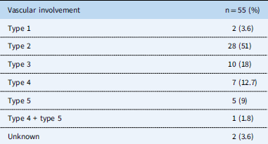
All patients underwent angiography and the patterns of vascular disease involvement were recorded according to an institutional classification created by Pantanowitz,Reference Pantonowitz 30 which is based on a combination of the Ueno classification modified by Lupi-HerreraReference Lupi-Herrera, Sanchez-Torres, Marcushamer, Misipreta, Horwitz and Vela 22 and the Nasu classification.Reference Nasu 31 Type 1 involves the aortic arch and branches; type 2 involves the thoraco-abdominal aorta and the renal arteries; type 3 is a combination of types 1 and 2; type 4 involves any part of the aorta with pulmonary artery involvement; type 5 has iliac artery involvement alone or in combination with types 1, 2, or 3.
Patients with active disease were treated with corticosteroids and chemotherapeutic agents including cyclophosphamide and methotrexate. An elevated erythrocyte sedimentation rate and/or a positive positron emission tomography scan indicating the presence of inflamed vasculature were considered as evidence of active disease. All patients received prednisone at a dose of 2 mg/kg, at a maximum dose of 60 mg, orally daily for 2 months and then changed to alternate days, followed by gradual weaning off of the medication by 5 mg/month. In addition, all patients were prescribed oral methotrexate once weekly at 0.15 mg/kg, at a maximum dose of 15 mg, with the addition of rescue oral folate of 5 mg for 2 days after the methotrexate, until no disease activity was detectable and for several years in some patients. Severe cases with vascular stenosis and aneurysms, end organ damage, and cardiomyopathy were treated with cyclophosphamide 500 mg/m2 × 3 doses as an intravenous infusion.
Those patients with severe unilateral renal artery stenosis and shown to have <8% function on a mercaptuacetyltriglycine radionucleide renal scan were referred for a nephrectomy.
All patients received anti-tuberculosis treatment for two reasons. First, there is a high prevalence of tuberculosis in the general South African population, and there is a concern that the administration of immunosuppressant therapy may result in re-activation of tuberculosis infection. The second reason is the tentative association of Takayasu’s arteritis with tuberculosis.
Statistics
Continuous variables are provided as a mean and standard deviation. The two-tailed paired t-test was used to analyse comparisons between values before and after treatment. Categorical variables were expressed as percentages and proportions. A p-value <0.05 was considered statistically significant.
Results
Demographic data
A total of 55 children were diagnosed to have Takayasu’s arteritis. Just over half of the patients originated from referral centres in and around Johannesburg, which is situated within the province of Gauteng (25/55). The majority of the remainder originated from the North West Province (24/55), which is a major referral area to the study institution. The other patients came from the Limpopo Province (1), the Northern Cape (1), the Eastern Cape (1), and Botswana (1); two patients did not have their addresses documented.
There were 42 female and 13 male patients, with a female:male ratio of 3.2:1. The mean age was 9.7±3.04 years. A positive purified protein derivative test suggestive of the presence of active tuberculosis was documented in 73% (40/55) patients. The result of the purified protein derivative was not recorded in the case files of the other 15 patients.
Presenting features
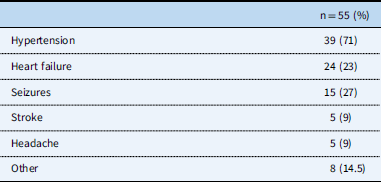
The majority of patients (Table 1) presented with hypertension. Other presenting features included heart failure, seizures, absent or reduced arm and leg pulses, strokes, headaches, and a variety of other manifestations such as chest pain in two patients, pulmonary tuberculosis in one, vomiting and abdominal pain in one, gangrene of the foot in one, blindness in one, a destroyed eye in one, and arthralgia in another.
Clinical presenting features.

Myocardial dysfunction
In all, 37 (67%) patients were documented to have myocardial dysfunction with a reduced mean fractional shortening of 15±5%, 24 (65%) of whom manifested with congestive cardiac failure.
Vascular involvement
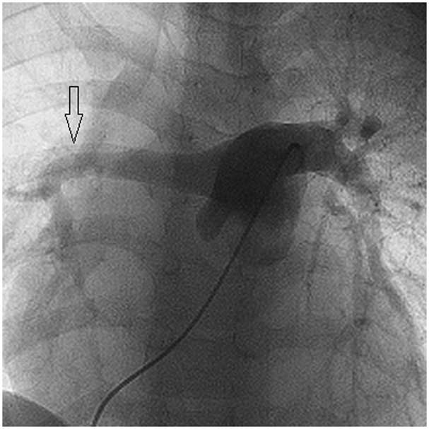
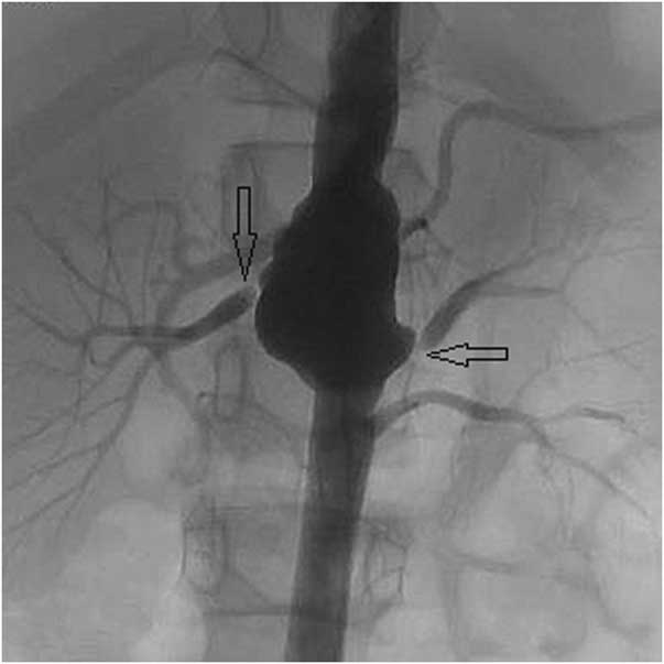
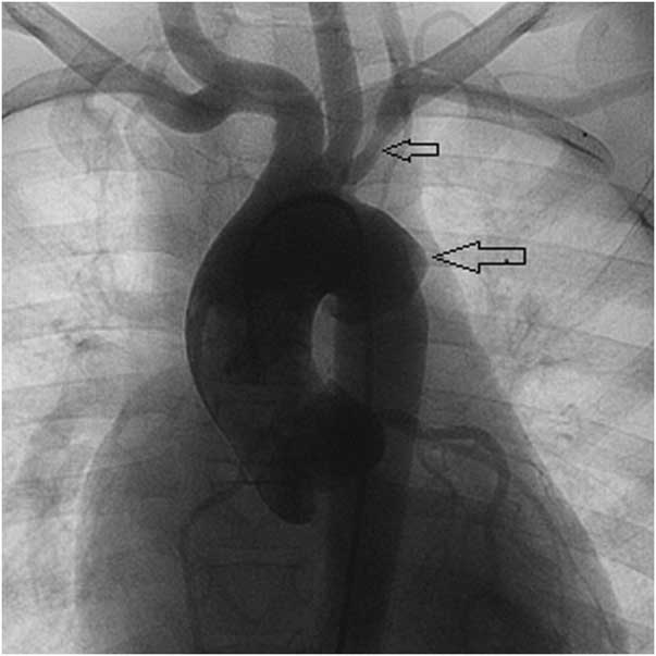
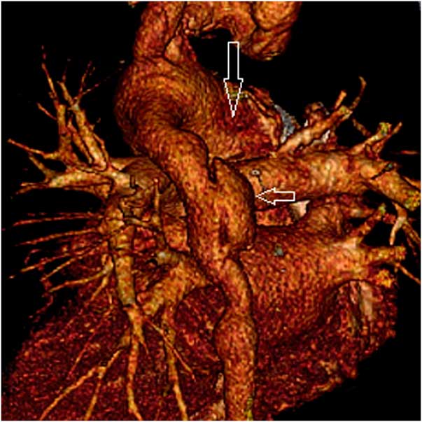
Angiography was the principal radiological modality used to diagnose the pattern of vascular involvement (Table 2). The majority – that is, 34/55 – were found to have stenotic areas of the abdominal aorta (62%), with a similar number (35/55, 64%) having renal artery stenosis. Bilateral renal artery stenosis was present in 12 patients. Branch pulmonary stenosis or attenuation was evident in eight patients (Fig 1). Aneurysms were found in 14 patients, eight involving the abdominal aorta (Fig 2), two involving the aortic arch (Figs 3 and 4), two affecting the aorta at the level of the diaphragm, and two involving the thoracic aorta. Coronary arteries were assessed during routine ascending aorta angiography and none were found to be affected.
Pulmonary angiogram showing narrowing of the right pulmonary artery distally with loss of the right upper lobe branch (arrow). The left pulmonary artery shows a normal branching pattern.

Abdominal angiogram showing an aneurysm of the aorta between lumbar vertebrae 1 and 2 associated with bilateral renal artery stenosis (arrows). Bilateral renal artery stents were inserted.

Ascending aortagram depicting an aneurysm of the transeverse arch (large arrow) and stenosis of the left subclavian artery (small arrow). A weak pulse was present in the left arm.

Three dimensional computed tomography scan reconstruction of the aorta viewed from the posterior aspect showing a very large aneurysmal ascending aorta (large arrow). Severe aortic regurgitation was present. A large aneurysm (small arrow) and irregularity of the thoracic aorta is also shown.

Vascular involvement according to the Pantanowitz Classification.Reference Pantonowitz 30

Type 1=aortic arch; type 2=descending aorta below the diaphragm including the renal arteries; type 3=type 1+type 2; type 4=pulmonary arteries+any part of the aorta; type 5=iliac arteries+any other part of the aorta
Interventions
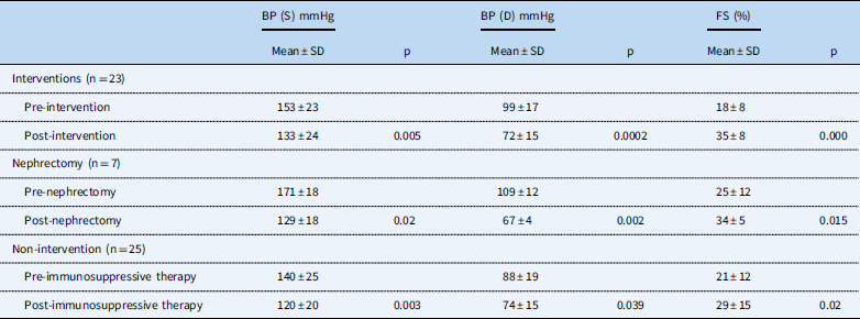
A total of 23 patients underwent 30 percutaneous interventions – eight stents: five aortic and three renal; 22 balloon angioplasties: 6 aorta, 15 renal artery, and one branch pulmonary artery; and seven had nephrectomies. All patients received immunosuppressive therapy. There was a significant reduction in both the systolic and diastolic blood pressures and an improvement in the fractional shortening in all patients undergoing percutaneous interventions, nephrectomies, and also the group receiving immunosuppressive therapy only (Table 3).
Blood pressure and myocardial dysfunction response to percutaneous intervention, nephrectomy, and immunosuppressive therapy.

BP=blood pressure; D=diastolic; FS=fractional shortening; n=number; mmHg=millimetre of mercury; p=p-value, level of significance; S=systolic
Follow-up
The mean follow-up period was 3.2 years, ranging from 0 to 16 years. Five patients – two from Gauteng, two from the North West Province, and one from the Northern Cape – were known to have died. Only 17 (31%) patients – seven with addresses from Gauteng and 10 from the North West Province – had follow-up to the end of 2015, 10 of whom have had interventions: three nephrectomies, two thoracic aortic stents, one abdominal aorta angioplasty, and four renal artery angioplasties. All 17 patients remained on anti-hypertensive medications and were documented to have blood pressure measurements below the 95th centile for age at the time of their last clinical review. The remainder, comprising 31 patients with known addresses and the two without addresses, have been lost to follow-up. One female patient had a successful term pregnancy born by Caesarean section at the age of 19 years, after which she was lost to follow-up. She manifested at the age of 11 years with heart failure, poor left ventricular function, and hypertension. Medical treatment during this admission included prednisone, methotrexate, cyclophosphamide, and anti-hypertensive medications. A percutaneous stenting procedure of a severe abdominal aortic stenosis was undertaken at the age of 16 years. She was maintained on amlodipine and metoprolol for blood pressure control thereafter, and her left ventricular function had normalised before she was pregnant.
Discussion
Takayasu’s arteritis is a rare acquired aortitis predominantly involving the aorta and its branches and mostly diagnosed in young females at our centre, with a female to male ratio of 3.2:1, which is in keeping with other studies.Reference Sekiguchi and Sazuki 17 Although it is mostly described in young adult females, it is diagnosed in children who frequently manifest with hypertension and heart failure.
The Southern African experience
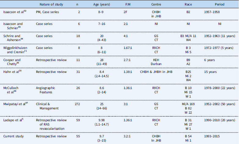
Several reviews and case reports on Takayasu’s arteritis in children have emerged from Southern Africa over the past 50 years since the first publication by Isaacson et al in 1959,Reference Isaacson, Klachko, Wayburne and Simson 32 the majority of which have emanated from Cape Town, South Africa. The last publication to document patients from Gauteng was 18 years ago in 1998Reference Hahn, Thomson, Kala, Beale and Levin 20 (Table 4).
Southern African experience.

A=Asian; B=Black; CHBH=Chris Hani Baragwanath Hospital; CT=Cape town; F=female; GS=Groote Schuur Hospital; JHB=Johannesburg; JHBH=Johannesburg Hospital; KEH=King Edward VIII Hospital; M=male; Mi=mixed race; n=number of patient; NI=no information; Period=period of study; PM=post mortem; RAS=renal artery stenosis; RXCH=Red Cross Children’s Hospital; W=white
Patterns of disease
The occurrence of stenotic lesions of the abdominal descending aorta and renal arteries (type 2) is the most common pattern in our study cohort. Patterns of disease vary from country to country. The dominant involvement of the abdominal aorta and renal arteries is similar to that found in India and Korea, but it is in contrast to studies from Japan, which show mainly aortic arch disease.Reference Yajima, Numano, Park and Sagar 12 A publication from Cape Town (South Africa) in 2005 showed that most patients had type 3 involvement of the aortic arch combined with the abdominal aorta. These patients were predominantly female and were older with a mean age of 25 years.Reference Mwipatayi, Jeffery and Beningfield 37 In comparison, an earlier study from Johannesburg (Gauteng Province, South Africa) in 1998 describing the characteristics of 31 children with Takayasu’s arteritis showed equal numbers of children with type 2 and type 3 patterns of disease.Reference Hahn, Thomson, Kala, Beale and Levin 20 Almost 25% of patients were found to have aneurysms of the aorta. This finding is similar to that of Kerr et al, who found 27% patients with aneurysms.Reference Kerr, Hallahan and Giordano 2
Geography and follow-up
Most patients gave addresses from Gauteng and the North West Provinces, which are areas in the central and northern parts of South Africa. However, because of the large migrant population frequenting our institution, their origins cannot be authenticated. The vast majority of patients have been lost to follow-up (33/55), most of whom gave addresses in areas adjacent to the hospital. Reasons for loss to follow-up include the possibility of death and return to neighbouring countries or provinces of origin not declared during their treatment. The ethnic group of our patients was mostly black African and representative of the referral population to our institution.
Clinical manifestations
Because the initial presenting features are mainly constitutional symptoms such as headaches, fever, weight loss, rashes, vomiting, abdominal pain, and musculoskeletal symptoms, the diagnosis of Takayasu’s arteritis at this early stage is difficult and needs a high index of suspicion. Most patients manifest with advanced disease after the development of arterial stenosis. Thus, the majority of study patients manifest late with hypertension secondary to aortic coarctation or renal artery stenosis, heart failure, and seizures, which may be caused by severe hypertension or cerebral arteritis or both. Similarly, the most common presenting features in the 1998 study from Johannesburg (Gauteng Province, South Africa) were hypertension and heart failure.Reference Hahn, Thomson, Kala, Beale and Levin 20 Not all of the patients with poor left ventricular function and heart failure were hypertensive despite having renal artery or aortic stenosis. The expected high blood pressure associated with Takayasu’s arteritis may be deceptively lowered in the presence of poor left ventricular function and reduced cardiac output.
Treatment
As the disease reaches an advanced stage with stenotic or aneurysmal vessel changes, treatment becomes more palliative than curative. Despite the severity of some lesions, patients do respond to interventional and immunosuppressive therapy, measured by a reduction in blood pressure levels and improvement in cardiac function. The majority of patients – that is, 100% of patients with complete records – similar to studies from Mexico (81% patients)Reference Lupi, Sanchez Torres and Castillo 4 and Gauteng in 1998 (90% patients)Reference Hahn, Thomson, Kala, Beale and Levin 20 were documented to have a positive purified protein derivative test. All the study patients received additional anti-tuberculosis treatment for 6 months. The clinical response to anti-tuberculosis treatment alone cannot be assessed because all patients received immunosuppressive and anti-hypertensive therapy simultaneously. Patients undergoing nephrectomy appear to have better control of their hypertension after surgery and anti-hypertensive treatment may be discontinued in some patients who become normotensive. The good response in the nephrectomy group may be owing to the complete removal of the poorly perfused kidney, which is deemed not to be salvageable by means of revascularisation or re-implantation. Percutaneous interventions were mainly undertaken in the quiescent phase of the disease in the majority of patients following several weeks to months of immunosuppressive therapy. If the blood pressure was uncontrollable medically, percutaneous interventions were undertaken in some patients even in the presence of inflammation, confirmed by the presence of a raised erythrocyte sedimentation rate or a positive positron emission tomography scan, which was invariably associated with a good short-term response. Inflammation at the time of revascularisation has been generally associated with increased vascular complications.Reference Saadoun, Lambert and Mirault 38
Cardiomyopathy
A high frequency of myocardial dysfunction (67%) was found in the study patients. Myocardial dysfunction in patients with Takayasu’s arteritis has been ascribed to various factors including hypertension, heart valve lesions, coronary artery involvement, and a myocardial factor. All the study patients recovered their myocardial function following treatment with combinations of immunotherapy, percutaneous intervention, nephrectomy, anti-failure, and anti-hypertensive therapy.
Pulmonary artery involvement
All eight patients with branch pulmonary artery stenosis or attenuation were found to have incidental pulmonary artery involvement during their routine angiographic work-up of Takayasu’s arteritis. All patients presented with systemic hypertension and heart failure. One patient had a pleural effusion thought to be due to tuberculosis. Although none of the patients had isolated pulmonary involvement, an isolated lesion in the absence of aortic lesions has been described. The diagnosis of Takayasu’s arteritis should be considered in patients with pulmonary artery stenosis, especially those manifesting with chest pain, a pleural effusion, haemoptysis, and signs of inflammation.Reference Nakabayashi, Kurata, Nangi, Miyane and Nagasawa 39
Takayasu’s arteritis in children
Most studies from South Africa describe Takayasu’s arteritis in children (Table 4). The age group of the study cohort at presentation is similar to the age groups described in the older publications emanating from other centres in South Africa. The youngest child presenting to our institution was a male child of 3 years who manifested with seizures, hypertension (blood pressure of 155/105mmHg), and a cardiomyopathy associated with heart failure. He was found to have type 4 disease involving the pulmonary arteries, associated with an aneurysm of the abdominal aorta and a left renal artery stenosis. He underwent a left nephrectomy and continues to be followed up 13 years later normotensive on anti-hypertensive treatment. His left ventricular dysfunction has normalised.
Conclusion
Takayasu’s arteritis in children at our institution is more common in female individuals, mostly involving the abdominal aorta and renal arteries. Most patients present with severe vascular complications and commonly manifest with hypertension and heart failure. Blood pressure control and myocardial dysfunction improved in all patients following treatment with immunosuppressive and/or percutaneous interventional therapy combined with anti-hypertensive medications.
Study limitations
The retrospective nature of the study with the inevitable problem of missing information and the long 22-year study period placed limitations on the availability of laboratory data such as erythrocyte sedimentation rates that may have added value to the analysis. Only data that were consistently available for all the patients have been used to illustrate disease patterns and treatment responses in the study. These data included demographic information, angiographic studies, documentation of clinical presentation, blood pressure, and echocardiographic measurements.
Acknowledgements
The authors posthumously acknowledge the contribution made by the late Dr Gcina Dumani in the treatment of children with Takayasu’s arteritis.
Financial Support
This research received no grants from any financial sector.
Conflicts of Interest
None.
Ethical Standards
Permission for publication was obtained from the Medical advisory Committee at the Chris Hani Baragwanath Academic Hospital and the Human Research Ethics Committee at the University of the Witwatersrand.