


Highlights
• STA–MCA bypass achieved 100% graft patency and no recurrent ischemic events in a Canadian Moyamoya disease/moyamoya syndrome (MMD/MMS) cohort.
• Favorable functional outcomes were observed in most patients, with complications highlighting the importance of strict perioperative management.
• Demonstrates the feasibility of safely re-establishing a cerebral bypass program in a contemporary North American center.
Introduction
Moyamoya disease (MMD) is a rare, chronic and progressive cerebrovascular disorder characterized by stenosis or occlusion of the terminal portion of the internal carotid arteries (ICAs), as well as the proximal middle cerebral arteries (MCAs) and anterior cerebral arteries. Reference Zhang, Zheng and Feng1 Progressive vascular narrowing triggers the development of a compensatory collateral network of fragile and dilated vessels, producing the angiographic “puff of smoke” appearance from which the disease derives its Japanese name “Moyamoya.” Reference Suzuki and Takaku2 The Suzuki staging system classifies MMD progression into six angiographic stages based on arterial stenosis severity and the development of collateral vessels, offering critical insights into disease progression and informing clinical decisions. Reference Suzuki and Takaku2,Reference Fujimura and Tominaga3 Although MMD is increasingly recognized worldwide, its prevalence remains highest in East Asian populations, with annual incidence rates ranging from 0.35 to 1.14 per 100,000 persons, Reference Zhang, Huang and Huang4–Reference Ahn, Park, Hann, Kim, Kim and Ahn6 compared to markedly lower rates reported in Europe (0.07–0.09 per 100,000) Reference Birkeland, Tharmabalan, Lauritsen, Ganesan, Bjarkam and von Weitzel-Mudersbach7 and the USA (0.086 per 100,000). Reference Uchino, Johnston, Becker and Tirschwell8 Genetic susceptibility also contributes to MMD pathogenesis, with the RNF213 p.R4810K variant representing the most well-established risk allele in East Asian populations. Reference Liu, Morito and Takashima9 Although this variant confers an increased risk of disease, its incomplete penetrance and variable expressivity suggest that additional genetic, environmental and vascular factors modulate disease development and clinical phenotype.
In contrast to MMD, Moyamoya syndrome (MMS) refers to a secondary cerebrovasculopathy exhibiting similar angiographic and pathophysiological features to MMD but arises in association with identifiable underlying conditions. Common etiologies of MMS include genetic conditions (e.g., Down syndrome, neurofibromatosis type 1), hematologic disorders (e.g., sickle cell disease), autoimmune or inflammatory diseases (e.g., systemic lupus erythematosus, antiphospholipid syndrome [APS] and primary angiitis of the central nervous system) or prior cranial irradiation. Reference Scott and Smith10 The distinction between MMD and MMS extends beyond their underlying etiology to encompass differences in clinical behavior. MMD typically manifests as a bilateral and symmetric arteriopathy, whereas MMS may present unilaterally or asymmetrically, depending on the precipitating condition. Reference Scott and Smith10 These distinctions carry important diagnostic and prognostic implications, as MMS often demonstrates a more heterogeneous clinical trajectory and requires a therapeutic approach that concurrently addresses both the primary systemic disorder and its secondary cerebrovascular consequences.
Clinically, adults with MMD/MMS commonly present with hemorrhagic rather than ischemic events, specifically intracranial hemorrhage, which often occurs due to the rupture of fragile collateral vessels. MMD/MMS is associated with a significant overall risk of ischemic and hemorrhagic strokes, estimated at approximately 5%, Reference Cho, Chung and Kim11–Reference Smith and Scott14 with studies reporting hemorrhage rates as high as 17.9% Reference Gross and Du15 and recurrent hemorrhage rates reaching up to 16.9% Reference Kim, Kim and Cho16 in non-surgically treated adults. While the natural history of MMD/MMS is not fully understood, its progressive nature, characterized by recurrent cerebrovascular events, underscores the importance of timely diagnosis and intervention. Reference Bagchi, Hore, Mandal and Pal17 Surgical revascularization is the cornerstone of MMD/MMS treatment, aiming to enhance cerebral perfusion and reduce ischemic and hemorrhagic risks. Reference Scott and Smith10 Revascularization techniques are broadly classified as direct, indirect and combined approaches. Among these, the superficial temporal artery to middle cerebral artery (STA–MCA) bypass is the most widely adopted direct approach, offering immediate blood flow restoration via an anastomosis between the spared extracranial and the affected intracranial vessels. Reference Newell18–Reference Newell and Vilela20 While it has demonstrated efficacy in improving outcomes for patients with MMD, Reference Kappel, Feroze, Torio, Sukumaran and Du21 ongoing research is essential to further refine surgical strategies Reference Powers, Clarke and Grubb22,Reference Ma, Wang and Wang23 and optimize outcomes across diverse patient populations, particularly those in non-Asian regions where the condition is less frequently studied. Reference Oliveira, Cieslak and Ferreira24
In North America and Europe, however, the practice landscape of bypass has evolved considerably over the past four decades. Reference Gallagher, Frantzias, Kailaya-Vasan, Booth and Tolias25 Following the results of the International EC–IC Bypass Trial 26 and later the Carotid Occlusion Surgery Study (COSS), Reference Powers, Clarke and Grubb22 the role of bypass for atherosclerotic cerebrovascular disease was questioned, leading to a substantial decline in procedural volume and fewer training opportunities in many neurosurgical centers. Reference Gallagher, Frantzias, Kailaya-Vasan, Booth and Tolias25,Reference Akbarian-Tefaghi, Kalakoti and Sun27 Although these trials did not address MMD/MMS where revascularization remains a well-established and guideline-supported therapy, Reference Scott and Smith10 their broader impact contributed to the progressive centralization of bypass expertise and the erosion of routine institutional experience. This historical contraction has created the contemporary challenge of ensuring the preservation and re-establishment of technically demanding microsurgical revascularization programs capable of treating these patients safely and effectively.
Perioperative management is a critical determinant of outcome after revascularization in MMD/MMS. Because cerebral perfusion may be pressure-dependent in chronically hypoperfused territories, hypotension, hypocapnia, dehydration and anemia can precipitate ischemic complications. Reference Parray, Martin and Siddiqui28,Reference Li, Zhang and Yu29 Conversely, postoperative hypertension and abrupt hemodynamic shifts may increase the risk of hyperperfusion syndrome, intracranial hemorrhage or graft-related complications. Accordingly, perioperative care generally emphasizes euvolemia, avoidance of prolonged fasting or dehydration, maintenance of normocapnia and individualized blood pressure targets based on baseline blood pressure, perfusion status, comorbidities and intraoperative findings. Reference Parray, Martin and Siddiqui28,Reference Li, Zhang and Yu29 These principles require close collaboration among neurosurgery, neuroanesthesia, vascular neurology and neurocritical care teams.
The present study describes the early experience of re-establishing a direct cerebral revascularization program at a Canadian university hospital, focusing on patients with MMD/MMS treated using STA–MCA bypass. By examining patient selection, operative strategy, perioperative management and short-term outcomes during the implementation phase of this program, this case series aims to provide practical insight into the feasibility, challenges and clinical results of rebuilding bypass capability in a contemporary North American setting.
Methods
Study design and setting
This retrospective case series was conducted at the Montreal Sacred Heart Hospital (Montreal, Quebec, Canada), a tertiary care center with advanced neurosurgical capabilities. The study included patients who underwent STA–MCA bypass for MMD or MMS by a single surgeon (PL) between December 2021 and March 2026. All patient data included in this case series were fully anonymized prior to analysis and publication. No identifiable information is disclosed. The study was conducted in accordance with institutional regulations.
Patient selection
Patients were included in the study if they had clinical and imaging evidence of MMD or MMS and an indication for STA–MCA bypass based on impaired cerebral perfusion or symptomatic presentation (e.g., ischemic or hemorrhagic events). Patients were excluded if they had incomplete medical records, were managed with indirect revascularization techniques alone or had primary conditions unrelated to MMD/MMS.
Surgical techniques
Preoperative imaging with high-resolution angiography was used to identify suitable superficial temporal artery (STA) donor branches and MCA recipient branches. The STA–MCA bypass was performed under general anesthesia using standard microsurgical techniques. For all cases, single-barrel bypasses were performed. A single STA branch, typically the parietal branch, was anastomosed to a single distal cortical MCA branch (M4). Intraoperative patency of the bypass grafts was meticulously assessed using indocyanine green angiography and Doppler ultrasonography. Postoperative management emphasized rigorous blood pressure control and vigilant monitoring in the intensive care unit (ICU) to prevent complications such as intracranial hemorrhage or ischemic events. Management was tailored according to intraoperative findings, postoperative imaging and each patient’s overall clinical risk profile.
Perioperative management
Perioperative management was coordinated by the neurosurgical, neuroanesthesia and neurocritical care teams. Patients were maintained euvolemic, with avoidance of prolonged preoperative fasting, dehydration, anemia, hypocapnia and hypotension. Intraoperatively, neuroanesthesia aimed to maintain normocapnia and blood pressure close to each patient’s preoperative baseline, recognizing that cerebral perfusion may be pressure-dependent in chronically hypoperfused territories. Reference Parray, Martin and Siddiqui28,Reference Li, Zhang and Yu29 Neuromonitoring was used in all cases to ensure that no changes occurred in the patient’s somatosensory evoked potential, electroencephalogram and motor evoked potential while the patient was under general anesthesia. Blood pressure targets were increased around 20 mmHg above the patient’s baseline when the MCA was clamped for the bypass and returned to the patient’s baseline when it was completed. Postoperatively, patients were monitored in the ICU with frequent neurological assessments and continuous hemodynamic monitoring. Blood pressure targets were individualized according to baseline blood pressure, perfusion status, intraoperative findings and comorbidities. The general objective was to avoid hypotension by maintaining systolic blood pressure above 120 mmHg while also preventing severe hypertension, particularly sustained systolic blood pressure elevations above approximately 160 mmHg unless a higher individualized target was required for perfusion. Early postoperative neuroimaging was obtained routinely within the immediate postoperative period or urgently in the event of neurological deterioration. Imaging consisted of CT/CT angiography or angiographic assessment when clinically indicated to evaluate for hemorrhage, infarction, mass effect and graft patency.
Data collection
Data collection was performed through the medical records. Specific details such as age, sex, demographics, clinical presentation, perioperative treatment, Glasgow Coma Scale (GCS) at presentation and anticoagulation therapy status were key variables for the study. Preoperative status and functional outcomes were evaluated using the Suzuki staging system and the modified Rankin Scale (mRS), respectively, recorded at both admission and discharge. The mRS is a widely used tool for assessing the degree of disability or dependence in daily activities following cerebrovascular events or other neurological impairments, with scores ranging from 0 (no symptoms) to 6 (death). Reference Nimbvikar, Panchawagh, Chavan, Ingole, Pargaonkar and Pai30
Outcomes and follow-up
Surgical details, perioperative complications and lengths of stay (LOS) in both the hospital and the ICU (ICU-LOS) were recorded. Follow-up assessments included evaluation of neurological function using mRS scores, confirmation of graft patency through postoperative imaging and documentation of any ischemic or hemorrhagic events. A favorable functional outcome was defined as a postoperative mRS score of 0–2 or no worsening from preoperative baseline at last clinical follow-up.
Results
Patient demographics and baseline characteristics
The cohort consisted of nine patients who underwent STA–MCA bypass for MMS (n = 4, 44%) and MMD (n = 5, 56%) between December 2021 and October 2025. Mean age of patients was 44 years (range 26–58), and 89% were females. Seven patients (78%) had hypertension, five (56%) had dyslipidemia, three (33%) had diabetes mellitus and one (11%) had peripheral vascular disease. Additionally, one (11%) young (26 years) female was diagnosed with APS (Table 1).
Clinical and demographic characteristics of patients undergoing STA–MCA bypass surgery

Table 1. Long description
The table presents clinical and demographic characteristics of patients undergoing STA-MCA bypass surgery. It has 9 rows and 8 columns. The columns are labeled as Case, Age, Sex, Diagnosis, Etiology/associated condition, Comorbidities, Suzuki score, Initial symptoms, Graft type, and Surgery side. The rows list individual patient data. Row 1: Case 1, Age 48, Sex M, Diagnosis MMS, Etiology/associated condition HTN, Comorbidities HTN, Dyslipidemia, Suspected atheroma, Suzuki score III, Initial symptoms RUL paresthesia with mild dexterity loss, Graft type STA-MCA, Surgery side L. Row 2: Case 2, Age 26, Sex F, Diagnosis MMS, Etiology/associated condition APS, Comorbidities APS, Suzuki score III, Initial symptoms Right hemiparesis, Graft type STA-MCA, Surgery side L. Row 3: Case 3, Age 56, Sex F, Diagnosis MMD, Etiology/associated condition NA (Idiopathic MMD), Comorbidities HTN, Dyslipidemia, Diabetes type II, Suzuki score III, Initial symptoms Severe LLL weakness, Ataxia, Graft type STA-MCA, Surgery side L. Row 4: Case 4, Age 44, Sex F, Diagnosis MMS, Etiology/associated condition HTN, Diabetes type II, Comorbidities HTN, Diabetes type II, Suzuki score II, Initial symptoms Right hemiparesis, Aphasia, Graft type STA-MCA, Surgery side L. Row 5: Case 5, Age 58, Sex F, Diagnosis MMD, Etiology/associated condition NA (Idiopathic MMD), Comorbidities HTN, PVD, Dyslipidemia, Suzuki score III, Initial symptoms Transient left-hand weakness, Graft type STA-MCA, Surgery side R. Row 6: Case 6, Age 43, Sex F, Diagnosis MMD, Etiology/associated condition NA (Idiopathic MMD), Comorbidities HTN, Suzuki score III, Initial symptoms Transient numbness of the left face, tongue and arm, Severe headaches, Graft type STA-MCA, Surgery side R. Row 7: Case 7, Age 46, Sex F, Diagnosis MMD, Etiology/associated condition NA (Idiopathic MMD), Comorbidities Dyslipidemia, Suzuki score III, Initial symptoms Left arm weakness and numbness, Graft type STA-MCA, Surgery side R. Row 8: Case 8, Age 32, Sex F, Diagnosis MMS, Etiology/associated condition Diabetes type II, HTN, Comorbidities Diabetes type II, HTN, Dyslipidemia, Myocarditis, Steatosis, Suzuki score II, Initial symptoms Left hemiparesis, Aphasia, Graft type STA-MCA, Surgery side R. Row 9: Case 9, Age 45, Sex F, Diagnosis MMD, Etiology/associated condition NA (Idiopathic MMD), Comorbidities HTN, Suzuki score III, Initial symptoms Right transient hemiplegia, Graft type STA-MCA, Surgery side L.
M = male; F = female; MMS = Moyamoya syndrome; MMD = Moyamoya disease; HTN = hypertension; APS = antiphospholipid syndrome; PVD = peripheral vascular disease; STA–MCA = superficial temporal artery to middle cerebral artery; L = left; R = right; RUL = right upper limb; LLL = left lower limb; NA = non applicable.
Preoperative assessment revealed that four (44%) patients exhibited no significant disability (mRS score of 1). Two (22%) patients had mild hemiparesis, resulting in mRS 2. Three (33%) patients had moderate functional impairment requiring technical assistance, indicating an mRS score of 3–4 (Table 1). Regarding angiographic staging, seven (78%) patients had a Suzuki score of III, characterized by marked stenosis of the internal carotid artery (ICA), while two (22%) patients demonstrated early initiation of moyamoya vessels compatible with stage II.
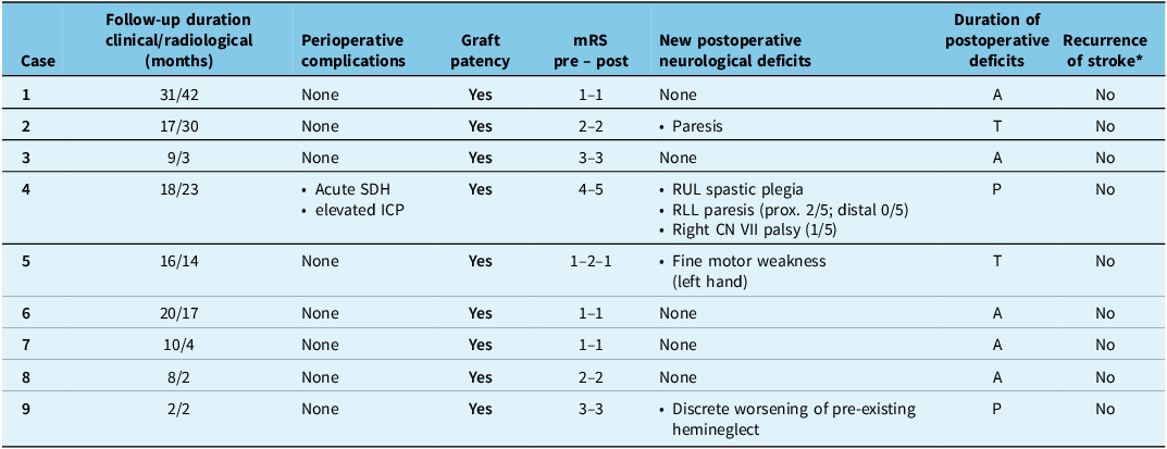
The median clinical follow-up duration was 16 months (range 2–31), while the median radiological follow-up period was 14 months (range 2–42) (Table 2).
Follow-up outcomes, complications and neurological evolution in patients following superficial temporal artery to middle cerebral artery bypass surgery

Table 2. Long description
A table summarizing follow-up outcomes and complications in patients after superficial temporal artery to middle cerebral artery bypass surgery. The table has 9 rows and 7 columns. The columns are labeled as follows: Case, Follow-up duration clinical/radiological (months), Perioperative complications, Graft patency, mRS pre-post, New postoperative neurological deficits, Duration of postoperative deficits, and Recurrence of stroke. Each row corresponds to a different case. Row 1: Case 1, Follow-up duration 31/42 months, No perioperative complications, Graft patency Yes, mRS pre-post 1-1, No new postoperative neurological deficits, Duration of postoperative deficits A, No recurrence of stroke. Row 2: Case 2, Follow-up duration 17/30 months, No perioperative complications, Graft patency Yes, mRS pre-post 2-2, New postoperative neurological deficit Paresis, Duration of postoperative deficits T, No recurrence of stroke. Row 3: Case 3, Follow-up duration 9/3 months, No perioperative complications, Graft patency Yes, mRS pre-post 3-3, No new postoperative neurological deficits, Duration of postoperative deficits A, No recurrence of stroke. Row 4: Case 4, Follow-up duration 18/23 months, Perioperative complications Acute SDH and elevated ICP, Graft patency Yes, mRS pre-post 4-5, New postoperative neurological deficits RUL spastic plegia, RLL paresis (prox. 2/5; distal 0/5), Right CN VII palsy (1/5), Duration of postoperative deficits P, No recurrence of stroke. Row 5: Case 5, Follow-up duration 16/14 months, No perioperative complications, Graft patency Yes, mRS pre-post 1-2-1, New postoperative neurological deficit Fine motor weakness (left hand), Duration of postoperative deficits T, No recurrence of stroke. Row 6: Case 6, Follow-up duration 20/17 months, No perioperative complications, Graft patency Yes, mRS pre-post 1-1, No new postoperative neurological deficits, Duration of postoperative deficits A, No recurrence of stroke. Row 7: Case 7, Follow-up duration 10/4 months, No perioperative complications, Graft patency Yes, mRS pre-post 1-1, No new postoperative neurological deficits, Duration of postoperative deficits A, No recurrence of stroke. Row 8: Case 8, Follow-up duration 8/2 months, No perioperative complications, Graft patency Yes, mRS pre-post 2-2, No new postoperative neurological deficits, Duration of postoperative deficits A, No recurrence of stroke. Row 9: Case 9, Follow-up duration 2/2 months, No perioperative complications, Graft patency Yes, mRS pre-post 3-3, New postoperative neurological deficit Discrete worsening of pre-existing hemineglect, Duration of postoperative deficits P, No recurrence of stroke.
A = absent; ICP = intracranial pressure; mRS = modified Rankin Scale; P = permanent; RLL = right lower limb; RUL = right upper limb; SDH = subdural hematoma; T = transient; CN = cranial nerve.
* At last clinical follow-up.
Outcomes
During the follow-up period, all (100%) bypass grafts demonstrated sustained patency, with no instances of occlusion or significant stenosis (Table 2). Additionally, no patients experienced recurrent ischemic events; however, one (11%) patient developed a subdural hematoma (SDH) during the immediate postoperative period. The median hospital stay was 4 days (range 3–42 days), and the median ICU stay was 3 days (range 19 hours–18 days). Postoperatively, most patients (n = 8; 89%) were discharged directly home, while one patient required transfer to a rehabilitation facility for extended recovery. In terms of functional outcomes, only three patients presented new postoperative neurological deficits. One of them experienced a one-point decline of the mRS score, characterized by transient left-hand fine motor weakness related to the non-operated hemisphere, which fully resolved by the 16-month clinical follow-up. Another patient showed worsening of 1 point on the mRS due to an SDH with documentation of mixed aphasia, right-sided facial palsy and right spastic hemiparesis (Table 2). A third patient exhibited a discrete worsening of pre-existing hemineglect, although the mRS score remained stable at 3.
Specific cases
Case 2: A 26-year-old female with APS and a history of multiple ischemic strokes was referred for revascularization due to a history of three ischemic strokes despite best medical management. Preoperative imaging confirmed significant hypoperfusion in the left MCA territory (Figure 1A), with a Suzuki score of III and an mRS score of 2. The patient underwent a STA–MCA bypass on the left side, performed without intraoperative complications.
(A) Preoperative angiography of Case 2, demonstrating significant hypoperfusion in the left middle cerebral artery (MCA) territory. Left internal carotid artery (ICA) injection demonstrating severe narrowing of the ICA, MCA and anterior cerebral artery. (B) Postoperative ICA and ECA angiography at the 5-month follow-up, confirming graft patency with no signs of stenosis or occlusion as well as complementary opacification of intracranial vessels from ICA and ECA through the bypass.

Immediately postoperatively, the patient experienced transient worsening of pre-existing right hemiparesis, including increased weakness in her right arm and leg, which resolved spontaneously within 12 hours. She returned to her preoperative baseline neurological status with an unchanged mRS score of 2, indicating mild functional impairment. Given the rapid resolution and absence of new neurological deficits, this transient deterioration was considered most consistent with recrudescence of prior stroke-related symptoms or transient perioperative hemodynamic vulnerability rather than a new ischemic event. Immediate postoperative neuroimaging demonstrated no new infarct, hemorrhage or other acute complications. Anticoagulation was resumed on postoperative day 3 after early postoperative imaging confirmed the absence of hemorrhagic or ischemic complications. During follow-up, angiography at 5 months confirmed graft patency with no evidence of stenosis or occlusion (Figure 1B). During 17 months of clinical follow-up, the patient remained neurologically stable with no recurrent ischemic events. This case illustrates the efficacy of STA–MCA bypass in young patients with MMS related to APS with recurrent cerebral ischemia despite adequate anticoagulation.
Case 4: A 44-year-old female with poorly controlled diabetes mellitus type II, refractory hypertension and a history of ischemic stroke secondary to MMS with bilateral carotid occlusion and resulting in aphasia and right hemiparesis (mRS score of 4) was urgently referred for surgical intervention. Imaging demonstrated severe hypoperfusion in the left MCA territory (Figure 2A) and a Suzuki score of III. She underwent a STA–MCA bypass on the left side, with no intraoperative complications.
(A) Preoperative angiography of Case 4, through the common carotid artery demonstrating severe hypoperfusion in the left middle cerebral artery (MCA) territory. (B) Lateral and (C) anteroposterior view of left ipsilateral vertebral injection showing filling of intracerebral arteries through the posterior communicating artery. (D) Postoperative CT scan, conducted immediately following bypass surgery, revealed an acute frontal-temporal-parietal subdural hematoma measuring 12 mm in thickness, associated with a 15-mm midline shift, which required urgent surgical evacuation. (E) Follow-up angiography with oblique left external carotid artery (ECA) injection at 1 year demonstrated sustained graft patency, with no evidence of bypass-related complications, such as occlusion or stenosis.

Figure 2. Long description
The image contains five separate panels showing medical imaging related to Moyamoya disease. Panel A: Preoperative angiography of Case 4, through the common carotid artery demonstrating severe hypoperfusion in the left middle cerebral artery (MCA) territory. Panel B: Lateral view of left ipsilateral vertebral injection showing filling of intracerebral arteries through the posterior communicating artery. Panel C: Anteroposterior view of left ipsilateral vertebral injection showing filling of intracerebral arteries through the posterior communicating artery. Panel D: Postoperative CT scan, conducted immediately following bypass surgery, revealed an acute frontal-temporal-parietal subdural hematoma measuring 12 millimeters in thickness, associated with a 15-millimeter midline shift, which required urgent surgical evacuation. Panel E: Follow-up angiography with oblique left external carotid artery injection at 1 year demonstrated sustained graft patency, with no evidence of bypass-related complications, such as occlusion or stenosis.
In the immediate postoperative period, the patient experienced severe hypertensive episodes lasting over 3 hours, with systolic blood pressure peaking at 230 mmHg. This was accompanied by significant neurological deterioration, including a rightward gaze deviation, drowsiness and a GCS score of 11. Moreover, the patient experienced vomiting and decerebrate posturing, prompting immediate interventions, including reintubation and administration of mannitol and hypertonic saline (NaCl 3%). A CT scan revealed preserved graft patency, as well as a left fronto-temporo-parietal SDH measuring 12 mm with a 15-mm midline shift, requiring emergent surgical evacuation (Figure 2B). Notably, no bleeding source was clearly identified intraoperatively, and the bypass graft was confirmed to be patent without evidence of leakage. Despite prompt intervention, the patient developed substantial neurological deterioration, including right spastic hemiplegia, mixed aphasia affecting both comprehension and expression and right-sided central facial paresis. Her mRS score worsened to 5 postoperatively, indicating severe disability. Follow-up imaging at 5 months confirmed graft patency, without bypass-related complications such as occlusion or stenosis (Figure 2C). Clinically, her hemiparesis showed slight improvement; however, she continued to experience significant aphasia and severe functional dependency. She was ultimately transferred to a long-term care facility because of ongoing care needs despite rehabilitation. This case highlights the importance of stringent postoperative blood pressure management and the potential for severe complications in high-risk patients, emphasizing careful patient selection.
Discussion
This case series provides insights into the outcomes and challenges of STA–MCA bypass as a revascularization strategy for MMD and MMS patients treated at a Canadian university center. The findings demonstrate the efficacy of this surgical approach in achieving high rates of graft patency and preventing recurrent ischemic or hemorrhagic events in appropriately selected patients. Moreover, while Case 2 demonstrated the potential for successful recovery even in the presence of high-risk comorbidities like APS, Case 4 underscored the importance of vigilant postoperative management to mitigate risks of complications such as hypertensive crises and intracranial hemorrhage. STA–MCA bypass serves as a cornerstone in the management of MMD and MMS, particularly in patients with progressive ischemia or recurrent strokes refractory to medical therapy. A comprehensive meta-analysis Reference Bagchi, Hore, Mandal and Pal17 of 17 studies including 2224 adult MMD patients found that bypass surgery significantly reduced recurrent stroke events compared with conservative treatment, with an odds ratio of 0.404 (95% CI 0.279–0.585; P < 0.001). In this case series, graft patency was achieved in all patients and maintained over follow-up periods ranging from 1 to 23 months. The absence of bypass occlusion or significant stenosis across the cohort aligns with existing evidence, Reference Kappel, Feroze, Torio, Sukumaran and Du21,Reference Miyamoto, Yoshimoto and Hashimoto31–Reference Liu, Su and Sun33 which underscores the durability of STA–MCA bypass in re-establishing and sustaining cerebral perfusion. These findings are consistent with outcomes reported in large-scale studies such as the Japan Adult Moyamoya Trial Reference Miyamoto, Yoshimoto and Hashimoto31,Reference Miyamoto34 and other revascularization studies, Reference El Naamani, Chen and Jabre35–Reference Ayyad, Al-Salihi, Ahmed, Hajali, Hammadi and Horn37 where bypass procedures have demonstrated significant reductions in the risk of future cerebrovascular events. Although recent trials, such as COSM Reference Ma, Wang and Wang23 and COSS, Reference Powers, Clarke and Grubb22 evaluating bypass surgery in symptomatic patients with atherosclerotic occlusion of the ICA or MCA without MMD/MMS have shown that combining bypass surgery with medical therapy does not significantly reduce the risk of recurrent ipsilateral ischemic stroke compared to medical therapy alone, bypass intervention remains a well-justified treatment option for carefully selected patients with radiological evidence of MMD and MMS.
Neurological outcomes in this series were favorable in the majority of cases, with postoperative mRS scores remaining relatively stable in eight of nine patients. However, the case of significant neurological deterioration highlights the potential for severe complications, such as intracranial hemorrhage, even in the setting of a technically successful procedure. In the patient who developed an acute SDH (Case 4), poorly controlled comorbidities, including diabetes mellitus and refractory hypertension, likely contributed to the adverse outcome observed. This emphasizes the importance of stringent perioperative monitoring, particularly of blood pressure, which is a known modifiable risk factor for postoperative complications in this patient population. Reference Parray, Martin and Siddiqui28,Reference Li, Zhang and Yu29 Prior studies have similarly highlighted the critical role of perioperative management in optimizing outcomes following revascularization surgery. Reference Kim, Oh, Bang, Kim and Cho38 This further supports the establishment of multidisciplinary care teams, including neurosurgical and neurocritical care specialists, in centers looking to develop a bypass program.
The inclusion of Case 2, a patient with MMS secondary to APS, highlights the heterogeneity of this patient population and the challenges associated with its management. APS is associated with significant vascular dysfunction, including endothelial injury, intimal and medial thickening and activation of inflammatory cascades. Reference Siddique, Risse, Canaud and Zuily39,Reference Knight and Kanthi40 It accounts for approximately 20–30% of ischemic strokes in young adults under 50 years of age. Reference Zedde, Grisendi, Assenza, Napoli, Moratti, Lara, Di Cecco, D’Aniello, Pavone, Pezzella, Candelaresi, Andreone, Valzania and Pascarella41 The recurrent thrombotic nature of the disease necessitates a precisely calibrated approach to anticoagulation and long-term management to effectively mitigate associated risks. In this instance, the absence of recurrent ischemic events following STA–MCA bypass underscores the procedure’s efficacy in improving cerebral hypoperfusion in complex syndromic conditions. This case further demonstrates that, even in high-risk individuals, favorable outcomes can be achieved through the implementation of carefully tailored and evidence-based surgical and medical strategies.
Since the publication of the COSS trial, Reference Powers, Clarke and Grubb22 bypass volumes have declined substantially across North America, contributing to reduced exposure during neurosurgical training and progressive centralization of technical expertise within a limited number of high-volume centers. Although MMD/MMS remain clear indications for revascularization, Reference Scott and Smith10 this historical shift has created a practical challenge for institutions seeking to maintain or re-establish proficiency in complex microsurgical bypass techniques. The present series reflects the early phase of rebuilding such capability within a Canadian academic center. From a programmatic standpoint, successful implementation required careful case selection in collaboration with vascular neurologists, concentration of cases within a dedicated surgical team comprising a fellowship-trained neurosurgeon, integration of experienced neuroanesthesia and neurocritical care support and standardized perioperative pathways emphasizing strict hemodynamic control and early graft surveillance. These elements are consistent with prior observations that outcomes in cerebral revascularization are closely linked to institutional experience, multidisciplinary coordination and procedural volume rather than technical factors alone. Reference Akbarian-Tefaghi, Kalakoti and Sun27,Reference Kim, Oh, Bang, Kim and Cho38 The single-surgeon model adopted in this study reduced inter-operator variability and allowed for consolidation of microsurgical expertise during the program’s development phase, an approach that may be particularly relevant for centers reintroducing low-frequency, high-complexity procedures. Nevertheless, the retrospective design and small cohort limit the generalizability of these findings, and longer-term follow-up is required to assess durability of clinical benefit and graft function. Future collaborative efforts across Canadian centers may help define referral networks, shared training pathways and prospective registries that ensure preservation of bypass expertise while maintaining procedural safety. Such initiatives may be essential to sustaining access to revascularization for patients with moyamoya in regions where historical declines in bypass utilization have reduced institutional familiarity with the operation.
Conclusion
In conclusion, this case series reinforces the role of STA–MCA bypass as an effective and durable revascularization strategy for Moyamoya disease and syndrome, which must be preserved within the surgical armamentarium of select surgeons for the benefit of the Canadian population. While the procedure demonstrates a high success rate in maintaining graft patency and preventing recurrent cerebrovascular events, the potential for severe complications highlights the necessity of meticulous patient selection, preoperative planning, technical proficiency and perioperative monitoring. These findings contribute to the growing evidence base supporting tailored surgical approaches to these complex cerebrovascular disorders.
Author contributions
P.L. and T.B.C. contributed to the study’s conception and provided supervision throughout the research process. Data curation and visualization were conducted by A.G. The initial draft of the manuscript was written by W.D., with W.D., A.G., T.B.C., K.E. and P.L. reviewing and editing subsequent versions. All authors gave final approval of the version to be published.
Funding statement
This research did not receive any specific grant from funding agencies in the public, commercial or not-for-profit sectors.
Competing interests
The authors have no personal, financial or institutional competing interests.



 Open access

