Introduction
The Kafue lechwe (Kobus leche kafuensis) is a unique antelope which is endemic only to the Kafue wetlands of Zambia and shares its grazing pastures with several species of other wildlife and domestic livestock. The number of these antelopes has declined as a result of many factors, ranging from illegal hunting and diminishing grazing lands as a result of encroachment by the weed Mimosa pigra, to a cycle of droughts that has increased idiopathic mortalities (Sheppe, Reference Sheppe1985; Kapungwe, Reference Kapungwe1993; Genet, Reference Genet2007). Under legal hunting, the Kafue lechwe is the most highly hunted animal (Siamudaala et al., Reference Siamudaala, Muma, Munang'andu and Mulumba2003). The population of lechwe on the Kafue wetlands is estimated to range between 40,000 and 45,000 (Kamweneshe et al., Reference Kamweneshe, Beilfuss and Simukonda2002).
Studies of the lechwe have mainly focused on bacterial diseases (Krauss et al., 1986; Ghirotti et al., Reference Ghirotti, Semproni, De Meneghi, Mungaba, Nannini, Calzetta and Paganico1991; Stafford, Reference Stafford1991; Pandey et al., Reference Pandey, Minyoi, Hasebe and Mwase1992, Reference Pandey, Mweene, Suzuki, Nambota and Kaji1994; Mwima, Reference Mwima1995; Zieger et al., Reference Zieger, Pandey, Kriek and Cauldwell1998). Transmission of parasites from livestock (goats and cattle) of the wetlands to antelopes, and vice versa, is possible (Phiri et al., Reference Phiri, Phiri, Chota and Monrad2007). This scenario could have a negative impact on the remaining antelopes, while the infected wildlife could form a stable and permanent reservoir for livestock infection. A number of diseases have been reported in the Kafue lechwe (Rottcher, Reference Rottcher1978; Pandey et al., Reference Pandey, Kobayashi, Nomura, Nambota, Mwima and Suzuki1999; Muma, Reference Muma2006). Some of these diseases, particularly brucellosis and tuberculosis (bovine TB), pose serious conservation and public health challenges. Studies on helminths have been restricted to a review (Stafford, Reference Stafford1991) and some case studies in the Lochinvar National Park involving a few helminths and animals.
The Kafue wetlands harbour large herds of cattle which are known to be predisposed to infections with Fasciola and other trematodes (Phiri et al., Reference Phiri, Phiri, Chota and Monrad2007). During the dry season, when there is limited pasture and water in the uplands, some cattle (transhumant) herds are moved to the plains around April or May and stay there until October or November. With the onset of the rains they are moved back to villages. However, some of the large-sized cattle herds, which do not find sufficient grazing land in the neighbourhood of the village, are permanently stationed in the plains (floodplain-resident herds) and share grazing land and water with wildlife throughout the year (Muma et al., Reference Muma, Samui, Oloya, Munyeme and Skjerve2007; Munyeme et al., Reference Munyeme, Muma, Skjerve, Nambota, Phiri, Samui, Dorny and Tryland2008). The risk of infection with helminths begins in June/July, when flood waters recede remarkably, up to November/December when the rains begin. During this period, cattle have unlimited access to potentially metacercariae/larvae-infested pastures.
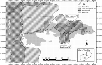
As a result of their large surface area and availability of nutritive pastures and water during drier periods of the year, the wetlands support a larger cattle population than most parts of the country. At least three-quarters of the cattle in the surrounding areas are driven into the wetlands to graze for about 6 months annually, a tradition that has been followed by many generations of inhabitants. Lechwe do not migrate seasonally but live within the wetlands, following the rise and fall of the flood waters. Blue Lagoon and Lochinvar national parks (NPs) and the surrounding game management areas lie within the Kafue wetlands and experience the same climatic conditions, including the seasonal flooding. However, the plains in Blue Lagoon hold more water after the floods and often stay waterlogged for a longer period compared to the plains in Lochinvar NP, which tend to get very dry and the water drains faster. These two parks lie adjacent to each other (see fig. 1) and are only separated from each other by the Kafue River.
Map of the Kafue flats showing the national parks (NP) and game management areas, with dots showing the sampling sites. Insert shows a map of Zambia, with B showing the study area.

In terms of animal activities on the plains, there is a lower lechwe density but a higher cattle population in Blue Lagoon than in Lochinvar. Cattle herds in Blue Lagoon are larger and there are more herds resident on the flood plain compared to Lochinvar (Muma et al., Reference Muma, Samui, Siamudaala, Oloya, Matope, Omer, Munyeme, Mubita and Skjerve2006; Munyeme et al., Reference Munyeme, Muma, Skjerve, Nambota, Samui, Rigouts and Tryland2009). Although the patterns of contact may be similar, interactions seem to be higher in Blue Lagoon than Lochinvar for the reasons stated above.
Disease transmission between wildlife and livestock can undermine conservation efforts, either by challenging the viability of threatened populations, or by eroding public tolerance of actual or potential wildlife disease reservoirs (Morgan et al., Reference Morgan, Lundervold, Medley, Shaikenov, Torgerson and Milner-Gulland2006). The relatively free movement of wild animals gives them the potential to act as vectors for the geographical spread of disease, even if infection doesn't persist for a long time in the wildlife population (Morgan et al., Reference Morgan, Lundervold, Medley, Shaikenov, Torgerson and Milner-Gulland2006).
This study aimed to evaluate the presence and distribution of helminths of lechwe and to compare these infection levels in lechwe of the Lochinvar and Blue Lagoon national parks. Higher animal numbers from both national parks than in previous studies were used to investigate helminths from gastrointestinal tract and internal organs.
Materials and methods
Study area
The Kafue wetlands (longitude 26–28°E and latitude 15°20′–15°55′S) are the second largest natural wetlands in Zambia, with massive flood plains drained by the Kafue River. They are situated in the Southern, Lusaka and Central provinces of Zambia. This study was conducted within the game management areas (GMAs) close to the Lochinvar and Blue Lagoon NPs (fig. 1). The Kafue wetlands cover an area of 600,400 hectares designated as a Ramsar site. Ramsar sites are wetlands of international importance designated under the Ramsar Convention. The Kafue wetlands contain the Blue Lagoon NP, with an area of 420 km2; Lochinvar NP, 410 km2; and Kafue GMA, covering 5175 km2. Game management areas are considered as buffer zones immediately surrounding NPs, where human settlement, limited agricultural activity and legal game hunting are allowed (Lewis et al., Reference Lewis, Kaweche and Mwenya1990). When livestock are present in these areas, there is an active interaction between livestock and wildlife, and hence GMAs are also referred to as ‘livestock–wildlife interface areas’.
The wetlands are also home to abundant bird life, some of which are migratory (e.g. the crane, Grus carunculatus), and wildlife that includes the unique Kafue lechwe, a semi-aquatic antelope. Other wildlife found are the wildebeest, zebra, buffalo, sitatunga, crocodile and hippopotamus.
Collection of lechwe
The Kafue lechwe is the predominant wildlife species of the Kafue wetlands (Sheppe, Reference Sheppe1985) and is confined to a relatively small area, particularly in and around Lochinvar and Blue Lagoon NPs.
The Zambia Wildlife Authority (ZAWA) provided 119 lechwe for the purpose of investigating various diseases affecting this species on the Kafue wetlands during the official annual cull. The lechwe were ethically hunted under special research licences. The survey was conducted during the 2008 hunting season (July–December) and as many as possible were shot in compliance with licence restrictions. Helminth sampling was only undertaken in 65 animals, due to the rigorous sampling techniques and sample processing which did not match with the rate of hunting.
The animals were subjected to a general post-mortem examination after culling. This consisted of visual inspection of each carcass and digital palpation of integument. Body condition scoring was determined for each animal slaughtered. Body condition score was performed, using the amount of abdominal and retroperitoneal fat, according to Morgan et al. (Reference Morgan, Shaikenov, Torgerson, Medley and Milner-Gulland2005) as: 1 (poor: almost no fat), 2 (fair/average: fair amount of fat) and 3 (good: plentiful fat, completely obscuring kidneys). Field assessment of the kidney fat index was also undertaken. The muscle conditions were also used to assist in scoring the nutritional body score. Although 65 animals were sampled for helminths, the body condition of all the 119 animals slaughtered was determined. All animals were eviscerated and the liver, lungs, trachea, abomasum, small and large intestines were processed separately within 24 h of slaughter. All viscera were also subjected to visual inspection and digital palpation. The liver and lungs were inspected for metacestodes and were incised for detailed examination.
Recovery and identification of helminths
In order to recover gastrointestinal helminths, the abomasum, small intestine and large intestine were ligated using a string and each examined separately.
The abomasum or rumen was opened along the greater curvature and its contents collected in a bucket. The empty abomasum or rumen was thoroughly washed with water paying particular attention to cleaning between the folds of the mucous membrane. The washings were thoroughly mixed and about 180 ml of the mixture were transferred to a 500 ml plastic bottle. Formalin (20 ml) was added and a lid screwed on securely. The plastic bottle was then inverted and shaken. The bottles were then transported to the laboratory at the University of Zambia for examination.
At the laboratory, contents of the bottles were separately emptied into sedimentation jars which were filled with water to about 2 cm from the brim. The contents in the jars were left to stand for 30 min. The supernatant was then decanted and the same process repeated until the sediment became clear. A small volume of the sediment at a time was added to a plastic Petri dish and examined using a stereomicroscope. All the detected helminths were picked out using fine forceps and stored in 10% formalin in a sample bottle until identified.
The procedure used for recovery of worms from the small and large intestines was similar to that for the abomasum. For the small intestines, the mesentery was separated from the intestines and these were opened lengthwise ensuring that the contents were collected in a bucket. Mucous membranes of both small and large intestines were scraped in order to recover smaller helminths. The opened intestines were then thoroughly washed and washings trapped in a bucket. Visible helminths were picked off with forceps as the intestines were opened and stored in 10% formalin in sample bottles. The procedure for sampling, washing and sub-sampling was the same as described above for parasites of the abomasum and rumen.
Liver inspection comprised examination of the liver and its associated gall bladder for the presence of liver flukes. After visual observation and palpation of the liver, sharp incisions were made into the parenchyma and major bile ducts. Exposed bile ducts were squeezed and examined for the presence of flukes.
Liver dissections were performed as described by Hansen & Perry (Reference Hansen and Perry1994) with minor modifications. Briefly, livers were cut into slices of about 5 mm in thickness in order to examine the hepatic parenchyma and to locate flukes in the bile ducts. Flukes that were seen while dissecting the livers were collected and temporarily stored in normal saline before being counted. After the whole organ had been sliced, the slices were immersed in a tray filled with water. In order to extract liver flukes, slices were individually removed from the water and simultaneously squeezed over a bucket so that any flukes so detected fell back into the water. The water was passed through a laboratory test sieve (size 500 μm; Endecotts Ltd, London, England) which trapped any flukes. Collected flukes were placed in a Petri dish and counted. Only the anterior ends of the flukes whose bodies were not intact after processing the livers were counted.
The trachea and main bronchi were opened using a pair of scissors and carefully checked for the presence of worms. Worms were picked out using fine forceps and stored in 10% formalin. In the laboratory they were processed and examined like the nematodes recovered from the gastrointestinal tract.
The opened abdominal cavity was checked for the presence of Setaria worms, with particular attention given to the serous surfaces. The worms were picked out with a pair of fine forceps, processed and examined like other nematodes.
Mesenteric veins were carefully inspected for the presence of Schistosoma sp., then they were cut and worms squeezed out. Schistosoma worms were picked out with a pair of fine forceps and stored in 10% formalin.
Recovered helminths from each animal were placed in lactophenol in plastic Petri dishes overnight so that they could be cleared. The cleared helminths were then placed on a glass slide and covered with a cover slip to facilitate microscopic examination. Standard identification keys by Soulsby (Reference Soulsby1982) and Gibbons et al. (Reference Gibbons, Jones and Khalil1994) were used to identify the helminths.
Data analysis
The results for worm infection were recorded as a binary outcome, present or absent, for different worm types, and were entered in Microsoft Excel® where data handling and cleaning was done. Similarly, the presence of worms in specific body organs was recorded as either present or absent. Statistical analysis was performed using STATA®/SE 10.0 for Windows (StataCorp, College Station, Texas, USA). The proportion of lechwe infected with a particular worm type, including the 95% confidence interval, was determined. Levels of infection for different worm types were compared between Blue Lagoon and Lochinvar using Fisher's exact test. Values of P < 0.05 were considered significant.
Results
Helminth species, prevalences and distribution
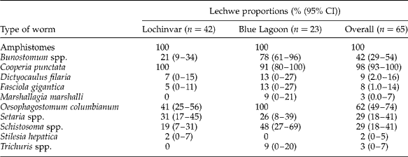
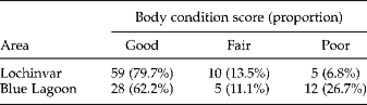
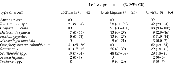
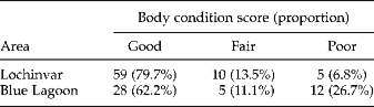
Worm infection was observed in all the lechwe examined after slaughter. Eleven helminth genera were found, namely Oesophagostomum, Bunostomum, Cooperia, Dictyocaulus, Marshallagia, Stilesia, Setaria, Trichuris, Fasciola, amphistomes and Schistosoma (table 1). All animals harboured amphistomes and the majority (97.9%) also had Cooperia punctata. The degree of worm infection was observed to vary between the two study areas, with Blue Lagoon recording higher infection levels compared to Lochinvar. Of the slaughtered animals from Blue Lagoon, 26.7% were in poor body condition, compared with only 6.8% from Lochinvar (table 2).
Worm infection in the Kafue lechwe (Kobus leche kafuensis) (n=65) in Lochinvar and Blue Lagoon national parks (2008).
Body condition scoring proportions of lechwe antelopes slaughtered (n=119) in Lochinvar and Blue Lagoon national parks.
Significant differences in worm infections between Blue Lagoon and Lochnivar were observed with Bunostomum spp. (P < 0.001) and Oesophagostomum spp. (P < 0.001). Marginal differences were observed with Marshallagia marshalli (P = 0.05) and Cooperia punctata (P = 0.05). However, no differences were observed for Trichuris (P = 0.12), Schistosoma spp. (P = 0.16), Fasciola gigantica (P = 0.231) and amphistomes (P = 1) as well as Setaria spp. (P = 1), Stilesia hepatica (P = 1) and Dictyocaulus filaria (P = 0.43).
The distribution of helminths recovered from each organ was as follows. Amphistomes (100%) in the abomasum and Oesophagostomum spp. (60.9%) in the large intestine were the most common, while Fasciola gigantica (7.8%) and Stilesia hepatica (1.6%) in the liver were the least common. Others were Trichuris (2%) and Shistosoma (29%) from the large intestine; Marshallagia marshalli (12.5% in the abomasum and 2% in the small intestine), Bunostomum (50% in the abomasum and 34% in the small intestine); Cooperia (51% in the abomasum and 6% in the small intestine); Setaria (29% in the abdomen) and Dictyocaulus filaria (9% in the lungs).
Discussion
This study reported the prevalence and distribution of helminth species affecting the Kafue lechwe. Eleven different types of helminths were identified in the animals studied. Stafford (Reference Stafford1991) reviewed a wide variety of parasites in the Kafue lechwe, including Schistosoma and Fasciola spp. More than 12 genera of helminths and different amphistome species have been reported. To the best of our knowledge, this is the first report of the presence and prevalence of Marshallagia marshalli in the lechwe. Marshallagia is a genus of intestinal worms in the family Trichostrongylidae but not known to have significant pathogenicity. Marshallagia marshalli has been reported in other antelopes that graze extensively on domestic livestock pastures (Morgan et al., Reference Morgan, Shaikenov, Torgerson, Medley and Milner-Gulland2005). The helminth is found in the abomasum of sheep, goats, antelopes, bighorn sheep and various wild ruminants.
The helminths reported in our study have also been found in domestic ruminants such as cattle, where Fasciola gigantica, Schistosoma spp. and amphistomes were the most prevalent (Phiri et al., Reference Phiri, Phiri and Monrad2006; Yabe et al., Reference Yabe, Phiri, Phiri, Chembensofu, Dorny and Vercruysse2008). Lechwe could, therefore, serve as a stable source of infection to domestic animals such as goats and cattle. The high level of interaction between lechwe antelopes (wildlife) and domestic animals (cattle) may intimate lechwe as a possible reservoir of infectious and parasitic diseases, given the spatial and temporal distribution of the diseases in lechwe (Munyeme et al., Reference Munyeme, Muma, Skjerve, Nambota, Phiri, Samui, Dorny and Tryland2008).
Sharing of grazing land and drinking water between cattle and wildlife is likely to facilitate bi-modal transmission of diseases (Muma et al., Reference Muma, Samui, Oloya, Munyeme and Skjerve2007; Munyeme et al., Reference Munyeme, Muma, Skjerve, Nambota, Phiri, Samui, Dorny and Tryland2008). Contact between wildlife and cattle has been reported to be a major source of concern in this ecosystem. Contamination of pasture by cattle herds that come to the plains, especially in Blue Lagoon, and also from lechwe which usually graze in large groups, could result in high egg densities in the environment. Moreover, the still waters in the plains along the waterfront and the moist soil of the flood plains are likely to promote the survival of both eggs and larval stages. However, since cattle are likely to be de-wormed periodically, the lechwe and other wildlife hosts are more likely to be a stable reservoir.
Even though other authors have reported the presence of helminths in the Kafue lechwe, their reports were mostly done 30–50 years ago and were based on cases involving a few individual animals from Lochinvar NP only (Krauss et al., 1986; Stafford, Reference Stafford1991) and on snail intermediate hosts (Wright, Reference Wright1966; Wright et al., Reference Wright, Southgate and Howard1979a, Reference Wright, Southgate and Howardb; Southgate et al., Reference Southgate, Howard, Rollinson, Brown, Ross and Knowles1985). The present study looked at the distribution of helminths in different regions of the gastrointestinal tract as well as lungs, liver and peritoneal cavity. Thus, our study involved more animals from both national parks than previously investigated. The 65 adult male lechwe we investigated may not be adequate for statistical comparison of intensities between host groups, but opportunities to sample larger numbers of these protected unique animals, especially females and young, have been rare and far between. On the other hand, Waid et al. (Reference Waid, Pence and Warren1985) reported that there was no apparent biological reason for age differentiation of animals less than 15 months of age. Examination of the liver, lungs, abomasi, small and large intestines and mesenteries was aimed at maximizing investigation of important trematodes, tapeworms and nematodes which are important for the livestock ruminants.
Actual transmission of helminths between antelopes and livestock is likely to depend on host abundance and patterns of contact, and not just on host specificity (Morgan et al., Reference Morgan, Milner-Gulland, Torgerson and Medley2004). In the Kafue wetlands, contact between lechwe and livestock generally occurs for about 6 months annually, depending on the seasonal flood cycle, with more months in periods of drought. The risk of infection with helminths begins in June/July, when flood waters recede remarkably, up to November/December when the rains begin (Phiri et al., Reference Phiri, Phiri, Chota and Monrad2007). During this period, cattle have unlimited access to potentially infested pastures.
The Kafue wetlands and their ecosystem have sustained wild and domestic animals for a long time. Ecological parameters have also been ideal for snail intermediate hosts for many of the reported trematode helminths (Phiri et al., Reference Phiri, Phiri, Chota and Monrad2007). Eliminating or reducing contact between the lechwe and cattle has been suggested before (Rottcher, Reference Rottcher1978; Phiri et al., Reference Phiri, Phiri, Chota and Monrad2007). This, however, would be feasible only with the cooperation and support of the local community. People would need to perceive the benefits of discontinuing the transhumant grazing system. This cooperation is most unlikely, however, given that the local communities have no alternative areas in which to graze or water their animals. The willingness of farmers and other agricultural stakeholders to support wildlife conservation efforts is likely to be affected by assessment of the risk of disease transmission to livestock, and on the control strategies available should this risk be high (Morgan et al., Reference Morgan, Lundervold, Medley, Shaikenov, Torgerson and Milner-Gulland2006).
Cattle densities are higher as a result of transhumance, while that of lechwe are lower, around Blue Lagoon NP. The opposite is true for the Lochinvar area. Blue Lagoon national park holds more water after the floods and it stays waterlogged for a longer period. In Lochinvar NP, it gets very dry and the water drains faster. Moisture is also critically important for the development, survival and movements of helminth larvae. Dry conditions can limit transmission and cause larvae to migrate into the soil, although excessive rainfall can wash away eggs and infective stages (Stromberg, Reference Stromberg1997). These factors may have an influence on sustainability of helminths and their gastropod intermediate hosts and could partly account for the differences observed in this study (Wright et al., Reference Wright, Southgate and Howard1979b). The cause–effect relationship between lechwe and domestic cattle in terms of parasitic diseases is very important given the levels of transhumance of cattle practised in this system, which may be deterministic in disease spread between these two animal species. Other factors include favourable climatic conditions for helminth reproduction, lack of veterinary services (especially anthelmintic treatment to cattle), and the presence of other wild ruminants, such as buffalo and wildebeest, whose epidemiological contributions to parasitism remain largely unknown.
Empirical evidence points to several biologically distinct mechanisms by which seasonality can impact host–pathogen interactions, including seasonal changes in host social behaviour and contact rates, variation in encounters with infective stages in the environment, annual pulses of host births and deaths, and changes in host immune defences (Altizer et al., Reference Altizer, Dobson, Hosseini, Hudson, Pascual and Rohani2006). Seasonal variations in temperature, rainfall and resource availability are ubiquitous and can exert strong pressures on population dynamics. For many intestinal parasites, infectious stages released into the environment are vulnerable to variation in temperature, rainfall and humidity before they encounter new hosts (e.g. Gordon et al., Reference Gordon, Davey and Peaston1934; Gillett, Reference Gillett, Taylor and Muller1974). Even though the incidence of many pathogens and parasites varies conspicuously by season (Altizer et al., Reference Altizer, Dobson, Hosseini, Hudson, Pascual and Rohani2006), the quality of the data we collected was not sufficient to determine seasonal effect pertaining in the wetlands, mainly because: (1) there exist licence and number restrictions; (2) lechwe do not migrate seasonally and sampling was only possible during the few months of the official culling; and (3) there are challenges in accessing the study area and lechwe during the flood (rainy) season. Similarly, the effect of body condition was difficult to elucidate, partly because Morgan et al. (Reference Morgan, Shaikenov, Torgerson, Medley and Milner-Gulland2005) reported that both parasitism and immunity impose energy costs, confounding relationships between parasite intensity and body condition, and largely because a single cross-sectional sample is unlikely to provide a sensitive test of the biologically important relationships. Besides, differences in infection intensity with age could be caused by variation in infection pressure between years. This is because the lag between maximum parasite intensity, peak body condition and effects on host rates may mean that the timing of observations can be crucial to detecting these effects (Stein et al., Reference Stein, Irvine, Ropstad, Halvorsen, Langvatn and Albon2002).
These limitations bring about knowledge gaps, as seasonal changes can affect hosts, pathogens and vectors in ways that alter components of the basic reproductive number that determines the rate at which infected hosts are produced (Altizer et al., Reference Altizer, Dobson, Hosseini, Hudson, Pascual and Rohani2006). Furthermore, these mechanisms include those that influence parasite transmission, in part by altering the behaviour of hosts, the biology of vectors or parasite infectious stages in the environment. Seasonality can further cause shifts in the base of susceptible hosts through annual variation in host births and deaths, or cause changes in underlying immunity to infection – and in natural systems it is likely that multiple seasonal drivers will interact in complex ways.
Variation in the weather within and between years is likely to influence risks of disease transfer between species (Morgan et al., Reference Morgan, Lundervold, Medley, Shaikenov, Torgerson and Milner-Gulland2006). Similarly, data that are restricted to short sampling periods might miss key events in disease dynamics, and not depict a true picture of cross-species infection.
The areas of our study were remote and resources for the study scarce. Collection of the washings and the initial volumes used varied and were not recorded for each animal, partly because of the increased workload from harvested carcasses. This lack of complete information was the reason for excluding data on worm burdens and emphasizing data on prevalence. This oversight is keenly being corrected in subsequent studies. Provided that the intestinal material is well mixed, the worms in the aliquot are a good reflection of the actual worm numbers (Reinecke, Reference Reinecke1984). Worm speciation was attempted as much as possible and where it was difficult only the genus was determined. For accurate speciation, molecular tools will need to be employed.
Some amphistomes were recovered from the abomasi of lechwe although the predilection site for adult amphistomes is in the fore stomachs (rumen and reticulum). Similarly, some Bunostomum spp. and Cooperia punctata adult worms, whose predilection site is the small intestine, were recovered from the abomasi; and some Marshallagia marshalli adult worms, which are normally found in the abomasi, were recovered from the small intestine. Despite the apparent overflow and misplacement of helminths from the predilection sites to other sites, the contrary may be a partial explanation. For example, Wright et al. (Reference Wright, Southgate and Howard1979b) have reported amphistome species in the abomasum of the Kafue lechwe. The impression may be the same for other helminths so reported, even though worms displaced from their usual locations during processing of samples can't be completely overlooked. Although the procedure for recovering helminths from the gastrointestinal tract was properly carried out, it is likely that worm misplacement could have occurred, especially in the few animals processed late after dusk.
Another limitation to this study is in agreement with Morgan et al. (Reference Morgan, Shaikenov, Torgerson, Medley and Milner-Gulland2005) – shooting individual lechwe after encountering them opportunistically may not be an ideal sampling method and can be prone to selection bias. The licences we were granted allowed us to harvest only adult males, which were considered as reproductively mature. Therefore, convexity in age-prevalence and age-intensity curves can be an artefact of aggregation in parasite populations, leading to typically small sample sizes from older hosts being more likely to underestimate the mean than large sample sizes from younger hosts (Morgan et al., Reference Morgan, Shaikenov, Torgerson, Medley and Milner-Gulland2005). However, Morgan et al. (Reference Morgan, Lundervold, Medley, Shaikenov, Torgerson and Milner-Gulland2006) reported potential benefits and limitations of models that can be applied successfully to disease transmission across the wildlife–livestock boundary.
In on-going research, quantitative helminth burden analysis is being undertaken to determine the level of parasitism. To fully understand the ecological and socio-economic implications of diseases in wildlife, future veterinary studies should be designed within the context of the ecosystem and the prevailing land-use practices.
Acknowledgements
The Norwegian Council for Higher Education's Programme for Development Research and Education (NUFU) funded part of this research. The Zambia Wildlife Authority (ZAWA) facilitated the harvest of the animals. Dr Hetron Munang'andu is thanked for permitting us to use the map of the Kafue wetlands. Comments and observations on the initial draft of the manuscript by Dr K.S. Nalubamba are acknowledged.