Introduction
The myxozoan genus Unicapsula Davis, 1924 (Myxosporea: Multivalvulida: Trilosporidae) is characterised as having a myxospore with three unequal shell valves (SVs) and polar capsules (PCs), of which one is prominent, while the two other PCs are rudimentary (Lom and Dyková, Reference Lom and Dyková2006; Fiala et al., Reference Fiala, Bartošová-Sojková, Whipps, Okamura, Gruhl and Bartholomew2015). The genus was erected by Davis (Reference Davis1924), when he found plasmodia of Unicapsula muscularis Davis, 1924 in the myofibres of Hippoglossus stenolepis Schmidt, 1904 (Pacific halibut) from the northeastern Pacific Ocean, off the coast of North America. Davis (Reference Davis1924) referred to infected fish as ‘wormy halibut’, referring to their heavy parasite load. The second species to be described, Unicapsula galeata Naidjenova et Zaika, 1970, was discovered in the trunk muscle of the whitesaddle goatfish Parupeneus ciliates (Lacepède, 1802) more than 40 years later after the erection of the genus (Naidjenova and Zaika, Reference Naidjenova and Zaika1970), and a total of only 13 species have been described to date (Schubert et al., Reference Schubert, Sprague and Reinboth1975; Alama-Bermejo et al., Reference Alama-Bermejo, Cuadrado, Raga and Holzer2009; Miller and Adlard, Reference Miller and Adlard2013; Tomochi et al., Reference Tomochi, Li, Tran, Yanagida and Sato2014; Yokoyama et al., Reference Yokoyama, Abe, Maehara and Suzuki2014; Al-Jufaili et al., Reference Al-Jufaili, Freeman, Machkevskyi, Al-Nabhani and Palm2016). As partially reflected in a limited number (only 13) of the currently recognized Unicapsula spp., Miller and Adlard (Reference Miller and Adlard2013) suspected that the richness of Unicapsula spp. in fish off the coast of Australia was lower than that of Kudoa or other speciose myxosporean genera. Indeed, Miller and Adlard (Reference Miller and Adlard2013) examined >4500 individuals of >500 species of teleost and elasmobranch fish collected from the water around Australia and found only three species, namely Unicapsula pyramidata (Naidjenova et Zaika, 1970), Unicapsula seriolae Lester, 1982, and Unicapsula andersenae Miller et Adlard, 2013, and only identified the parasites in seven teleost species.
In the present study, we report seven Unicapsula spp. from 15 commercial fish species in southern China and Japan, grown in the periphery of Pacific Ocean (South China Sea off southern China, Philippine Sea off southern Japan and Bering Sea off Alaska) and the East Atlantic Ocean off the coast of western Africa, on the basis of myxospore morphology and ribosomal RNA gene (rDNA) sequences. In addition to providing the first molecular-genetic characterization of U. muscularis and U. galeata, which are the type and the second-described species of the genus, respectively, the present study also provides multiple new hosts and geographical records for several species. This integrated taxonomic approaches could contribute to an improved understanding of the genus' global biodiversity and prevalence.
Materials and methods
Fish samples and parasitological examination
In total 22 fish species were examined in the present study. Between November 2015 and January 2019, 317 individual fish from 20 species were purchased from wet markets in Zhanjiang City, Guangdong Province, China and were examined parasitologically in Guangdong Ocean University. Of these 20 species, two frozen imported fish species, the royal threadfin Pentanemus quinquarius (Linnaeus, 1758) and West African goatfish Pseudupeneus prayensis (Cuvier, 1829), had been captured from the East Atlantic Ocean, off the West African coast between Senegal and Angola (based on the species' known distribution), although exact locations were unknown. In addition to those 20 species, two individuals of the golden threadfin breams Nemipterus virgatus (Houttuyn, 1782) were purchased from a local fish market in Kochi, Japan, in August 2016 and were transported on ice to Yamaguchi University, and several frozen fillets of the arrow-tooth flounder Atheresthes stomias (Jordan et Gilbert, 1880), which had been imported from the Bering Sea, off the coast of Alaska, USA, were purchased in February 2017 from a local fish market near Yamaguchi University.
The 22 putative host fish species were identified using DNA barcoding of the mitochondrial cytochrome c oxidase subunit I gene (cox-1), as described by Ward et al. (Reference Ward, Zemlak, Innes, Last and Hebert2005) and Zhang and Hanner (Reference Zhang and Hanner2011). Details of amplification and sequencing of cox-1 fragments are provided in the following section. The current taxonomic status of the fish was consulted to Prof. Hiroyuki Motomura from Kagoshima University Museum, Japan.
In the Guangdong Ocean University and Yamaguchi University laboratories, the collected fish were thawed (if necessary) and dissected, and the gills, viscera and brain were removed for microscopic examination. The fish fillets were either examined on the day of their arrival or frozen until examination. To check for the presence of myxosporean cysts or pseudocysts, thin slices of the trunk muscle were placed in physiological saline, pressed between glass plates and examined under a dissection microscope. The commercially packaged fish fillets were examined in a similar manner. The tissue-embedded myxosporean plasmodia were then divided into two groups, and fixed in either 10% neutral-buffered formalin solution, for morphological examination or 70% ethanol, for molecular analysis.
For morphological examination, the myxospores were released from the formalin-fixed tissues using fine forceps, observed using a microscope equipped with differential interference contrast imaging, and photographed at a magnification of ×800. Measurements were conducted using digital photographs and the guidelines of Lom and Arthur (Reference Lom and Arthur1989). All measurements are expressed in μm unless otherwise stated, and are reported as ranges with means in parentheses.
In addition, the parasite specimens in fixatives were deposited in the Meguro Parasitological Museum, Tokyo, Japan (collection nos. 21381, and 21500–21514).
DNA extraction, amplification and sequencing
To determine the species identities of the putative host fish, the mitochondrial cox1 region was sequenced for each specimen, according to Ward et al. (Reference Ward, Zemlak, Innes, Last and Hebert2005) and Zhang and Hanner (Reference Zhang and Hanner2011). Briefly, small pieces of the ethanol-preserved fish tissue were washed thrice in sterile pure water, and DNA was extracted using an Illustra™ tissue and cells genomicPrep Mini Spin Kit (GE Healthcare UK, Buckinghamshire, UK) according to manufacturer instructions. Polymerase chain reaction (PCR) amplification of the cox1 region was performed in a 20-μl reactions that contained DNA polymerase, Blend Taq-Plus- (TOYOBO, Dojima Hama, Osaka, Japan), and either of two primer pairs, FishF1 (5′- TCA ACC AAC CAC AAA GAC ATT GGC AC-3′) and FishR1 (5′- TAG ACT TCT GGG TGG CCA AAG AAT CA-3′) (Ward et al., Reference Ward, Zemlak, Innes, Last and Hebert2005) or FishF2_t1 (5′- TGT AAA ACG ACG GCC AGT CRA CYA AYC AYA AAG AYA TYG GCA C-3′) and FR1d_t1 (5′- CAG GAA ACA GCT ATG ACA CYT CAG GGT GWC CGA ARA AYC ARA A-3′) (Zhang and Hanner, Reference Zhang and Hanner2011). The PCR was performed using the following conditions: 3 min at 94°C; followed by 40 cycles of 30 s at 94°C, 1 min at 52°C, and 1 min at 72°C; and 7 min at 72°C.
Parasite DNA was extracted from the plasmodia of each source as outlined above, and overlapping rDNA fragments were PCR amplified as described by Tomochi et al. (Reference Tomochi, Li, Tran, Yanagida and Sato2014). The resulting PCR products were purified using a FastGene Gel/PCR Extraction Kit (NIPPON Genetics Co., Tokyo, Japan) and sequenced directly using the primers described by Li et al. (Reference Li, Sato, Tanaka, Ohnishi, Kamata and Sugita-Konishi2013) and Tomochi et al. (Reference Tomochi, Li, Tran, Yanagida and Sato2014). For the arrow-tooth flounder isolate (i.e. U. muscularis), two new forward primers were designed for the 5′-terminus of the 28S rDNA using a partially sequenced fragment of the isolate: KUDOA28S3F (5′- CAG GTA AGA TAA CCC GCT GAA C -3′) and KUDOA28S4F (5′- GGC TTT GAC AGA CGT ACG TG -3′). These two primers worked better for the U. muscularis isolate than KUDOA28S2F (5′- AGG CAA GAC TAC CTG CTG AAC -3′), which was suitable for amplifying 28S rDNA from the other isolates. When direct sequencing results were not satisfactory, purified PCR products were cloned into the pTA2 plasmid vector (TArget Clone™; TOYOBO), as described previously (Tomochi et al., Reference Tomochi, Li, Tran, Yanagida and Sato2014). Following propagation, the plasmid DNA was extracted using a FastGene Plasmid Mini Kit (NIPPON Genetics Co.) and inserts from multiple independent clones, at least three, were sequenced using universal M13 forward and reverse primers.
The resulting nucleotide sequences were submitted to the DDBJ/EMBL/GenBank databases under the accession nos. LC484867–LC484883 (fish cox-1) and accession nos. LC316969 and LC474121–LC474141 (Unicapsula spp.).
Phylogenetic analysis
For phylogenetic analysis, the newly obtained 18S and 28S rDNA sequences of Unicapsula spp. and related sequences from the DDBJ/EMBL/GenBank databases were aligned using CLUSTAL W (Thompson et al., Reference Thompson, Higgins and Gibson1994), with subsequent manual adjustment. Two sets each of nucleotide sequences of the 18S and 28S rDNA were prepared. The first set of short sequences was used to cover as much data as possible from different Unicapsula spp., and the second set of long sequences was used to more accurately estimate phylogenetic relationships of different Unicapsula spp. The accession numbers of the sequences analysed are provided in the figures showing phylogenetic trees. Regions that were poorly aligned and characters that were missing from any of the sequences were excluded from subsequent analyses. A total of 722 and 1621 characters, of which 150 and 367 were variable, respectively, remained for subsequent analysis for the 18S rDNA regions, and 540 and 2156 characters, of which 257 and 660 were variable, respectively, remained for subsequent analysis for the 28S rDNA regions. Maximum likelihood (ML) analysis was performed using PhyML (Guindon and Gascuel, Reference Guindon and Gascuel2003; Dereeper et al., Reference Dereeper, Guignon, Blanc, Audic, Buffet, Chevenet, Dufayard, Guindon, Lefort, Lescot, Claverie and Gascuel2008), which is available on the ‘phylogeny.fr’ website (http://www.phylogeny.fr/). The probabilities of the inferred branch were assessed by using approximate likelihood-ratio test (aLRT), which is an alternative to the non-parametric bootstrap estimation of branch support (Anisimova and Gascuel, Reference Anisimova and Gascuel2006).
Results
Parasitological examination
The microscopic examination of trunk muscle slices from 317 fish samples, representing 22 potential host species, revealed plasmodia and myxospores from several myxosporean genera (i.e. Unicapsula, Kudoa, Myxobolus and Myxidium) in 55 fish samples (15 species from 10 families), 83 fish samples (nine species from eight families), 42 fish samples (one species from one family) and 19 fish samples (three species from three families), respectively. Most of the fish species (n = 18) were only infected by a single myxosporean species. However, there were four exceptions: six myxosporean species were detected in the longfinned mullet Moolgarda perusii (Valenciennes, 1836), and two myxosporean species in the dotted gizzard shad Konosirus punctatus (Temminck et Schlegel, 1846), royal threadfin, and arrow-tooth flounder. In the latter two cases, the infection was due to different combinations of Kudoa spp. and Unicapsula spp.
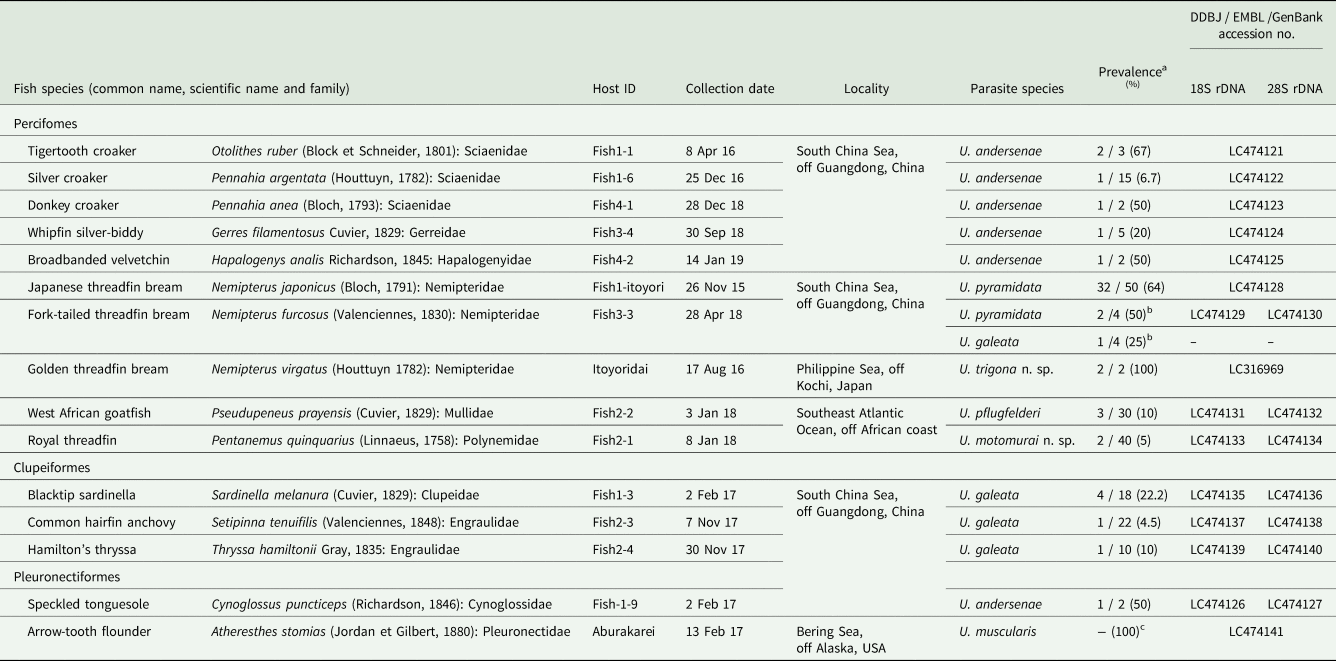
As mentioned above, pseudocyst-forming plasmodia of Unicapsula spp. were found in the trunk muscle of 55 fish samples (Table 1), including 32 samples of the Japanese threadfin bream Nemipterus japonicus (Bloch, 1791), which were infected with U. pyramidata. The fish samples varied in the prevalence and infection intensity of Unicapsula plasmodia.
Fish samples examined and detected Unicapsula spp.

a No. of positive fish samples / no. of examined fish samples (%).
b Coinfection by U. pyramidata and U. galeata was observed for a single fish. The rDNA sequences were not obtained for U. galeata.
c One package with several fillets.
Morphological and genetic myxospore characterization
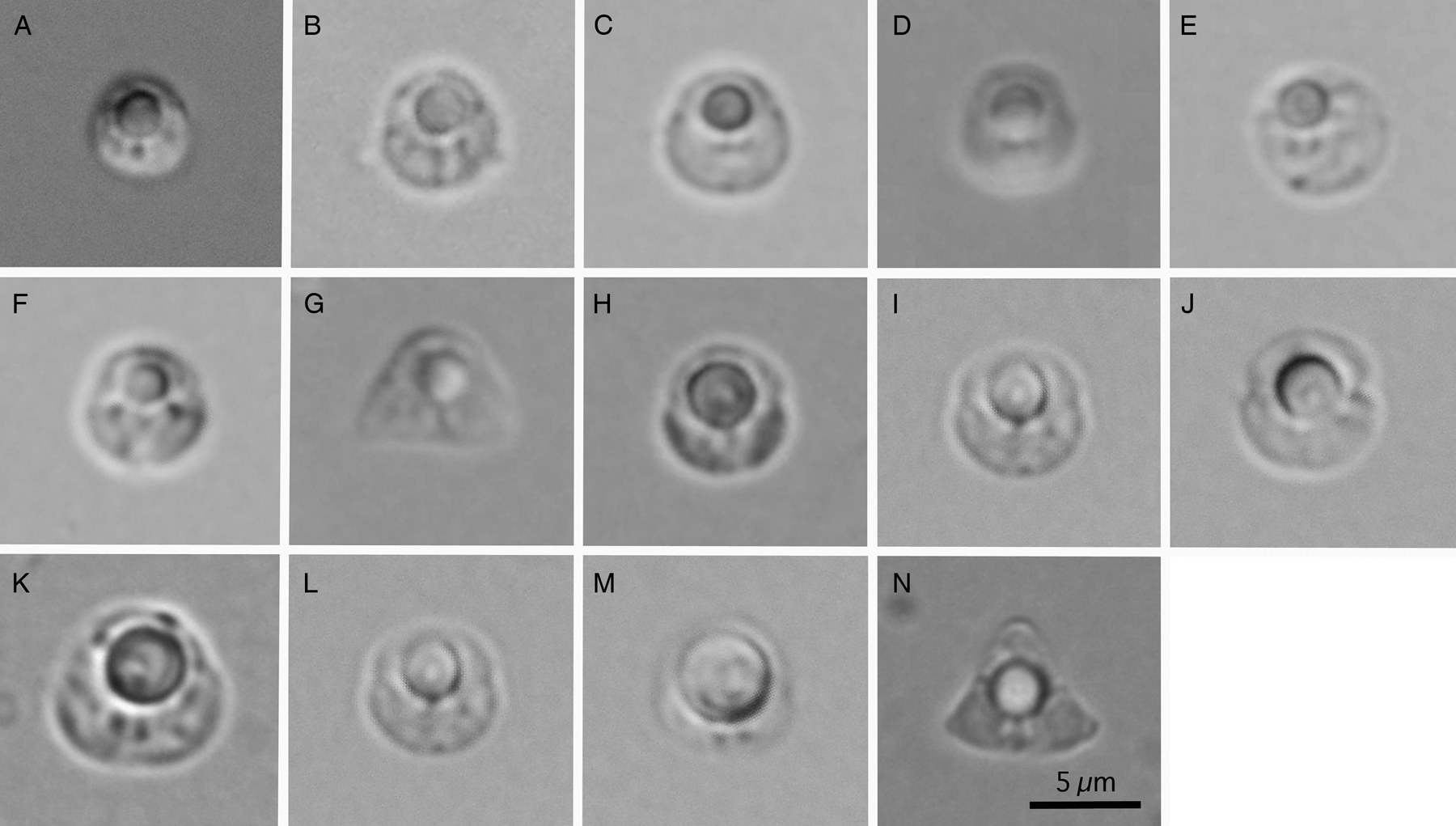
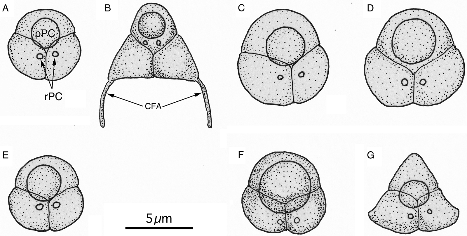
Myxospores in pseudocysts from the trunk muscles of the 15 fish host species had three unequal SVs and three PCs, of which two were rudimentary and one was prominent and functional with a polar filament. These observed morphological features of the myxospores were well coincident with the definition of the genus Unicapsula (Lom and Noble, Reference Lom and Noble1984; Lom and Dyková, Reference Lom and Dyková2006; Alama-Bermejo et al., Reference Alama-Bermejo, Cuadrado, Raga and Holzer2009; Fiala et al., Reference Fiala, Bartošová-Sojková, Whipps, Okamura, Gruhl and Bartholomew2015). Myxospore formation in plasmodia in all host fish was well synchronized. Myxospores observed in the present study were classified according to the shape and dimension of myxospores, localization and dimension of prominent PCs, and myxospore ornamentation (Figs 1 and 2). Lastly, seven Unicapusla spp., including two new species, were differentiated by sequencing the 18S and 28S rDNA (Table 1).
Microscopic view of myxospores of Unicapsula spp. collected in the present study. (A–F) Unicapsula andersenae (A, isolate Li1-1; B, Li1-6; C, Li4-1; D, Li3-4; E, Li4-2; and F, Li1-9); (G) Unicapsula pyramidata (Li3-3); (H–J) Unicapsula galeata (H, Li1-3; I, Li2-3; and J, Li2-4); (K) Unicapsula muscularis (Aburakarei); (L) Unicapsula pflugfelderi (Li2-2); (M) Unicapsula motomurai n. sp. (Li2-1); and (N) Unicapsula trigona n. sp. (Itoyoridai). All photographs at the same magnification (scale bar in N).

Stylized diagrams of Unicapsula spp. collected in the present study. (A) Unicapsula andersenae: (B) Unicapsula pyramidata; (C) Unicapsula galeata; (D) Unicapsula muscularis; (E) Unicapsula pflugfelderi; (F) Unicapsula motomurai n. sp.; and (G) Unicapsula trigona n. sp. Unicapsula spores consist of three shell valves with one prominent PC (pPC) and two rudimentary PCs (rPC) as examples are shown in (A). Unicapsula pyramidata myxospores have caudal filamentous appendages (CFA).

Unicapsula andersenae (Miller et Adlard, 2013)
This species was detected in six fish species: tigertooth croaker (Sciaenidae), silver croaker (Sciaenidae), donkey croaker (Sciaenidae), whipfin silver-biddy (Gerreidae), broadbanded velvetchin (Hapalogenyidae) and speckled tonguesole (Cynoglossidae), as shown in Table 1. Plasmodia in the myofibres were elongated, like fine threads, had tapering ends and ranged from 0.5 to 2.5 mm in length.
Bilaterally symmetrical, almost subspherical myxospores (n = 33 from three fish hosts), which measured 4.6–7.0 (5.4) in length by 4.3–6.5 (5.2) in width, with a functional semi-spherical PC, which measured 1.9–2.8 (2.3) in diameter, and two smaller PCs, which measured 0.4–1.0 (0.6) in diameter (Figs 1A–F and 2A). Turns of polar filaments in prominent PCs were not discernible. The dimensions of myxospores, functional PCs and other morphological features were consistent with the known morphology of U. andersenae (Table 2).
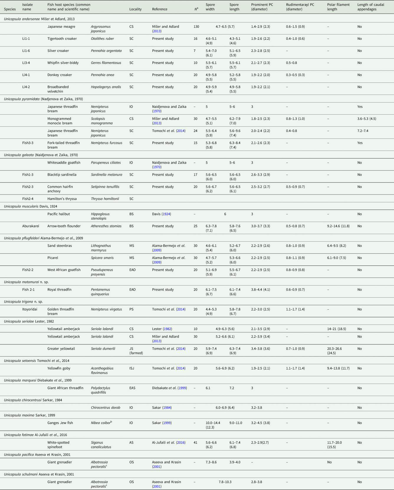
Morphological characteristics of Unicapsula spp. myxospores recorded from this and previous studies (measurements in μm)

AS, Arabian Sea; BS, Bering Sea; CS, Coral Sea (Australia); EAO, East Atlantic Ocean; IO, Indian Ocean; ISJ, Inland Sea of Japan; JS, Japan Sea; MS, Mediterranean Sea; OS, Okhotsk Sea; PS, Philippine Sea. off Kochi, Japan; and SC, South China Sea.
PC, polar capsule; PF polar filament; and CA, caudal appendages.
a Number of spores measured. ‘–’ means no data.
b Nibea coibor (Hamilton, 1822); Syn. Pseudosciaena coibar (Hamilton, 1822)
c Albatrossia pectoralis (Gilbert, 1892); Syn. Coryphaenoides pectoralis (Gilbert, 1892)
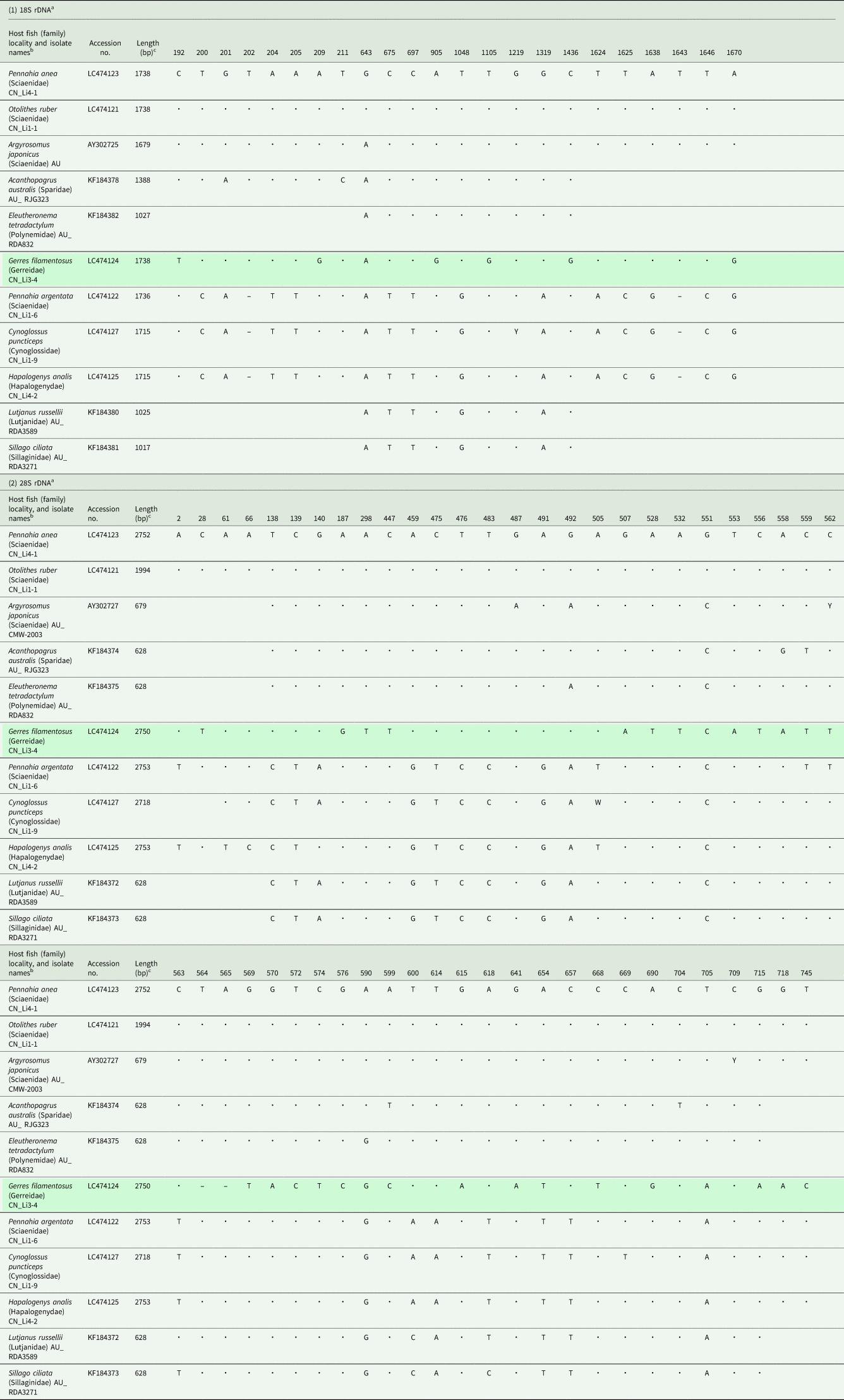
Both 18S and 28S rDNA sequences were successfully obtained for six isolates (DDBJ/EMBL/Genbank accession nos. LC474121–LC474127). These sequences were sorted into three genotypes: (A) two isolates (Li1-1 and Li4-1) from the tigertooth croaker and donkey croaker; (B) one isolate (Li3-4) from the whipfin silver-biddy; and (C) three isolates (Li1-6, Li1-9 and Li4-2) from the silver croaker, speckled tonguesole and broadbanded velvetchin (see Table 3). Pairwise comparisons revealed that the 18S rDNA of the three groups differed by 0.69% (7/1736)–0.98% (17/1736) nucleotide substitutions with/without two insertion/deletions (indels) and that the 28S rDNA of the three groups differed by 1.78% (49/2752)–3.35% (92/2750) nucleotide substitutions with one to three indels. Similar genetic segregation was previously reported for the 18S and 28S rDNA sequences of Australian U. andersenae isolates (Table 3). The genotype A 28S rDNA sequences (Li1-1, Li4-1, CMW-2003, RJG323 and RDA832) exhibited 99.04–99.68% similarity over 628-bp length, whereas the genotype C sequences (Li1-6, Li1-9 and Li4-2, RDA3589 and RDA3271) exhibited 99.52–99.84% similarity. However, the two genotypes were less similar to each other, ranging 96.34% (605/628)–97.77% (614/628) similarity.
Nucleotide differences in the 18S and 28S rDNA of Unicapsula andersenae isolates from different locations

a Nucleotide position relative to the 5′-terminus of the 18S and 28S rDNA sequences of Unicapsula andersenae Li4-1 (LC474123). Dots denote that a base was identical to the base of the uppermost nucleotide sequence and blanks indicate no available data.
“– “ indicates a nucleotide deletion.
b Locality is expressed by country name (CN, China; and AU, Australia).
c Lengths of the 18S and 28S rDNA sequences from the DDBJ/EMBL/GenBank databases are expressed in base pairs (bp).
Remarks: This species was described by Miller and Adlard (Reference Miller and Adlard2013) from five fish species in the families Sciaenidae, Sparidae, Polynemidae, Lutjanidae and Sillaginidae (all in the Order Perciformes) and was reported to form pseudocysts in host trunk muscles. Although the significance of genetic segregation is unknown, this phenomenon was again observed in Chinese isolates of U. andersenae (present study), which exhibited the morphological and genetic characteristics of the species described by Miller and Adlard (Reference Miller and Adlard2013). Six fish species of the families Sciaenidae, Gerreidae, Hapalogenyidae and Cynoglossidae are new host records, and South China Sea, off Guangdong, China, is a new geographical record.
Unicapsula pyramidata (Naidjenova et Zaika, 1970)
This species was highly prevalent in the Japanese threadfin bream Nemipterus japonicus (Bloch, 1791) and fork-tailed threadfin bream Nemipterus furcosus (Valenciennes, 1830) samples that originated from the South China Sea, off the coast of Guangdong, China. The species intensively formed pseudocysts (0.8–2 mm in length with tapering ends) in host myofibres (Table 1). One of four fork-tailed threadfin bream samples was parasitised by both U. pyramidata and U. galeata, which had plasmodia of 1.43–1.91 (1.67) mm × 0.06–0.08 (0.07) mm (n = 7) and 0.69–1.41 (1.01) mm × 0.05–0.10 (0.07) mm (n = 10), respectively.
Bilaterally symmetrical triangular myxospores, which each possessed a blunt apical SV (n = 15 from a fork-tailed threadfin bream sample), measured 5.3–6.8 (5.8) in length by 6.3–8.4 (7.4) in width, with a functional semi-spherical PC, which measured 2.1–2.6 (2.3) in diameter, and two smaller PCs, which measured 0.6–0.8 (0.7) in diameter (Figs 1G and 2B). Two posterior SVs had pointed distal ends with caudal filamentous appendages. These morphological characters were consistent with descriptions of U. pyramidata from Scolopsis monogramma in the Coral Sea, off the coast of Lizard Island, Great Barrier Reef, Australia (Miller and Adlard, Reference Miller and Adlard2013); Nemipterus japonicus in Ha Long Bay, South China Sea (Tomochi et al., Reference Tomochi, Li, Tran, Yanagida and Sato2014); and Nemipterus japonicus in the Indian Ocean (Naidjenova and Zaika, Reference Naidjenova and Zaika1970) (Table 2).
The 18S and 28S rDNA nucleotide sequences of the U. pyramidata isolates from Japanese threadfin bream and fork-tailed threadfin bream (LC474128–LC474130) were highly similar to the deposited sequences of Australian and Vietnamese isolates, with few nucleotide substitutions and indels. Interestingly, a serial rDNA nucleotide sequence (including the internal transcribed spacer (ITS) regions, 5246-bp) of an isolate from a Japanese threadfin bream only differed by four substitutions and three indels and was comparable in lengths to the Vietnamese isolates (AB971675 and AB971676).
Remarks: The new U. pyramidata isolates from the Japanese threadfin bream and fork-tailed threadfin bream from the South China Sea were highly similar, in terms of myxospore morphology and rDNA nucleotide sequence identity, to isolates collected from the waters around Australia and Vietnam (Miller and Adlard, Reference Miller and Adlard2013; Tomochi et al., Reference Tomochi, Li, Tran, Yanagida and Sato2014). The fork-tailed threadfin bream is a new host record for U. pyramidata.
Unicapsula galeata (Naidjenova et Zaika, 1970)
The plasmodia of this species were found in the trunk muscles of four of 18 (22.2%) blacktip sardinella samples, one of 22 common (4.5%) hairfin anchovy samples and one of 10 (10.0%) Hamilton's thryssa samples fished from the South China Sea, off the coast of Guangdong, China. As mentioned above, one of the four fork-tailed threadfin bream samples fished in the same sea area harboured plasmodia of both U. galeata and U. pyramidata.
Thread-like plasmodia, which had tapering ends, were found in trunk muscle myofibres and measured 1.06–1.73 (1.34) mm in length and 0.04–0.18 (0.10) mm in width (n = 14). Bilaterally symmetrical subspherical myxospores (Figs 1H–J and 2C), which were 5.6–6.7 (6.1) in length and 5.6–6.5 (6.1) in width, without caudal filamentous appendages (n = 37). A single functional and semi-spherical PC was 2.5–3.3 (2.8) in diameter and two smaller PCs were 0.5–0.9 (0.7) in diameter.
The 18S and 28S rDNA nucleotide sequences of isolates obtained from a common hairfin anchovy sample (Li2-3) and Hamilton's thryssa sample (Li2-4) were identical over 1692-bp and 2713-bp, respectively (LC474137–LC474140), whereas the 28S rDNA nucleotide sequence of an isolate from a blacktip sardinella sample (Li1-3) exhibited two nucleotide substitutions (LC474135 and LC474136). These sequences were distinct from the six Unicapsula spp. for which nucleotide data is available from DNA databases, as well as from three species that are characterized by the present study, based on the frequent occurrence of nucleotide substitutions and indels.
Remarks: This species was originally described from the trunk muscle of the whitesaddle goatfish Parupeneus ciliates (Lacepède, 1802) (syn. Pseudopeneus pleurotaenia and Parupenes ciliates), which was collected from the Indian Ocean by Naidjenova and Zaika (Reference Naidjenova and Zaika1970). The myxospore morphology, dimensions (5 × 5–6 μm), and prominent PC diameter (3 μm) of that isolate are consistent with those of myxospores obtained in the present study (Figs 1H–J and 2C; Table 2). The whitesaddle goatfish is widely distributed in the Indo-Pacific Ocean, which ranges from the western Indian Ocean to Japan and to northern Australia. The marine distributions of the fish host species identified by the present study (i.e. blacktip sardinella, hairfin anchovy and Hamilton's thryssa) overlap with that of the U. galeata type host. Although it is necessary to genetically confirm the conspecificity of U. galeata from its type host at the type locality and the U. galeata isolates analysed by the present study, it is possible (1) that U. galeata is capable of using fish hosts from different families (e.g. Mullidae, Clupeidae, Engraulidae and Nemipteridae), as demonstrated for U. andersenae, which was recorded from members of the Sciaenidae, Sparidae, Polynemidae, Lutjanidae, Silaginidae, Cynoglossidae, Hapalogenyidae and Gerreidae (Miller and Adlard, Reference Miller and Adlard2013; present study), and (2) that U. galeata is widely distributed in the Indo-Pacific Ocean, along with its host fish, as demonstrated for U. pyramidata (Miller and Adlard, Reference Miller and Adlard2013; Tomochi et al., Reference Tomochi, Li, Tran, Yanagida and Sato2014; present study).
Unicapsula muscularis (Davis, 1924)
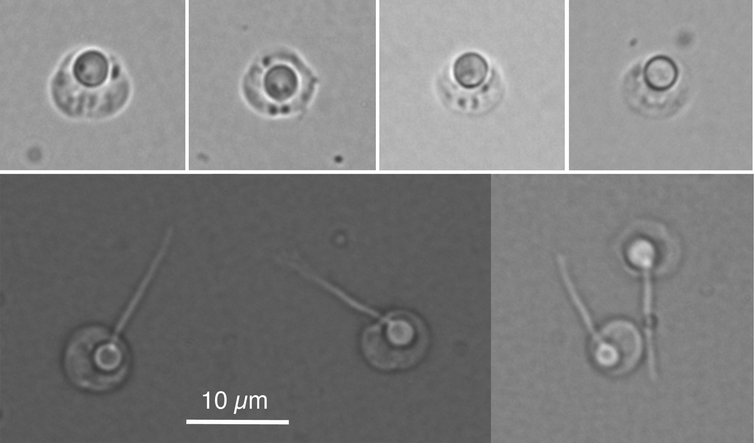
This species was found in imported arrow-tooth flounder, Atheresthes stomias (Jordan et Gilbert, 1880) that was purchased from a fish market near Yamaguchi University. The fish originated in the Bering Sea, off the coast of Alaska, USA. Fish that were packed similarly and purchased on different occasions were consistently infected with plasmodia of Unicapsula and Kudoa species. Then, we supposed a high prevalence of these two multivalvulid species in the trunk muscles of arrow-tooth flounder, as recorded by Davis (Reference Davis1924) in the trunk muscles of the Pacific halibut Hippoglosus stenolopis Schmidt, 1904, from the Pacific Ocean off the coast of North America. In the myofibres of arrow-tooth flounder, Unicapsula plasmodia were 2.71–9.94 (6.65) mm in length (n = 15), whereas the Kudoa plasmodia were 5.25–13.78 (7.87) mm in length (n = 15). No host responses, such as leukocyte accumulation or tissue degeneration, were observed.
Bilaterally symmetrical, almost subspherical myxospores (n = 25) measured 5.8–7.6 (6.5) in length by 6.3–7.8 (7.1) in width, with a functional semi-spherical PC measuring 3.0–3.7 (3.3) in diameter, and two smaller PCs measuring 0.5–0.8 (0.7) by 0.5–0.6 (0.5) (Figs 1K, 2D and 3). Turns of polar filaments in prominent PCs were not discernable, and the polar filaments (n = 10) were 9.2–14.6 (11.8) in length. The dimensions of the myxospores and functional PCs of the present specimens were consistent with those of U. muscularis (Table 2), which is the type species of the genus. However, the species' original description was concise, and no additional description has been made on the species' myxospore morphology.
Microscopic view of myxospores of Unicapsula muscularis (isolate Aburakarei) collected in the present study.

A serial rDNA nucleotide sequence (5117-bp) was obtained from the U. muscularis plasmodia recovered from the arrow-tooth flounder. The sequence contained 1706-bp partial 18S rDNA, 300-bp ITS-1, 157-bp 5.8S rDNA, 253-bp ITS-2 and 2701-bp partial 28S rDNA sequences (DDBJ/EMBL/GenBank accession no. LC474141). The 18S rDNA nucleotide sequence of U. muscularis was most similar to sequences of Unicapsula trigona n. sp., which was erected in the present study [LC316969; 93.13% (1586/1703) with three indels], U. pyramidata [AB971675 and AB971676; 92.78% (1580/1703) with three indels] and U. setoensis [AB971679; 92.72% (1580/1704) with two indels], followed by sequences from another Unicapsula spp. The 28S rDNA nucleotide sequence of U. muscularis was similar to that of the Unicapsula spp. mentioned above, but by less than 91%.
Remarks: Although U. muscularis is well-known as the type species of the genus (Lom and Dyková, Reference Lom and Dyková2006) and is the cause of ‘wormy halibut’ (Davis, Reference Davis1924), little is known about its parasitism and biology. After our first observation of multivalvulid parasitism in commercially available arrow-tooth flounder, we examined several packages of the imported fish on different occasions and constantly found the trunk muscle myofibres of the host species contained Unicapsula and Kudoa plasmodia. This infection status is very similar to the infection of these two species in the Pacific halibut reported by Davis (Reference Davis1924). Morphological characteristics of Unicapsula and Kudoa myxospores observed in the arrow-tooth flounder are also similar to those of these two species found in the Pacific halibut reported by Davis (Reference Davis1924). Based on these circumstantial supports, the Unicapsula species found in the arrow-tooth flounder is speculated to be U. muscularis described from the Pacific halibut Hippoglosus stenolopis Schmidt, 1904, from the Pacific Ocean off the coast of North America. The rDNA sequences of U. muscularis was similar to but distinct from, those of congeners with available rDNA sequences, such as U. andersenae, U. fatimae, U. pflugfelderi, U. pyramidata, U. seriolae, U. setoensis and Unicapsula spp., for which rDNA nucleotide sequences were newly obtained in the present study (U. galeata, U. trigona n. sp. and U. motomurai n. sp.).
Unicapsula pflugfelderi (Alama-Bermejo et al., Reference Alama-Bermejo, Cuadrado, Raga and Holzer2009)
This species was found extensively in the trunk muscle myofibres of three imported samples of the West African goatfishes Pseudupeneus prayensis (Cuvier, 1829). The pseudocysts ranged 0.5–2 mm in length and exhibited tapering ends.
Bilaterally symmetrical, almost subspherical myxospores (n = 20), which measured 5.1–6.9 (5.9) in length by 5.5–6.7 (6.1) in width, with a functional semi-spherical PC, which measured 2.2–2.9 (2.5) in diameter and two smaller PCs, which measured 0.8–0.9 (0.8) in diameter (Figs 1L and 2E). Turns of polar filaments in prominent PCs were not discernable.
Partial 18S rDNA (1690-bp) and partial 28S rDNA (2702-bp) sequences were obtained for the present isolate (DDBJ/EMBL/GenBank accession nos. LC474131 and LC474132). Parts of these sequences were highly similar to the partial 18S and 28S rDNA nucleotide sequence of U. pflugfelderi; 100% over 726-bp (AM931470) and 722-bp (AM931471), excluding primer-annealing sequences and 98.65% (586/594) with the 28S rDNA sequence (AM831468).
Remarks: As indicated by Miller and Adlard (Reference Miller and Adlard2013), it seems impossible to differentiate U. pflugfelderi and U. andersenae on the basis of morphology when using light microscopy. However, the species can be easily distinguished on the basis of their 18S and 28S rDNA nucleotide sequences. These two species are currently distributed in different oceans. The West African goatfish is the new host record for U. pflugfelderi.
Unicapsula motomurai n. sp. (Myxozoa: Myxosporea: Multivalvulida)
This species was found in the trunk muscles of two of 40 (5%) samples of the royal threadfins Pentanemus quinquarius (Linnaeus, 1758), which had been imported from the Southeast Atlantic Ocean, off the coast of West African, between Senegal and Angola. However, the exact fishing point of the examined fish was unknown. The species possessed remarkably large functional PCs, relative to the myxospore dimensions.
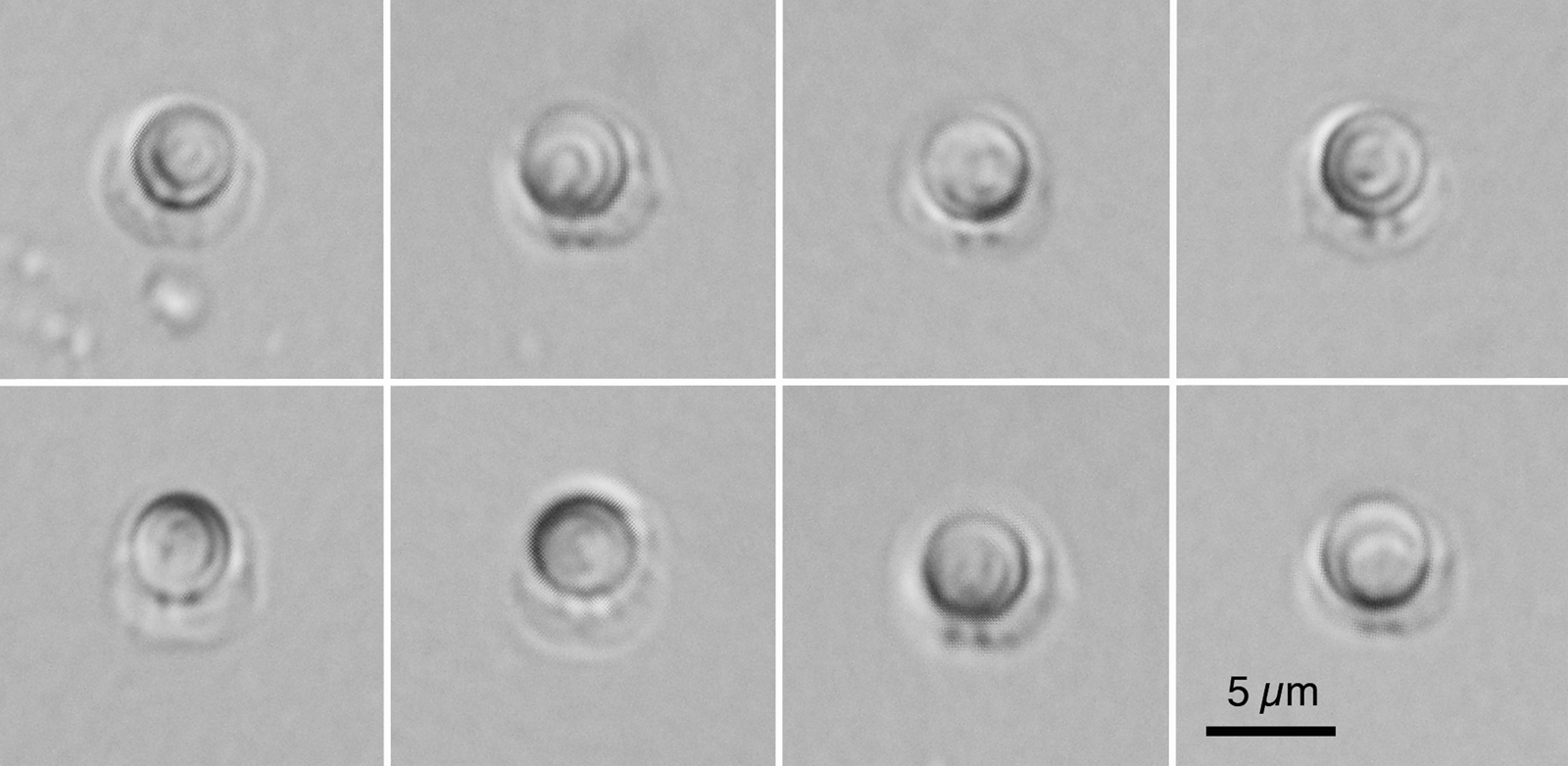
Description (Figs 1M, 2F and 4; Table 2)
Thread-like plasmodium with tapering ends in the myofibres of trunk muscles, measuring 0.66–1.42 (1.01) mm in length, and 0.04–0.09 (0.06) mm in width (n = 9). Bilaterally symmetrical subspherical myxospores (n = 20), measuring 6.1–7.5 (6.7) in length by 6.1–7.4 (6.6) in width, with a functional semi-spherical PC measuring 3.8–4.4 (4.1) in diameter and two smaller PCs measuring 0.6–0.9 (0.7) in diameter.
Microscopic view of myxospores of Unicapsula motomurai n. sp. (isolate Li2-1) collected in the present study.

The partial 18S rDNA nucleotide sequence of 1709-bp length and partial 28S rDNA of 2702-bp length (DDBJ/EMBL/GenBank accession nos. LC474133 and LC474134, respectively) were most similar to the 18S and 28S rDNA sequences of U. pfugfelderi: 99.10% (1654 / 1669) and 96.15% (2596 / 2700) with two indels, respectively.
Taxonomic summary
Host: Pentanemus quinquarius (Linnaeus, 1758), royal threadfin (Actinopteryii: Perciformes: Polynemidae).
Locality: Southeast Atlantic Ocean, off the coast of West Africa.
Site of infection: Pseudocysts in trunk muscle myofibres.
Materials deposited: Hapantotype no. 21510, Meguro Parasitological Museum, Tokyo, Japan.
Prevalence: Two of 40 examined fish, but the high intensity in infected fish.
Etymology: The species name is dedicated to Prof. Hiroyuki Motomura (Kagoshima University Museum), who helped identify the fish samples used in the present study, especially threadfins, based on his comprehensive research and review of family Polynemidae (Motomura, Reference Motomura2004).
Remarks: The most conspicuous traits of this new species are its relatively large dimensions and the ratio of its prominent PCs [3.8–4.4 μm; average 4.1 μm (n = 20)] to its myxospores. Similar feature is observed for U. chirocentrusi Sarkar, Reference Sarkar1984 (prominent PCs, 3.2–3.8 μm) from the trunk muscle of Chirocentrus dorab (Forsskål, 1775) in the Indian Ocean; for U. maxima Sarkar, Reference Sarkar1999 (prominent PCs, 3.2–4.5 μm; average 3.8 μm) from the renal parenchyma of Nibea coibor (Hamilton, 1822) [syn. Pseudosciaena coibar (Hamilton, 1822)] in the Indian Ocean; and for both U. pacifica Aseeva et Krasin, Reference Aseeva and Krasin2001 (prominent PCs, 3.9–4.0 μm) and U. schulmani Aseeva et Krasin, Reference Aseeva and Krasin2001 (prominent PCs, 2.8–3.8 μm) in the urinary bladder of Albatrossia pectoralis (Gilbert, 1892) [Syn. Coryphaenoides pectoralis (Gilbert, 1892)] from the Okhotsk Sea (Table 2). All these species mentioned above were recorded from fish hosts in the Indo-Pacific Ocean and Okhotsk Sea, different from the royal threadfin with U. motomurai n. sp., which were found to be distributed in the Eastern Atlantic Ocean. The organ/tissue specificity of U. maxima and U. schulmani is distinct from other species that form pseudocysts in trunk muscle myofibres. Furthermore, Unicapsula spp., except for U. chirocentrusi, possess myxospores that are larger than those of U. motomurai n. sp. (⩾7.3 μm vs ⩽7.5 μm), which reduces the ratio of prominent PCs to myxospores (Table 2). The myxospore dimensions of U. chirocentrusi are almost similar to those of U. motomurai n. sp. However, the dimensions of prominent PCs are smaller (3.2–3.8 μm vs 3.8–4.4 μm). Since the present study generated sufficiently long 18S and 28S rDNA nucleotide sequences for U. motomurai n. sp., molecular-genetic characterization, along with an intensive morphological comparison with Unicapsula spp. (which possess morphologically similar myxospores), enables us to identify the new species.
The new species described here, U. motomurai n. sp., is the third Unicapsula species, after U. pflugfelderi and U. marquesi Diebakate et al., Reference Diebakate, Fall, Faye and Toguebaye1999, to be reported from fish distributed in the Eastern Atlantic Ocean.
Unicapsula trigona n. sp. (Myxozoa: Myxosporea: Multivalvulida)
The thorough examination of almost all trunk muscles of two golden threadfin bream samples, with body weights of 917 g and 1174 g, revealed five and 19 plasmodia, respectively. The shortest plasmodium measured 0.73 mm in length and 0.14 mm in width, whereas the longest one measured 6.98 mm in length and 0.17 mm in width. Triangular myxospores resembled the myxospore morphology of U. pyramidata. However, the two species could be differentiated by morphological and genetic differences.
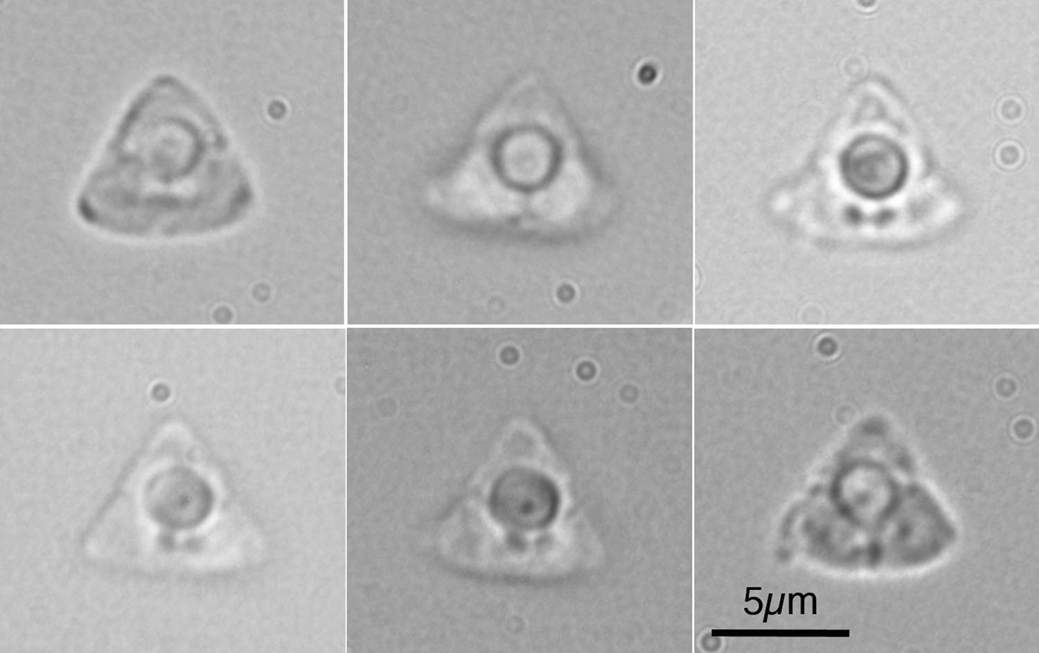
Description (Figs 1N, 2G and 5; Table 2)
Thread-like plasmodium with tapering ends in trunk muscle myofibres, measuring 0.73–6.98 (2.56) mm in length and 0.09–0.29 (0.16) mm in width (n = 11). Bilaterally symmetrical triangular myxospores with a fairly pointed apical corner and rounded caudal corners, formed by an apical SV and two caudal SVs, respectively; 4.4–5.3 (4.9) in length and 5.8–7.8 (6.7) in width, without caudal filamentous appendages (n = 20). A functional semi-spherical PC measuring 2.2–3.0 (2.5) in diameter, and two smaller PCs measuring 1.1–1.7 (1.4) in diameter, located closely at the centre of myxospores.
Microscopic view of myxospores of Unicapsula trigona n. sp. (Itoyoridai) collected in the present study.

The rDNA sequence of the present isolate was 5240-bp in length, including 1708-bp partial 18S rDNA, 404-bp ITS-1, 156-bp 5.8S rDNA, 270-bp ITS2 and 2702-bp partial 28S rDNA sequences (DDBJ/EMBL/GenBank accession nos. LC316969). On the basis of both morphological and genetic characteristics, this new species was most similar to U. pyramidata: 18S rDNA, 99.24% (1695/1708); 5.8S rDNA, 99.36% (155/156); and 28S rDNA, 98.74% (2664/2698), when compared with a deposited rDNA sequence of U. pyramidata (AB971676; 5249-bp). Identical nucleotides occurred at less than 70% of sites for both the ITS-1 and ITS-2 regions, with multiple indels over 52-bp and 38-bp lengths, respectively.
Taxonomic summary
Host: Nemipterus virgatus (Houttuyn, 1782), golden threadfin bream (Actinopteryii: Perciformes: Nemipteridae).
Locality: Philippine Sea, off the coast of Kochi Prefecture, Japan.
Site of infection: Pseudocysts in the trunk muscle myofibres.
Materials deposited: Hapantotype no. 21381, Meguro Parasitological Museum, Tokyo, Japan.
Prevalence: Two of two fishes examined, with low intensity (several plasmodia/fish) as far as examined.
Etymology: The species name refers to its triangular myxospore shape.
Remarks: Of the 13 known and two novel Unicapsula spp., only U. pyramidata and Unicapsula trigona n. sp. possess triangular myxospores. There are, however, several morphological differences between them, and U. trigona n. sp. is characterized by its lack of caudal filamentous appendages, pointed apical SV corner, rounded posterior SV corners and the localization of prominent PCs at the centre of myxospores. This morphological differentiation is supported by genetic differences in the rDNA nucleotide sequences: long 28S rDNA sequences showed 98.74% similarity over 2698-bp, and short 28S sequences, limited to the initial 800-bp from the 5′-terminus, showed 96.63% similarity. In addition, the myxospores of U. marquesi exhibit a mitre-like shape with two prominent posterior SVs that lack caudal filamentous appendages and the species forms elongated cysts in the gill filaments of the giant African threadfin Polydactylus quadrifilis (Cuvier, 1829) in the East Atlantic Ocean, off the coast of Senegal. Even though molecular data for U. marquesi have yet to be deposited at DNA databases, the unique myxospore morphology and tissue specificity differentiate the species from the rest of Unicapsula spp., including U. trigona n. sp.
Phylogenetic analyses
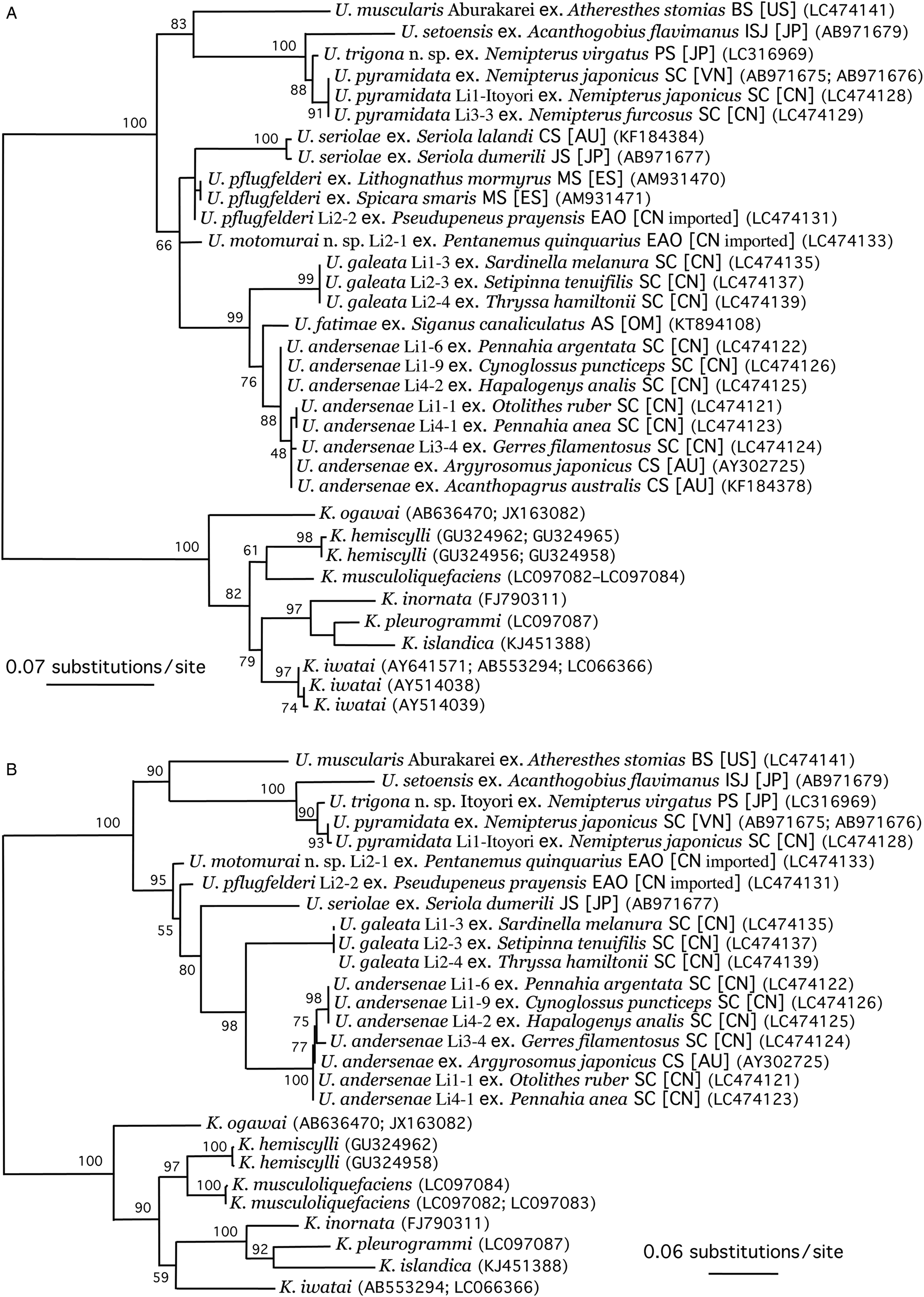
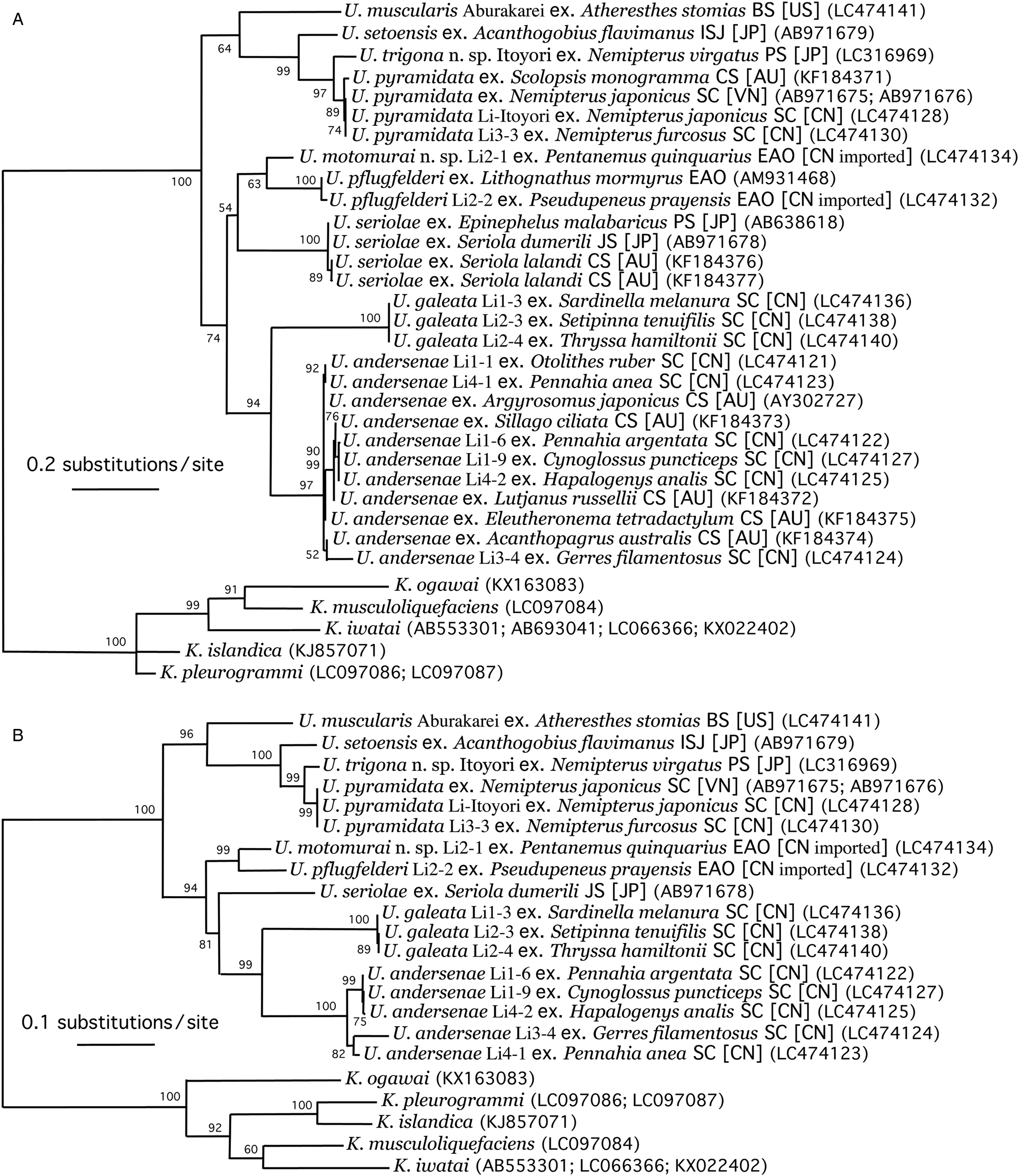
In the present study, long nucleotide sequences of the 18S and 28S rDNA were obtained for all 15 of the new Unicapsula isolates. The phylogenetic trees constructed from short sequences (722 and 540 characters of the 18S and 28S rDNA, respectively; Figs 6A and 7A) depict the phylogenetic relationships of almost all the Unicapsula nucleotide sequences available from DNA databases, and the newly obtained rDNA sequences coincide well with the previously deposited ones. However, the phylogenetic trees constructed from long sequences (1621 and 2156 characters of the 18S and 28S rDNA, respectively; Figs 6B and 7B) might reflect more accurate molecular relationships among the Unicapsula spp. Indeed, the topologies of the phylogenetic trees based on the long 18S rDNA sequences and long 28S rDNA sequences were almost identical, whereas those of phylogenetic trees based on the short 18S rDNA sequences and short 28S rDNA sequences were different. Based on the data analysed in the present study, it appears that there is little intraspecific genetic diversity in Unicapsula spp., except for U. andersenae, which exhibited frequent nucleotide variation in both the 18S and 28S rDNA (Table 3). We could not find currently phylogenetic clustering of species based on myxospore shape (spherical vs. triangular), host classification or geographical distribution.
Maximum likelihood phylogenetic tree of 18S rDNA sequence (A: 722 characters, and B: 1621 characters) from Unicapsula spp. Each species name is followed by the name of its fish host, the sea [country] from which it was collected and DDBJ/EMBL/GenBank accession number in parentheses. Abbreviation of sea names: AS, Arabian Sea; BS, Bering Sea; CS, Coral Sea around Australia; EAO, East Atlantic Ocean; ISJ, Inland Sea of Japan; JS, Japan Sea; MS, Mediterranean Sea; PS, Philippine Sea off Kochi, Japan; and SC, South China Sea. Abbreviation of country names: AU, Australia; CN, People's Republic of China; ES, Spain; JP, Japan; OM, Oman; US, United States of America; and VN, Vietnam.

Maximum likelihood phylogenetic tree of 28S rDNA sequence (A: 540 characters, and B: 2156 characters) from Unicapsula spp. Labelling of each isolate and abbreviations of sea and country names are similar to those described in the legend for Fig. 6.

Discussion
The type species for the genus Unicapsula Davis, Reference Davis1924 is U. muscularis specified by Davis (Reference Davis1924) and accepted by Lom and Noble (Reference Lom and Noble1984) and Lom and Dyková (Reference Lom and Dyková2006). The lack of additional taxonomic study, such as morphological observation, of the species after its original description by Davis (Reference Davis1924) is evident. However, the present study successfully characterised U. muscularis, both morphologically and molecular-genetically, that was detected in the myofibres of arrow-tooth flounder from the northeastern Pacific Ocean, off the coast of North America, although the isolation source was not from the type host for U. muscularis.
As shown in Table 2, 13 Unicapsula spp. have been recorded to date, and the present study adds two new species (i.e. U. trigona n. sp. and U. motomurai n. sp.). According to Lom and Noble (Reference Lom and Noble1984), the genus Unicapsula is defined as a group of multivalvulid species with subspherical myxospores with three unequal SVs; one small SV that covers a single spherical PC, and two larger, bilaterally and symmetrically arranged SVs that contain two PC rudiments. The distinct morphology of Unicapsula myxospores enabled the easy identification of the genus. However, species-level identification based mainly on myxospore morphology was rather difficult. This taxonomic status might be reflected in a limited number of specific descriptions in the genus, as well as in a limited number of reports of fish infected with Unicapsula spp. in the world. Specific differentiation of the morphologically simplest myxospores of Unicapsula spp. emphasizes the importance of molecular taxonomic approaches (Whipps et al., Reference Whipps, Grossel, Adlard, Yokoyama, Bryant, Munday and Kent2004; Alama-Bermejo et al., Reference Alama-Bermejo, Cuadrado, Raga and Holzer2009; Miller and Adlard, Reference Miller and Adlard2013; Tomochi et al., Reference Tomochi, Li, Tran, Yanagida and Sato2014; Al-Jufaili et al., Reference Al-Jufaili, Freeman, Machkevskyi, Al-Nabhani and Palm2016). In this sense, the present study successfully provides sufficient molecular data (i.e. 18S and 28S rDNA sequences) for 15 isolates of seven Unicapsula spp.
For the amplification of 18S and 28S rDNA sequences from of Unicapsula spp., primer combinations designed for Kudoa spp., which are closely related multivalvulid myxosporeans, are useful with a few exceptions: the primer for the 5′-terminus of the 18S rDNA was not useful for any Unicapsula spp. (Tomochi et al., Reference Tomochi, Li, Tran, Yanagida and Sato2014), and the primer for the 5′-terminus of the 28S rDNA of U. muscularis (present study). The molecular characterisation of 15 new isolates of seven Unicapsula spp. and of the deposited rDNA sequences of several other Unicapusla spp. (i.e. U. andersenae, U. fatimae, U. pflugfelderi, U. pyramidata, U. seriolae, and U. setoensis), allowed us to analyse the interspecific and intraspecific phylogenetic relationships of 10 Unicapsula spp. (using 18S rDNA; Fig. 6) and 9 Unicapsula spp. (using 28S rDNA; Fig. 7). As suspected, the phylogenetic trees based on the shorter 18S and 28S rDNA sequences exhibited different topological relationships among certain species (e.g. those of U. seriolae and U. motomurai n. sp. with other species). Meanwhile, the phylogenetic trees that were based on the longer 18S and 28S rDNA sequences possessed nearly identical topologies. Although the monophyly of all described Unicapsula spp. has been demonstrated in the previous studies (Miller and Adlard, Reference Miller and Adlard2013; Tomochi et al., Reference Tomochi, Li, Tran, Yanagida and Sato2014; Fiala et al., Reference Fiala, Bartošová-Sojková, Whipps, Okamura, Gruhl and Bartholomew2015), the genus has at least two major clades: (I) U. muscularis, U. setoensis, U. trigona n. sp. and U. pyramidata; and (II) U. motomurai n. sp., U. pflugfelderi, U. seriolae, U. galeata, U. fatimae and U. andersenae. The division of the genus into two these major clades is not related to either myxospore shape (spherical vs triangular) or to geographical distribution. The present study segregated U. andersenae isolates from South China Sea fish into three genetic groups (Table 3), as previously reported by studies in the Coral Sea around Australia (two genotypes; Miller and Adlard, Reference Miller and Adlard2013). With the exception of U. andersenae, which may actually be a species complex, like Kudoa thyrsite (Whipps and Kent, Reference Whipps and Kent2006; Burger and Adlard, Reference Burger and Adlard2011; Kasai et al., Reference Kasai, Li, Mafie and Sato2016), little intraspecific variation was observed in the rDNA sequences of the 15 isolates. This may be partly due to the limited number of isolates analysed. However, U. galeata and U. pyramidata from different fish hosts or geographical distribution exhibited few to zero nucleotide substitutions.
Studies of the economic impacts of Unicapsula infection to commercial or recreational fish are currently limited to knowledge on U. muscularis and U. seriolae. Unicapsula muscularis is known to form macroscopic pseudocysts in the trunk muscle of the Pacific halibut, which is called ‘wormy halibut’ when infected (Davis, Reference Davis1924), and U. seriolae is known to cause postmortem myoliquefaction in yellowtail amberjack Seriola lalandi Valenciennes, 1833, fished from the waters around Australia (Lester, Reference Lester1982). As far as could be observed by the microscopic analysis of wet specimens, no inflammatory cell accumulation was observed around myofibres with Unicapsula pseudocysts, nor was postmortem myoliquefaction in fish samples examined in the present study. As mentioned in Tomochi et al. (Reference Tomochi, Li, Tran, Yanagida and Sato2014) and Ohnishi et al. (Reference Ohnishi, Obara, Arai, Yoshinari and Sugita-Konishi2018), the parasitism of aquacultured greater amberjack Seriola dumerili (Risso, 1810) by U. seriolae is considered responsible for sporadic outbreaks of food-borne disease, which is characterized by clinical diarrhoea and vomiting after the consumption of raw greater amberjack. Greater yellowtail and other Seriola spp. are popular with Japanese people, and raw muscle slices are often served as sashimi or sushi. Consequently, Japanese aquaculture is expanding dramatically (Nakada, Reference Nakada, Lovatelli and Holthus2008). Like Kudoa food poisoning outbreaks, which are caused by K. septempunctata in aquacultured olive flounders (Kawai et al., Reference Kawai, Sekizuka, Yahata, Kuroda, Kumeda, Iijima, Kamata, Sugita-Konishi and Ohnishi2012; Sugita-Konishi et al., Reference Sugita-Konishi, Sato and Ohnishi2014), aquaculture provides intensive infection in fish (exceeding a certain threshold number of myxospores for food poisoning), which might be responsible for the increase in food-borne diseases associated with the consumption of raw greater amberjack during the last decade. Even though the infection of arrow-tooth flounder (and probably Pacific halibut) by U. muscularis is intensive enough to exceed such threshold myxospore numbers per gram muscle, commercially available fish in Japan are mainly frozen and imported from North America and are absolutely free from the risk of food-borne myxosporean diseases, since myxospores are inactivated by freezing (Kawai et al., Reference Kawai, Sekizuka, Yahata, Kuroda, Kumeda, Iijima, Kamata, Sugita-Konishi and Ohnishi2012; Sugita-Konishi et al., Reference Sugita-Konishi, Sato and Ohnishi2014).
The present study has added two new species, U. trigona n. sp. and U. motomurai n. sp., to the list of Unicapusla spp., and the genus now contains 15 species, of which the rDNA nucleotide sequences of 10 species have been characterised. Additional morphological and genetic characterisation of U. chirocentrusi, U. maxima, U. pacifica, U. schulmani and U. marquesi is needed to better understand the biodiversity and evolutionary history of the genus.
Acknowledgements
We are indebted to Prof. Hiroyuki Momomura (Kagoshima University Museum), for his help with fish-specific identification and Prof. Emeritus Shuhei Tanaka (Faculty of Agriculture, Yamaguchi University), for his help with the scanning electron microscopy of Unicapsula myxospores (not shown here).
Financial support
This study was supported in part by Grant-in-Aid for Scientific Research 2017 from The Towa Foundation for Food Science and Research (HS), Grant-in-Aid for Scientific Research 2019 from The TOYO SUISAN FOUNDATION for Food Science and Research (HS), Grant-in-Aid for International Collaboration Research in Asia 2016 from the Heiwa Nakajima Foundation (HS), Grant-in-Aid for International Collaboration Research 2019 from the Goho life Sciences International Fund (JYZ), and JSPS KAKENHI grant number 18K05995 (HS). Special thanks are extended to the United Graduate School of Veterinary Science, Yamaguchi University and Institute of Hydrobiology, Chinese Academy of Science, for supporting international collaborative research (travel grants for YCL and JYZ, respectively).
Conflict of interest
None.
Ethical standards
Not applicable.