Drug-refractory atrial and ventricular arrhythmias in children without structural heart disease are one of the major challenges faced by physicians. It is not always feasible to identify the underlying disease causing these arrhythmias. Usually, these arrhythmias are diagnosed as idiopathic. According to Roggen et al,Reference Roggen, Pavlovic and Pfammatter 1 ventricular tachycardia is diagnosed as idiopathic in 50% of cases. Distinguishing idiopathic ventricular tachycardia from potentially life-threatening conditions including myocarditis is recommended by current guidelines.Reference Crosson, Callans and Bradley 2
On the other hand, myocarditis may be suspected in young patients with symptomatic ventricular and supraventricular arrhythmias not consistent with a congenital substrate. It can clinically present as arrhythmia only.Reference Epstein, DiMarco and Ellenbogen 3 Although endomyocardial biopsy remains the gold standard for the diagnosis of myocarditis, it is not used routinely because of lack of specificity and risk of complications.Reference Kindermann, Barth and Mahfoud 4 The prognostic benefits of immunohistological endomyocardial biopsy were shown recently in adult patients with suspected myocarditis.Reference Kindermann, Kindermann and Kandolf 5
The aim of this prospective study was to assess the diagnostic value of endomyocardial biopsy in children with drug-refractory arrhythmia.
Materials and methods
Study population and protocol
We carried out a prospective single-centre study including 19 consecutive young patients with drug-refractory arrhythmia, from 2010 to 2013.
The primary end point of this study was to determine the underlying possible causes of drug-refractory arrhythmia using contemporary diagnostic tools including endomyocardial biopsy. The secondary end point was assessment of the role of endomyocardial biopsy in predicting non-pharmacological management outcomes.
The inclusion criteria were as follows:
-
∙ aged < 18 years;
-
∙ use of antiarrhythmic drug(s) for refractory arrhythmia;
-
∙ structurally normal heart or changes in a structure of the heart initially diagnosed as arrhythmia-induced cardiomyopathy.
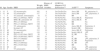
Drug-refractory arrhythmia was defined as symptomatic recurrent arrhythmia recorded by electrocardiography or Holter monitoring after regular use of prescribed antiarrhythmic drug(s) for >3 months (Table 1).
Patients demographic characteristics at baseline.

AAD=antiarrhythmic drug; AF=atrial fibrillation; Amio=amiodarone; AT=atrial tachycardia; BB=beta blocker; F=female; HRD=heart rhythm disorder; IC = flecainide, propafenone, moricizine; LA=left atrium; LV=left ventricle; M=male; NSVT=non-sustained ventricular tachycardia; PT=physical tolerance; PVCs=premature ventricular captures; RV=right ventricle; S=syncope; SB=sinus bradycardia; SSNS=sick sinus node syndrome; VT=ventricular tachycardia; W=weakness
* Percentile values (95th) were used for the assessment of dilatation by echocardiography
** LV dysfunction was considered as a reduction of LV ejection fraction <60% (Teichholz method). LV ejection fraction was estimated during sinus rhythm in all cases, except in the 18th and 19th patient
*** Arrhythmia was defined as drug refractory if symptomatic recurrent arrhythmia was recorded by electrocardiography or Holter monitoring after regular use of prescribed AAD(s) listed below for >3 months
Only one patient had neither radiofrequency ablation nor implantable cardioverter defibrillator implantation (5th in Table 1).
Baseline characteristics (Table 1)
Patients underwent a complete history investigation, physical examination, laboratory studies including thyroid function, human immunodeficiency virus, creatinine kinase, creatinine kinase-MB (MB-Creatine kinases are dimeric molecules composed of M and B subunits and exist as the isoenzymes MM, MB, and BB), lactate dehydrogenase, Troponin I, and pro-brain natriuretic peptide, echocardiography, electrocardiography, treadmill test, and Holter monitoring. Cardiac MRI (Table 2) and genetic tests were performed according to indications and physician decisions. Indications for radiofrequency ablation were either class I or IIa, and indication for implantable cardioverter defibrillator implantation was class I, according to current guideline recommendations.Reference Epstein, DiMarco and Ellenbogen 3
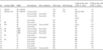
The results of cardiac MRI, endomiocardial biopsy, radiofrequency ablation, and follow-up.
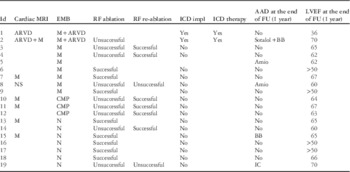
AAD=antiarrhythmic drugs; Amio=amiodarone; ARVD=arrhythmogenic right ventricular dysplasia; BB=beta blocker; CMP=cardiomyopathy; EMB=endomyocardial biopsy; FU=follow-up; ICD=implantable cardioverter defibrillator; impl=implantation; LVEF=left ventricular ejection fraction; M=myocarditis; N=normal; RF=radiofrequency
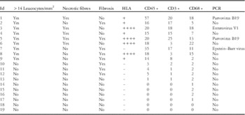
Endomyocardial biopsy (Table 3)
The American Heart Association/American College of Cardiology/European Society of Cardiology consensus statement indicates 14 clinical scenarios for endomyocardial biopsy.Reference Cooper, Baughman and Feldman 6 In our study, endomyocardial biopsy was performed according to the 8th – unexplained cardiomyopathy in children, indication class IIa, and level of evidence C – and the 13th – unexplained ventricular arrhythmias, indication class IIb, and level of evidence C – scenarios. According to these guidelines, myocarditis may be suspected and endomyocardial biopsy considered in heart failure with haemodynamic compromise of <2 weeks’ duration (class I recommendation) or <3 months’ duration if associated with heart block or new ventricular arrhythmias (class I recommendation).Reference Cooper, Baughman and Feldman 6
Samples were obtained from >1 region of the right ventricle apex and septum. The number of samples ranged from five to eight. The samples were 1–2 mm3 in size, as the current statement requires. A Cordisbioptome (Johnson & Johnson, Miami, FL, United States of America) was used for biopsy. Access was obtained via the right internal jugular vein. Both echocardiographic and fluoroscopic guidance were used for endomyocardial biopsy.
A histopathological analysis carried out following the Dallas criteria, immunohistochemistry, and polymerase chain reaction were used for the interpretation of endomyocardial biopsy.
Histopathological examination: the samples were stained with haematoxylin and eosin (van Gieson stain or Masson’s trichrome stain). Light microscopy morphometric analysis of cells and cell patterns was carried out (LeicaQWin Plus v3, Leica Microsystems Imaging Solutions, Cambridge, United Kingdom).
For the immunohistological analysis of the myocardium, the following antibodies were used: rabbit polyclonal antibodies to CD3 (DAKO, Hamburg, Germany), mouse monoclonal antibodies to CD45 (LCA; clones 2B11+PD7/26; DAKO), mouse monoclonal antibodies to HLA-DR (clone LN-3; Leica), and mouse monoclonal antibodies to CD68 (PG-M1, DAKO).
Active myocarditis was defined as the presence of more than 14 infiltrating leucocytes/mm2 (CD45+), up to 4 monocytes/mm2, CD3+ T-lymphocytes ⩾7 cells/mm2, and myocyte damage and/or fibrosis, in addition to enhanced HLA-DR expression in professional antigen-presenting immune cells and the endothelium.Reference Caforio, Calabrese and Angelini 7
Polymerase chain reaction was used for viral genome detection – enterovirus, cytomegalovirus, herpes simplex virus, parvovirus, and Epstein–Barr virus. Quantitative polymerase chain reaction and identification of viral infection by antibodies were performed.
Data collection and follow-up (Table 2)
All data were collected using a clinical electronic database from 2010 to 2013. Of 19 patients, 17 had radiofrequency ablation, and two patients had implantable cardioverter defibrillator implantation. Significant complication was defined as an event related to the endomyocardial biopsy. Echocardiography was performed to evaluate valve insufficiency or pericardial effusion at baseline, also at the end of the endomyocardial biopsy and before discharge after 6–12 months. Radiofrequency ablation was defined as successful based on symptoms, Holter monitoring, and electrocardiography during 1 year of follow-up.
Results
The mean age of the patient population was 14.1±2.9 years (from 7 to 17 years). All these patients had a history of drug-refractory arrhythmia for >5 months (mean 30 months). Patients had palpitation (42%), syncope (21%), weakness, and reduction of physical tolerance (21%). Mild dilatation of at least one of the heart chambers – left/right ventricles or the left atrium – was reported in all patients at baseline (Table 1). Arrhythmia-induced cardiomyopathy was the initial diagnosis in 13 of these patients. The remaining six had a structurally normal heart. Laboratory analyses showed results in the normal range. None of the patients had previous history of HIV, viral, or other infections.
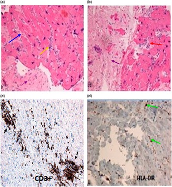
Overall 86 biopsies were performed in 19 patients. Myocarditis was diagnosed based on histopathological and immunohistological analyses in nine (47.4%) patients. Polymerase chain reaction was positive for viral genome in four of them; five patients had active myocarditis. The results of the endomyocardial biopsy are shown in Table 3. The images of the endomyocardial biopsy (4th patient) are illustrated in Figure 1. No complication was reported during the endomyocardial biopsy.
Samples of the endomyocardial biopsy of the 4th patient. ( a and b ) Active myocarditis: endomyocardial biopsy shows infiltration of aggressive attack of lymphocytes and macrophages, cardiomyocyte necrosis, and fibrosis. Infiltration of lymphocytes with their introduction into cardiomyocytes (yellow and red arrows), perimuscular fibrosis (blue arrow; each cardiomyocyte is surrounded by fibrous tissue), endocardial fibrosis (purple arrows). Haematoxylin and eosin stain, 200×. ( c ) Cellular infiltration of T-lymphocytes (infiltrates indicated by black arrows). Antibodies to the CD3 T-lymphocytes, DAKO, 200×. ( d ) HLA-DR expression on the vascular endothelium and infiltrate of the cells. Antibodies of the company DAKO, 200×. ( d ) Active myocarditis. Expression of CD3+on T-lymphocytes. HLA-DR expression on the vascular endothelium and infiltrate of the cells (green arrows). Antibodies of the company DAKO, 200×.

Results of the endomyocardial biopsy.

PCR=polymerase chain reaction
Cardiac MRI was performed in eight patients. According to the results, seven of these patients had myocarditis. In two patients, arrhythmogenic right ventricular dysplasia was diagnosed by endomyocardial biopsy, MRI, and genetic testing (PKP2). Both of them had an implantable cardioverter defibrillator implantation with further appropriate successful device shocks. In patients with MRI results, there was a correlation with changes on MRI and the location of the arrhythmia. Radiofrequency ablation was performed in 17 patients. In all, 13 of them had sustained or non-sustained ventricular tachycardia, originating from the right ventricle in eight patients and the left ventricle in five patients, and four patients had atrial tachyarrythmia, with atrial tachycardia in three patients and atrial fibrillation in one patient; three of 12 patients with myocarditis/cardiomyopathy and four of seven patients without myocarditis/cardiomyopathy had normal left ventricles. All three with myocarditis and only one of four without myocarditis and normal left ventricular function had sustained ventricular tachycardia. The overall success rate of radiofrequency ablation in patients with no myocarditis on endomyocardial biopsy was 90%, and five out of 10 (50%) underwent re-ablation; five out of six (83%) patients with endomyocardial biopsy-proved myocarditis had successful radiofrequency ablation, and three of six underwent re-ablation. No significant complication was reported during ablation. Atrial tachycardia in three patients was focal and successfully ablated. Monomorfic ventricular tachycardias and premature ventricular captures were predominantly from the outflow tract. In three patients with polymorphic ventricular tachycardia, there was a predominant premature ventricular capture trigger identified that led to the decision to pursue ablation. The approach in those in whom frequent premature ventricular captures (at least 15,000 during 24 hours ambulatory Holter monitoring) were the dominant arrhythmia, ablation of the main premature ventricular captures were performed.
At the end of follow-up, four patients were still taking antiarrhythmic drugs. A symptomatic and echocardiographic improvement was reported in all patients except in one with arrhythmogenic right ventricular dysplasia (Table 2).
Discussion
Previous endomyocardial biopsy studies have been conducted in paediatric populations with clinical presentation suggestive of myocarditis.Reference Martin, Webber and Fricker 8 In this descriptive study, patients had no clear clinical findings or history suggestive of active or residual myocarditis before endomyocardial biopsy. Initially, the echocardiographic and clinical findings were interpreted as an arrhythmia-induced cardiomyopathy based on Fenelon’s criteria in most of these patients: dilatation of the heart or heart failure in the presence of chronic or very frequent cardiac arrhythmia.Reference Fenelon, Wijins, Andries and Brugada 9 They further pointed out that if chronic tachycardia continued for >10–15% of the day, with a rate of >150% of that predicted for age, tachycardiomyopathy ensues.
As recently reported by Gopinathannair et al,Reference Gopinathannair, Etheridge, Marchlinski, Spinale, Lakkireddy and Olshansky 10 the specific pathophysiological mechanism and therapeutic strategies of arrhythmia-induced cardiomyopathy remain unclear. We suggested that endomyocardial biopsy can be a useful tool for solving some of the uncertainties of arrhythmia-induced cardiomyopathy. Cardiac MRI was performed in eight patients for detecting arrhythmogenic right ventricular cardiomyopathy with left ventricular involvement and infiltrative disease when clinically suspected. According to the results, seven of these patients had myocarditis, whereas only two of them had arrhythmogenic right ventricular dysplasia. No doubt that current practice in developed countries shows high sensitivity of advanced imaging modalities, which could change our results, but this is a real-life experience from the developing world.
This prospective study shows that 47% of paediatric and young adult patients with drug-refractory arrhythmia have myocarditis based on endomyocardial biopsy. Some studies reported that endomyocardial biopsy is a useful diagnostic tool for unexplained cardiac dysfunction in adults and also for paediatric populations with arrhythmia without structural heart disease. More than 30% of adults had myocarditis based on endomyocardial biopsy.Reference Ardehali, Qasim and Cappola 11 Wiles et al showed that 50% of children with arrhythmias may have subclinical cardiomyopathy or unsuspected myocarditis.Reference Epstein, DiMarco and Ellenbogen 3 On the other hand, Webber et alReference Webber, Boyle, Jaffe, Pickering, Beerman and Fricker 12 performed 76 endomyocardial biopsies in 63 children for evaluating arrhythmias: only one endomyocardial biopsy showed myocarditis.
It is interesting that some patients with positive right ventricular biopsy had atrial arrhythmias. We hypothesised that there are multiple factors that could increase the incidence of atrial arrhythmias during inflammatory processes. First of all not only local structural changes such as inflammatory processes, fibrosis, necrosis, scarring, secondary hypertrophy, and atrophy in the cardiac myocytes and interstitium can lead to arrhythmias, but also pro-arrhythmic effects of cytokines and inflammatory mediators can cause arrhythmias possibly through a modulation of ion channel function, parameters of ventricular dynamics and vascular changes, the disturbance of microvascular and macrovascular perfusion, and the resulting myocardial changes; two of these patients had MRI before ablation, and both had atrial inflammation. Only one patient had atrial hypertension from ventricular diastolic dysfunction by echocardiography.
Interpretation of the endomyocardial biopsy is one of the challenges faced by pathologists, which is not consistent.Reference Baughman 13 Moreover, it is related to the stage of myocarditis, as arrhythmia may persist after resolution.Reference Friedman, Kearhey, Moak, Fenrich and Perry 14 Endomyocardial biopsy criteria for myocarditis are not very sensitive: the sensitivity of the Dallas criteria is 66% if more than five samples are used.Reference Hauck, Kearney and Edwards 15 In this study, five to eight samples were obtained. Another study reported that the sensitivity of immunohistochemistry is higher compared with using the Dallas criteria only (48 versus 5%, respectively).Reference Kühl, Noutsias, Seeberg and Schultheiss 16 All these tools were used in this study to ensure the maximum possible accuracy.
A combined AHA/ACC/ESC statement does not support routine clinical use of endomyocardial biopsy for the diagnosis of myocarditis as it is a non-benign procedure.Reference Cooper, Baughman and Feldman 6 A study on children concluded that endomyocardial biopsy can be performed safely with overall 1.9% morbidity and mortality, and the only risk factor for perforation was inotropic agent dependency;Reference Pophal, Sigfusson and Booth 17 however, some studies reported up to 6% rate of complication during echocardiography-guided endomyocardial biopsy.Reference Han, Park and Lee 18 Absence of complications in this study can be explained by the lack of inotropic agent-dependent patients, by using both echocardiographic and fluoroscopic guidance for endomyocardial biopsy. Although there was no complication in this study, assessment of the risk–benefit ratio is important.
The concept that unsuspected myocarditis can present with life-threatening or refractory arrhythmia is not new.Reference Hosenpud, McAnulty and Niles 19 In our study, all three patients with myocarditis and only one of four without myocarditis and normal left ventricular function had sustained ventricular tachycardia. As the number of our patient sample is very small, further large research is needed to evaluate the role of endomyocardial biopsy in optimal therapeutic strategies and sudden death prevention in patients with unsuspected myocarditis and life-threatening or refractory arrhythmia.
The results of catheter ablation in patients with myocarditis have been published before and are unclear.Reference Chauhan, Hameedullah, Nanthakumar and Downar 20 , Reference Kettering, Kampmann, Mollnau, Kreitner, Munzel and Weiss 21 A recently published study including 20 patients with drug-refractory ventricular tachycardia and viral myocarditis proven by endomyocardial biopsy showed the high effectiveness of radiofrequency ablation.Reference Russo, Casella and Pieroni 22 The previous studies were performed in adult populations and some of them used epicardial approach for radiofrequency ablation.Reference Russo, Casella and Pieroni 22 In this study, the overall success rate of endocardial radiofrequency ablation was 83% in patients with myocarditis and 90% in patients with no myocarditis on endomyocardial biopsy.
Nevertheless, the limitations of this study are the small sample size and multiple types of drug-refractory arrhythmias. The results of the electrophysiology study are not in detail because of small sample size. In those patients in whom MRI was performed, none had areas of hyperenhancement, which is specific for patchy distribution of fibrosis and can result in false-negative biopsy results. MRI was not performed in all patients, and thus we cannot rule out false-negative results of the biopsy, which is a limitation of this study.
Larger studies in future may be helpful in identifying the value of endomyocardial biopsy for paediatric and young patients before radiofrequency ablation and to assess how extensive the scar burden on electroanatomical mapping is and how it may correlate with the location of arrhythmias.
Conclusion
Approximately half of the children with drug-refractory arrhythmia had an unsuspected myocarditis according to the results of the endomyocardial biopsy. We suggest that myocarditis can be one of the possible mechanisms of refractoriness in some cases. Endomyocardial biopsy can be safely performed with the fluoroscopic and echocardiographic guidance. The overall success rate of radiofrequency ablation in patients with and without myocarditis proven by endomyocardial biopsy was 83 and 90%, respectively. Further studies may be helpful to identify the role of endomyocardial biopsy in patients with drug-refractory arrhythmias to evaluate the effectiveness of cause-specific therapy and ablation in this population.
Acknowledgements
None.
Financial Support
This research received no specific grant from any funding agency or from commercial or not-for-profit sectors.
Conflicts of Interest
None.
Ethical Standards
All patients had a written parental consent form for inclusion. The ethics committee at the Federal Almazov Heart Blood Endocrinology approved the study protocol.