INTRODUCTION
Efforts to describe and document the occurrence of marine invertebrate larvae in coastal waters have largely been limited to European investigations. The classic work of Thorson (Reference Thorson1946) is best known for the large number of species treated and illustrations that still serve to assist with identification of larvae, including polychaetes, collected from meroplankton. Other European investigators including Smidt (Reference Smidt1944), Hannerz (Reference Hannerz1956), Rasmussen (Reference Rasmussen1956, Reference Rasmussen1973), Cazaux (Reference Cazaux1968, Reference Cazaux1969, Reference Cazaux1972, Reference Cazaux1982), Bhaud (Reference Bhaud1966, Reference Bhaud1967), Bhaud & Cazaux (Reference Bhaud and Cazaux1987), Wilson (Reference Wilson1928, Reference Wilson1929, Reference Wilson1932a, Reference Wilsonb, Reference Wilson1933, Reference Wilson1936a, Reference Wilsonb, Reference Wilsonc, Reference Wilson1948, Reference Wilson1968, Reference Wilson1970a, Reference Wilsonb, Reference Wilson1977, Reference Wilson1982) and Plate & Husemann (Reference Plate and Husemann1994) described the larval morphology of numerous species of polychaetes. Elsewhere Blake (Reference Blake1969) described spionid larvae from New England and in the 1970s conducted a large-scale project to describe invertebrate larvae from northern California; Carrasco (Reference Carrasco1976) described spionid larvae from Chile.
The present paper concludes descriptions of polychaete larvae from the vicinity of Tomales Bay, California. This project was initiated in 1971 and continued through 1977 with additional casual observations through 1978. Funded by the US National Science Foundation (NSF), it was intended to develop data and observations on the larvae of marine invertebrates from the coastal waters of northern California in an effort to better understand the ecology, life history and systematics of a rich marine fauna. Previous publications of data on polychaete larvae arising from this project were included in Blake (Reference Blake1972, Reference Blake1975a, Reference Blakeb, Reference Blakec, Reference Blake1980, Reference Blake and Reish1991, Reference Blake, Rouse and Pleijel2006), Blake & Arnofsky (Reference Blake, Arnofsky, Dorresteijn and Westheide1999), Blake & Lapp (Reference Blake and Lapp1974), Blake & Woodwick (Reference Blake and Woodwick1975) and Day & Blake (Reference Day and Blake1979). There are also several important unpublished student theses on reproductive biology and larvae of polychaetes arising from the same project including Armitage (Reference Armitage1979), Hillyard (Reference Hillyard1979), McEuen (Reference McEuen1979) and Parke (Reference Parke1973).
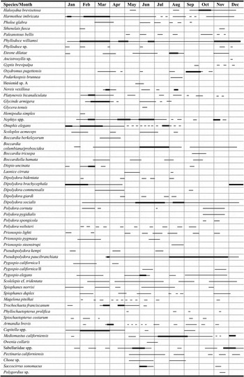
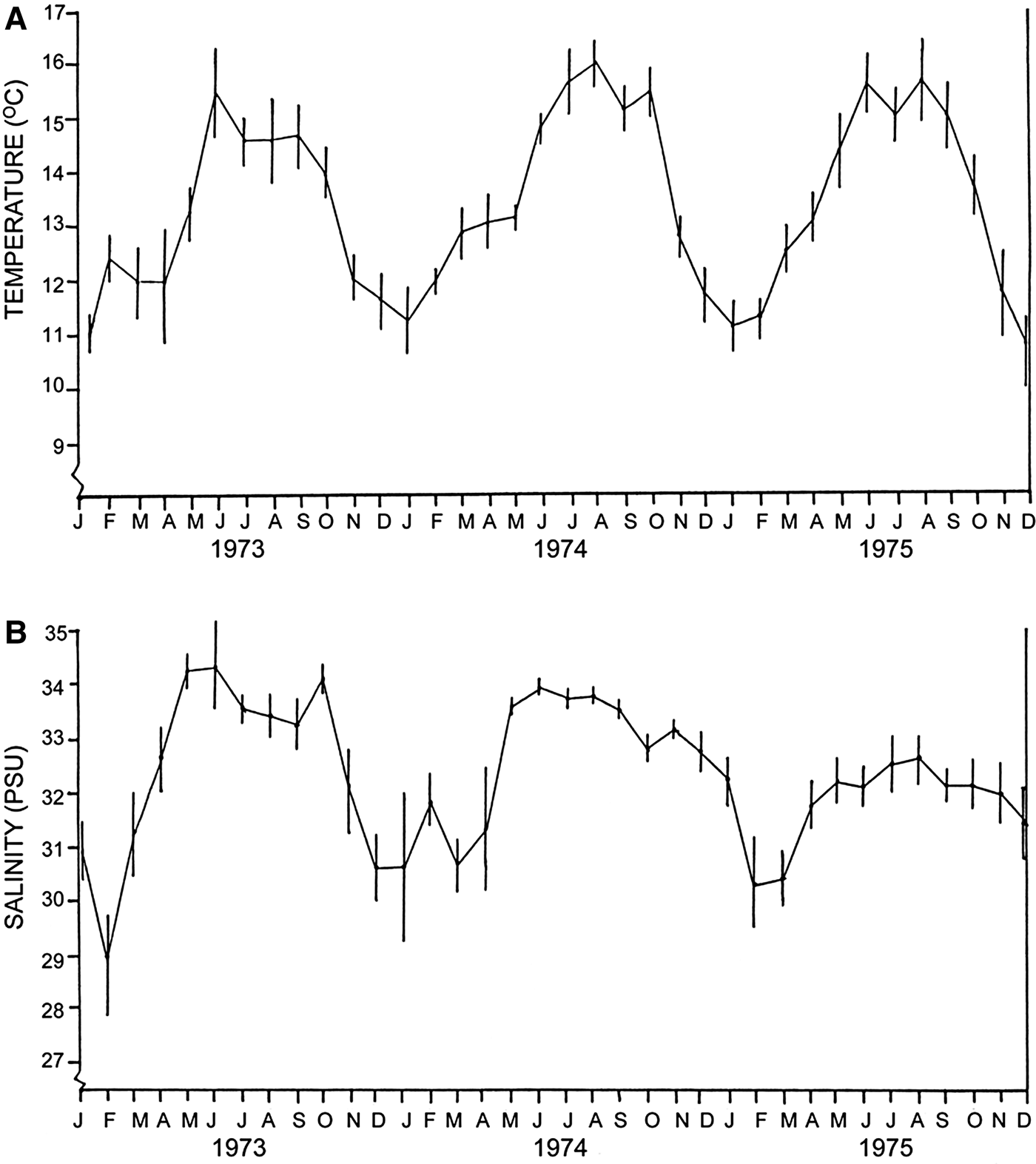
Included in the present study are descriptions of larvae of 14 species of polychaetes, several of which were reared in the laboratory from egg through to metamorphosis. Other descriptions are based entirely on larvae obtained from plankton of Tomales Bay. A summary of seasonal occurrence of larvae of 60 polychaetes species collected from the plankton over a 5-year period is reported.
MATERIALS AND METHODS
Field methods
Field collections of adult polychaetes were normally made on various protected mud and sand flats and eel grass habitats of Tomales Bay, and rocky intertidal sites north of Dillon Beach on Bodega Bay that were variously protected from wave action. On several occasions, benthic infauna was collected offshore in Bodega Bay in water depths of 50–60 m using a 0.1 m2 Van Veen grab. A few samples were collected from various intertidal sites in nearby Bodega Harbor. Plankton was collected from two localities in Tomales Bay. The main site was at Lawson's Landing, which is located near the entrance to Tomales Bay and has free tidal exchange with the adjacent Bodega Bay and the Pacific Ocean. Here the plankton net was simply lowered into the incoming or outgoing current and oblique 10 min qualitative tows were made. The second locality was about 8 miles down the bay at the town of Marshall where salinities were lower and currents were negligible. At this site the net was lowered into the water and towed as the technician walked back and forth along the pier. Nets used were either a No. 6 (0.210 mm mesh) or a No. 10 (0.158 mm mesh). The latter net was not used during phytoplankton blooms.
Laboratory and culture methods
In the laboratory, larvae were sorted by species from plankton samples and placed into finger bowls where they were cultured in refrigerated culture chambers at constant temperatures of 10 and 20°C. Other finger bowls were placed in tables where running seawater provided temperatures similar to the ambient environment at that time of year. Larvae were fed from cultures of several phytoplankton species including Dunaliella tertiolecta, Phaeodactylum tricornutum and Skeletonema costatum. Cultures were obtained from Dr Robert Guillard of the Woods Hole Oceanographic Institution and maintained using his culture medium (Guillard & Ryther, Reference Guillard and Ryther1962). In addition, a dried and powdered supply of Chlorella was also obtained and when mixed with seawater was added to cultures with some success. Commercial aquarium foods such as Tetramin® were also used.
Microscopes included the Wild M-5 stereomicroscope equipped with photographic attachments and a Zeiss RA research microscope equipped with Phase contrast and Nomarksi differential interference optics; photographic adapters and drawing arms were available for both microscopes. Larvae were observed on defined schedules with selected stages of development removed from the cultures for observation. Individual larvae were carefully observed using a hanging-drop technique where larvae were confined to a drop on a coverslip suspended over a depression on a slide. This method allowed the observers to photograph and illustrate the larvae in real time. Photographs were taken with a Polaroid Land Camera providing an instant black and white positive image. This image was then traced with the drawing arm on the stereomicroscope to obtain an accurate shape of the larva. The sketch was then used to insert details of the same larva that was still available on the hanging drop preparation. High-quality 35 mm colour or black-and-white photographs were also made using a Pentax SLR camera with Tri-X and Ektachrome Professional film.
Maintenance of cultures included daily feeding schedules, water changes, and checks of general health of the larvae. In many cases, certain species were easy to culture and were reared to settlement and metamorphosis. For others, however, health of the cultured larvae degraded over time and they perished. These failures were usually traced to bacterial build up in the cultures, inability of the larvae to develop due to an inappropriate food source, or possibly an inappropriate temperature/salinity environment. However, the success rate was high, as evidenced by the larvae of more than 100 species of polychaetes and other invertebrates being maintained and identified in cultures.

RESULTS
Larval morphology and development
Family NEREIDIDAE
Micronereis nanaimoensis Berkeley & Berkeley, Reference Berkeley and Berkeley1953
(Figures 1 & 3A)
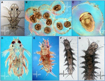
Development of Micronereis nanaimoensis Berkeley & Berkeley, Reference Berkeley and Berkeley1953: (A) egg mass attached to Zostera blade with a female worm on the outside; (B–C) metatrochophore within the egg mass: (B) dorsal view; (C) ventral view; (E–F) 3-setiger planktic nectochaete: (E) dorsal view; (F) ventral view; (G) homogomph notosetae; (H) larval jaws; (I) adult jaws.

INTRODUCTION
The genus Micronereis is relatively small, with only 11 species currently recognized as valid (World Register of Marine Species, WoRMS, accessed 12 June 2016). The only species recognized in the north-eastern Pacific is M. nanaimoensis originally described by Berkeley & Berkeley (Reference Berkeley and Berkeley1953) from British Columbia. Phyllodocella bodegae Fauchald & Belman, Reference Fauchald and Belman1972, described from Bodega Bay, California, is a junior synonym.
There are relatively few records of this species since the original description. Micronereis nanaimoensis was reported from Bodega Harbor by Fauchald & Belman (Reference Fauchald and Belman1972) (as Phyllodocella bodegae) and is included in keys of polychaetes from California (Blake & Ruff, Reference Blake, Ruff and Carlton2007) and from Washington and British Columbia (Banse & Hobson, Reference Banse and Hobson1974; Banse, Reference Banse, Reish and Fauchald1977). Berkeley & Berkeley (Reference Berkeley and Berkeley1953) recorded the species as laying egg masses on blades of Zostera, Ulva and other seaweeds; Blake & Ruff (Reference Blake, Ruff and Carlton2007) recorded the species as laying egg masses on blades of eel grass in Tomales Bay and on Obelia sp. in Bodega Harbor. The species is sexually dimorphic.
On 7 June 1978, during a field trip with students to eel grass beds (Zostera marina) in Tomales Bay, small greenish egg masses were observed on blades of Zostera. Some of these were collected, still attached to the eel grass blades, and transported to the laboratory. The egg masses were being incubated by one or two females and the species was identified as Micronereis nanaimoensis. After separation of the egg masses, they were individually set up in culture dishes and studied. Embryos and larvae were carefully removed from the egg masses at different stages of development. The following account describes the nature of the egg masses, the role and activities of the female worms that appeared to be incubating them, and details of the early larval development. Berkeley & Berkeley (Reference Berkeley and Berkeley1953) gave a similar account of the early larvae. The present study provides additional details of the larval morphology and rate of growth.
DEVELOPMENT WITHIN THE EGG MASS AND INCUBATION BY FEMALES
The egg masses ranged from 1.0–1.5 cm in diameter (Figure 1A) and were covered with detritus and diatoms to varying degrees. Each had an associated female worm that moved continuously over the surface; one egg mass had two females. Male worms were not collected.
The contents of the egg masses were white in colour and consisted of 200–300 eggs and embryos in various stages of development. Eggs measured ~180–200 µm in diameter. There was no movement of the eggs or embryos within the capsules on the day of collection (Day 1); some embryos were undergoing cleavage, but there was no evidence of cilia. By Day 2, individual embryos were observed to have vibratory movement within the egg masses and cilia were observed. These embryos were most likely at the stereoblastula stage and development was expected to proceed rapidly from that point.
By Day 3, development had progressed to a metatrochophore stage, still within the egg mass (Figure 1B, C). Several specimens were extracted from the egg mass and examined in hanging drop preparations. These early larvae are ~240 µm long and 190 µm wide with considerable morphology already developed. The interior of each larva is filled with 10 or more large yolky cells, each with numerous lipid droplets; some of this yolk is visible ventrally protruding from the oral opening (Figure 1C). Posterior to the mouth is an internal heart-shaped structure that represents the anlage of the pharynx. The anterior margin of the body is broadly rounded and bears a pair of orange-coloured eyespots; the largest is lateral and cup-shaped; the smallest is more medial and irregular in shape (Figure 1B, C). The posterior end bears two protuberances, the anlage of pygidial cirri. Three sets of internal setal sacs are present representing the anlage of noto- and neurosetae of setigers 1–3; no setae are emergent, however. A prominent prototroch composed of 16–20 separate cells or groups of cilia completely surrounds the anterior end of the larva. Three additional lateral patches of cilia are present posterior to the prototroch and likely represent dorsal nototrochs and ventral gastrotrochs. A distinct neurotroch and telotroch are not present at this stage of development.
HATCHING AND DEVELOPMENT IN THE PLANKTON
By Day 7 the larvae were emerging from the egg masses and swimming in the culture dishes. An example of an early 3-setiger nectochaete (Figure 1D) is ~375 µm long and 160 µm wide; these larvae are widest at setiger 2. The coelomic space is still packed with large yolky cells. The anterior end or head is about 1.3× as wide as long and triangular-shaped, narrowing to a broadly rounded apex. The four eyespots are now red in colour with the larger pair anterior and lateral to the smaller pair; both are cup-shaped and with a distinct clear lens surrounded by a pigmented cell. The two posterior protuberances have developed into distinct anal or caudal cirri; each has a few internal bacillary glands and stiff sensory cilia. Long compound homogomph noto- and neurosetae (Figure 1G) are present on setigers 1–3; these number about 4–6 per fascicle. The prototroch surrounds the anterior end posterior to the eyespots and consists of numerous cells or groups of cilia. Further ciliary development is associated with setigers 1–3. Ciliary bands are incomplete on setigers 1–2, but completely encircle the body on setiger 3. As with the prototroch, each band of cilia is composed of separate cells or groups of cilia.
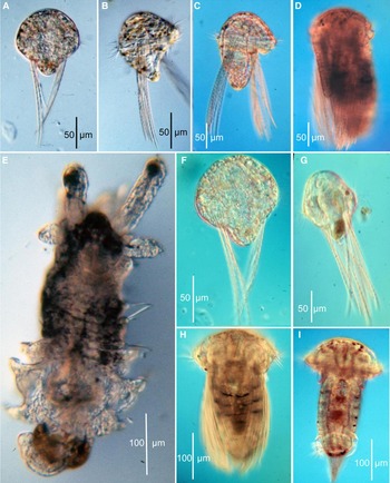
Free swimming 3-setiger nectochaetes were collected from the cultures on Day 8 (Figures 1E, F & 3A). These larvae are ~410 µm long and 165 µm wide. The overall shape is narrower and there is less yolk, but there are still yolky cells in the coelom. Internally, the pharynx is prominent both dorsally and ventrally and a pair of elongate jaws are present (Figure 1H). These were termed ‘jaw-supports’ by Berkeley & Berkeley (Reference Berkeley and Berkeley1953) because they differed from the jaws found in adults (Figure 1I); however, these jaws were observed to project through the mouth on one occasion. Rullier (Reference Rullier1954) determined that these jaws developed in both young males and females of Micronereis variegata and Paxton (1975) found that these jaws were present in juvenile females of M. minuta and M. piccola. The mouth has a pair of muscular anterior lips. In addition, with growth and modification of the pre-setiger region the eyespots shift to a position posterior to the prototroch. The anterior end or head is now as wide as long, narrowing anteriorly to a rounded apex; rust-coloured pigment spots are present dorsally anterior to the eyespots and prototroch. The anterior pair of eyespots is larger, oblong in shape and anterior to the smaller rounded posterior pair; both are red in colour and have a distinct clear lens. There are no antennae or tentacular cirri present. The two anal cirri are well developed and have several internal bacillary glands and stiff external sensory cilia. The noto- and neurosetae are long and there are 5–8 setae per fascicle. The cilia of the prototroch, while still encircling the anterior end dorsally and ventrally, are now absent from the mid-dorsum. Dorsally, nototrochs cross the body posterior to each parapodium; each consists of about 7–8 cells or groups of cilia. Ventrally, the ciliary bands or gastrotrochs are complete on setigers 2 and 3, but incomplete mid-ventrally on setiger 1. Ventrally, a distinct and unusual neurotroch is present extending posteriorly to the end of setiger 2 (Figure 1F). This neurotroch initially consists of paired cells that merge at setiger 1 and form oval-shaped patches of numerous short cilia; these patches become smaller and are absent by setiger 3.
Experiments with directed light demonstrated that these 3-setiger nectochaetes are phototactic upon entering seawater. Over the next 2 days, they continued to swim and lost most of their yolk reserves. Several cultures of these larvae were established and efforts were made to supply food that would support continued growth. Dunaliella terteolecta, in culture at the time, was added with a pipette, drop by drop. In addition, aquarium supplements such as Tetramin® and Chlorella were used when it became apparent that the larvae were not feeding. However, these larvae did not feed on any of the substances provided, and eventually all larvae died by Day 29. However, one larva that had developed four setigers, tentacular cirri, large black eyes, and larger, more obvious jaws was observed on Day 16. Unfortunately, this one larva was not studied in detail because it was expected that many more would develop to this stage and beyond.
REMARKS
As part of their original description of Micronereis nanaimoensis, Berkeley & Berkeley (Reference Berkeley and Berkeley1953) also reported on the natural history, including pairing of males and females, spawning, egg mass formation, and early development of the larvae. Although both sexes swarm into the water column upon sexual maturity, there is no mass swarming as characterizes some other nereidids. These authors determined that mature females were stimulated to begin spawning and form an egg mass by the presence of a male that attaches to her briefly by means of specialized male hooks. As this process is underway the male separates from the female and releases sperm. In contrast Racovitza (Reference Racovitza1894) and Rullier (Reference Rullier1954) observed that the male of M. variegata remained attached to the female for 3 days, not leaving her until egg laying was complete.
Berkeley & Berkeley (Reference Berkeley and Berkeley1953) described embryos and larvae similar to those described here. However, our larvae progressed to a more advanced nectochaete stage and more details of larval ciliation and general morphology were observed. Rullier (Reference Rullier1954) reared larvae of M. variegata to a more advanced stage of nine setigers where the tentacular cirri and parapodia were fully developed. He did not map or describe ciliary patterns.
The 3-setiger nectochaete larvae of M. nanaimoensis differ significantly from those of other nereidids because tentacular cirri and antennae are not present at this stage; tentacular cirri begin to develop at the 4-setiger stage, while antennae never develop in any species of Micronereis (Paxton, Reference Paxton1983).
The development of egg masses is not common in nereidids. Apart from Micronereis species, similar egg masses were reported for Neanthes sp. from Japan by Okuda (Reference Okuda1946). However, this species exhibited no evidence of incubation by adults and the larvae developed antennae and tentacular cirri by an early 3-setiger stage. Another type of encapsulation and development is reported for Nereis vexillosa (see below).
Nereis vexillosa Grube, Reference Grube1851
(Figures 2 & 3B–G)
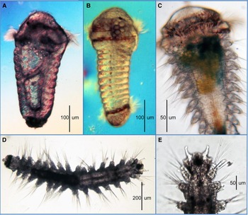
Development of Nereis vexillosa Grube, Reference Grube1851: (A) encapsulated trochophore; (B) encapsulated 3-segment metatrochophore; (C–D) hatching 3-setiger larvae: (C) ventral view; (D) dorsal view; (E) 4-setiger planktic larva, dorsal view; (F) 5-setiger planktic larva, dorsal view.

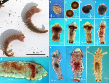
Photomicrographs of larvae and juveniles: (A) Micronereis nanaimoensis, 3-setiger nectochaete, dorsal view; (B–G) Nereis vexillosa: (B) group of nine encapsulated 3-segment metatrochophores; (B) encapsulated 3-segment metatrochophore, dorsal view; (C) early 5-setiger planktic larva, dorsal view; (E) late 5-setiger larva ready to settle; (F) 6-setiger juvenile; (G) 10-setiger juvenile.

INTRODUCTION
Nereis vexillosa is a common nereidid along the Pacific coast of North America. The species is also a characteristic large nereidid in shallow waters of the north-western Pacific including the shores of Russia, Japan and China. In northern California the species is typically found among mussel beds and barnacles in the rocky intertidal on semi-exposed shores (Blake & Ruff, Reference Blake, Ruff and Carlton2007). An early paper on the life history of N. vexillosa was published by Johnson (Reference Johnson1943) who was able to identify the species as responsible for egg masses of about 3–7 cm that were often found along the shore or washed up on beaches.
Johnson (Reference Johnson1943) determined that male and female heteronereids of Nereis vexillosa spawned an hour or two before midnight. In the laboratory, isolated females were induced to spawn when males discharged sperm. During spawning the eggs coalesced into the distinctive egg masses.
As part of regular meroplankton monitoring in Tomales Bay, distinctive nereidid larvae having large red eyespots were often encountered. These were linked to smaller encapsulated pre-trochophores and trochophores also found in the plankton, first encountered on 8 March 1973. The encapsulated larvae were surrounded by a thin transparent membrane that was likely derived from a fertilization membrane (Figure 3B). These embryos were moving within the ‘capsule’.
The identification of these early planktic encapsulated larvae and larger red-eyed nectochaetes were confirmed following collection of sexually mature heteronereids of N. vexillosa from among rocks north of Dillon Beach on 10 March 1974. Females spawned in the laboratory and the resulting fertilized eggs produced a large surrounding fertilization membrane that encapsulated the embryos. These embryos rapidly developed into a metatrochophore with three rudimentary segments with enclosed setal sacs. The large band of red pigment nearly surrounding the anterior end represents the precursor of the eyespots and confirmed the identity of other larvae with large red eyespots collected from the plankton.
The following account presents descriptions of selected stages of development of larvae obtained from both plankton and the laboratory fertilizations and cultured at 15°C.
DEVELOPMENT WITHIN THE EGG MASS
The earliest larval stages were spherical pre-trochophores 190–200 µm long observed one day after fertilization. Each contained four large spherical oily lipid droplets and numerous smaller yolk granules; cilia were not organized into any bands.
The earliest larvae recognized as trochophores were 2 days old; they measured ~200 µm in diameter and exhibited a definite polarity with a circular band of cilia in the anterior half of the embryo (Figure 2A). No eyespots or other cilia are apparent. The four oil or lipid droplets are conspicuous. The smaller yolk granules are coalesced into four or five greenish coloured yolk masses. The remainder of the cytoplasm is relatively smooth.
After 5 days, 3-segment metatrochophores were present. These larvae were 270 µm long and 170 µm wide (Figures 2B & 3C). At this stage the surrounding capsule is about 400 µm in diameter. The body is more or less oval in shape with lateral bulges where the setae will emerge. The anterior end is broadly rounded and bears a pair of large transverse red patches, the anlage of the large red eyespots of later larvae; two smaller red eyespots are also present; there are no antennae. Three body segments are demarcated by three pairs of internal setal sacs representing noto- and neurosetae, but setae were not yet protruding through the cuticle. Bands of ciliated cells surround the body posterior to each setal sac; these are amphitrochs. At the posterior end two short rounded lobes represent anlage of the anal cirri. A rudimentary mouth is developing. Internally 3–4 large oily lipid droplets are conspicuous; the rest of the yolk reserves are relatively fine-grained and light green in colour.
HATCHING AND DEVELOPMENT IN THE PLANKTON
Hatching occurred at the early 3-setiger stage, about 4 days after fertilization. These nectochaetes measured about 310 µm long. Two views of these hatching larvae are shown: ventral (Figure 2C); dorsal (Figure 2D). Their bodies are still rather compact, but are elongating and narrowing; upon emergence they swim by use of segmental ciliary bands. There are still four large oil or lipid spheres in the gut as well as smaller, fine-grained blue-green coloured yolk particles. The mouth is a narrow vertical ciliated groove. The anterior end or head is broadly rounded and two very short lobes are developing, best seen dorsally; these are the anlage of two antennae. Three pairs of eyespots are developing; the large transverse patches of red pigment seen earlier are largest and most prominent dorsally with narrow ventral extensions; medial to these are two small eyespots; the anteriormost pair of these smaller eyespots has a distinctly clear lens. The ciliation is extensive and includes a prominent prototroch that extends entirely across the ventral side and part way across the dorsum. A few cilia anterior to the prototroch on the ventral side may represent an akrotroch. Ciliary bands are present dorsally and ventrally on each of the three body segments. On setiger 1, these are limited to lateral locations both ventrally and dorsally; on setigers 2–3, however, they extend across both the venter and dorsum as gastrotrochs and nototrochs, respectively. There is no separate telotroch and a neurotroch was not observed. The setae are all compound spinigers. A band of reddish pigment encircles the posterior end. Two anal cirri are present, each with stiff sensory cilia.
By the Day 6 (2 days after hatching) the nectochaetes have three fully developed setigers with the fourth developing (Figures 2E & 3D). These larvae are about 375 µm long and feeding on phytoplankton. There are still lipid droplets in the gut but the remaining granular material is gone, replaced by yellow material believed to represent phytoplankton. Anteriorly, the three pairs of eyespots are well developed; the large pair is dorsolateral and conspicuous at low magnifications, the two smaller pairs are dorsal and posterior to the large ones. Body ciliation is similar to the previous stage with a distinct prototroch on the head and noto- and gastrotrochs dorsally and ventrally, respectively, with those of setiger 4 developing. The pharynx is well-developed and muscular, but distinct jaw parts are not evident. Anteriorly, the head has narrowed and is tapered toward the anterior end; the antennae and first pair of tentacular cirri are present, both with stiff sensory cilia; posteriorly, the anal cirri have elongated and also bear stiff sensory cilia. There is reddish pigment on the pygidial segment.
By the Day 8, the larvae have five setigers and measure about 450 µm long (Figures 2F & 3D). These larvae are at the settling stage. The head has elongated and is narrower anteriorly with an anterior medial notch; the two antennae are long and bear several stiff sensory cilia. The one large and two smaller pairs of red eyespots are fully developed. The body segments are ciliated similar to the previous larvae. Internally, the pharynx has a pair of jaws, each with five teeth. The gut is a uniform yellow-green colour from ingested phytoplankton. Posteriorly, the pygidial segment bears two long anal cirri, each with stiff sensory cilia; reddish pigment crosses the pygidial segment. All setae are homogomph spinigers.
METAMORPHOSIS AND JUVENILE MORPHOLOGY
Metamorphosis into crawling juveniles occurs between the 5- and 6-setiger stage. Figure 3E is a 5-setiger pre-settling stage that is about 470 µm long; Figure 3F is a well-developed 6-setiger juvenile that is about 1.15 mm long and has fully transitioned to the benthos. Figure 3G is a 10-setiger juvenile about 1.4 mm long. Key changes are the loss of cilia required for existence in the plankton, elongation of the anterior margin of the prostomium, lengthening of the antennae, formation of the palps, elaboration of the tentacular cirri, and development of functional and eversible jaws required for feeding. At the same time, the large pair of red eyespots remains at the level of the tentacular segment while the prostomium lengthens and enlarges. The parapodia assume adult form with noto- and neurosetae. Unfortunately, the cultures were not maintained in a manner that allowed for a more complete description of changes during and immediately after metamorphosis.
REMARKS
The occurrence of isolated individuals or groups of encapsulated embryos and early larvae of Nereis vexillosa in the meroplankton samples indicates that they had either not been coalesced into the egg masses produced by the adults at the time of spawning or that they had broken loose or been abraded from the original egg masses while subjected to wave action or other physical disturbance. In one respect, these isolated larvae were subject to an earlier phase of dispersal in the plankton than the larvae that were retained in the original egg mass. These observations suggest that Nereis vexillosa has a form of ‘dispersal polymorphism’ because the solitary or grouped larvae become planktic earlier than their counterparts that are retained in the egg mass and will presumably disperse farther from their point of origin than larvae that are released at a later date. Interesting reviews of dispersal polymorphism are by Hadfield & Strathmann (Reference Hadfield and Strathmann1996), Toonen & Pawlik (Reference Toonen and Pawlik2001) and Nanninga & Berumen (Reference Nanninga and Berumen2014).
As part of this project, the larval development of three species of Nereididae has now been described: Platynereis bicanaliculata (Blake, Reference Blake1975c) and Micronereis nanaimoensis and Nereis vexillosa (this study). These three species represent different strategies of spawning and larval development: (1) P. bicanaliculata represents the classic pattern of male and female heteronereids that swarm into the water column as part of a lunar cycle and spawn, with the larvae developing entirely in the water column; (2) N. vexillosa represents a modification of this pattern in which the male heteronereid releases sperm, eggs are spawned by the females, and both coalesce into an egg mass that is free on the substrate, with individual embryos and early larvae protected by a capsule produced by the fertilization membrane; (3) M. nanaimoensis also produces an egg mass, but this is attached to eel grass, algae, or other substrates and protected or incubated by one or two females.
Despite these obvious differences in spawning and patterns of development, the actual morphology of the larvae produced is very similar among the three species. The early trochophores and metatrochophores of each species have a similar appearance. Platynereis bicanaliculata larvae have apical cilia that are absent in N. vexillosa and M. nanaimoensis; however, all three species have multiple eyespots, a well-developed prototroch, and segmental bands of nototrochs and gastrotrochs. In P. bicanaliculata and N. vexillosa, much of the yolk is converted to oily lipid droplets that persist until the larvae begin to feed; in M. nanaimoensis, the yolk is retained in large granular yolk cells. Both P. bicanaliculata and N. vexillosa undergo metamorphosis into a crawling juvenile at the 5 to 6-setiger stage and have adult jaws; details of the metamorphosis of M. nanaimoensis are not known, however, the jaws that initially develop differ from those of adults.
As part of a recent review of annelid phylogeny, Weigert & Bleidorn (Reference Weigert and Bleidorn2016) identified selected model annelids that were candidate species for evolutionary developmental studies (evo-devo). Included among these candidates was Platynereis dumerilii, a European species that has been successfully cultured in the laboratory and the subject of numerous investigations (Fischer & Dorresteijn, Reference Fischer and Dorresteijn2004; Fischer et al., Reference Fischer, Henrich and Arendt2010). However, for other workers not in European waters, P. bicanaliculata, common in the north-eastern Pacific, has a nearly identical life history and is easily cultured in the laboratory (Blake, Reference Blake1975c).
Family SIGALIONIDAE
Sthenelais fusca Johnson, Reference Johnson1897
(Figure 4)
Sthenelais fusca Johnson, Reference Johnson1897: (A) 7-setiger planktic nectochaete, dorsal view, elytra removed from the left side; (B) 8-setiger post-larva, dorsal view, elytra removed from the left side; (C) capillary neuroseta from (A); (D) compound notoseta from (A); (E) compound neurosetae from (A–B).

INTRODUCTION
Larvae of the polychaete family Sigalionidae are poorly known. The only study of note is on Sthenelais boa Claparède, 1868 by Cazaux (Reference Cazaux1968), who described planktic metatrochophores and nectochaetes from Arcachon Bay, France. Sigalionid larvae were rare in our samples, but on 26 November 1974 a plankton sample from Tomales Bay contained several large 7-setiger nectochaetes that were later identified as Sthenelais fusca; on 12 Feb 1975 an 8-setiger newly metamorphosed post-larva was collected from the same location at the entrance to Tomales Bay. The morphology of these specimens is presented and compared with S. boa as described by Cazaux (Reference Cazaux1968). For these observations, where the tentacular segment is setigerous, it is treated as setiger 1.
7-SETIGER NECTOCHAETE
These larvae are planktotrophic and have a shape characteristic of most other scale-bearing polychaetes at this stage of development: the anterior end is broadly curved, hemispherical in shape, and bears a broad prototroch consisting of multiple rows of long cilia (Figure 4A). The specimen measured 450 µm long and 400 µm wide across the prototroch. The body tapers from the enlarged anterior episphere to a narrow posterior end that bears two anal or caudal cirri; each of these is broad basally and tapers to a pointed tip with stiff sensory cilia present on the sides and tips. The venter of the larva has a gold-coloured pigment scattered over the surface. Internally, the large circular part of the gut is dark green from feeding on phytoplankton; the narrow posterior part of the gut is generally clear.
The anterior end has two pairs of medial dark reddish eyespots and another pair located in a lateral position. The medial eyespots are cup-shaped and have a lens; the lateral eyespots are irregular in shape (Figure 4A). Some dusky black pigment is present on the anterior end of the broad anterior episphere; rusty granular pigment is present in parts of the prototroch. A low, rounded protuberance is the anlage of the medial antenna and bears several stiff sensory cilia (Figure 4A). A few small cells of cilia are present near the lateral eyespots. The prototroch consists of 4–5 rows of long and short rapidly beating cilia.
The elytra have been removed from the left side of the illustration in order to depict the segmental morphology. Rudiments of the tentacular cirri are present on setiger 1; these are short, narrow and tapered. Each of the following segments has a short notopodium and an elongate and triangular-shaped neuropodium. Elytra are present on setigers 2, 4, 6 and 7; each is oval in shape and with a relatively smooth surface, a single papilla on the posterior margin, and 3–4 additional short papillae or tubercles variously positioned (Figure 4A). The tentacular segment bears only serrated capillary setae. Segments 2–6 have short serrated capillary notosetae (Figure 4C); the neurosetae are all compound spinigers with pointed, serrated shafts and bearing either (1) a long narrow blade with a distinctive hood (Figure 4D) or (2) with a single jointed blade (Figure 4E).
8-SETIGER POST-LARVA
The specimen is 600 µm long not including the medial antenna or anal cirri and 450 µm wide including mid-body parapodia. There are no ciliary bands evident and the specimen is effectively a juvenile (Figure 4B). The gut is greenish in colour due to food resources, presumably phytoplankton. The anterior end is broadly rounded and bears a single medial antenna; the posterior end bears two long anal cirri. There are two pairs of reddish cup-shaped eyes; a more lateral third pair that was evident in the 7-setiger nectochaete was not apparent. Black pigment is present at the base of the medial antenna. The first segment is the tentacular segment and bears both noto- and neurosetae and elongate, clavate-shaped tentacular cirri (Figure 4B). The notopodia of segments 2–8 are short and rounded.
There are four fully developed elytra on setigers 2, 4, 6, 7 and one developing on setiger 8. On segments lacking elytra, a short dorsal lobe is present. The neuropodia are elongate and taper to a pointed tip; cirri and bracts are not evident on the parapodia at this stage of development. The elytra are relatively smooth on the dorsal surface and have short conical or flattened papillae on the margins; the flattened papillae have three or more notches and each papilla has at least one stiff sensory cilia (Figure 4B, inset). The tentacular segment (setiger 1) has all serrated capillary setae; the notosetae of setigers 2–7 are all short serrated capillaries and the neurosetae are all compound spinigers with single- or double-jointed blades (Figure 4E). Stout neuropodial falcigers that occur in some anterior setigers in adults were not observed.
REMARKS
These larvae were identified as Sthenelais fusca largely based on the nature of the relatively smooth elytra with simple marginal papillae and compound neurosetae with only 1–2 joints; the species is the only sigalionid known to occur commonly in the study area (Blake, Reference Blake, Blake, Hilbig and Scott1995; Blake & Ruff, Reference Blake, Ruff and Carlton2007).
The metatrochophore and nectochaete stages described here differ from those of S. boa described by Cazaux (Reference Cazaux1968) from Arcachon Bay on the Atlantic coast of France. At what appear to be the same stage of development, the larvae of S. boa have fewer segments, but greater development of parapodial cirri, tentacular cirri and antennae. The elytra of S. boa are heavily papillated over the entire surface and numerous papillae are present on parts of the body. The morphology of the setae was not reported for S. boa. Nectochaete larvae of S. boa and S. limicola were described by Plate & Husemann (Reference Plate and Husemann1994) from off Helgoland, Germany. The larvae of S. boa reported by these authors were similar to those described by Cazaux (Reference Cazaux1968); whereas the larvae of S. limicola had fewer papillae on the body and elytra. Setae of these species were not described by these authors.
Family NEPHTYIDAE
Nephtys caecoides and N. californiensis are two of the most common species of Nephtyidae in California coastal waters occurring from the intertidal to shallow shelf depths (Blake & Ruff, Reference Blake, Ruff and Carlton2007). Nephtys caecoides is found in sandy muds of tidal flats, whereas N. californiensis is more typical of clean sandy beaches. Both species are commonly found in the vicinity of Tomales Bay and Bodega Bay.
Larval nephtyids were taken from meroplankton tows from Tomales Bay regularly from January through August 1972. They were absent in October and December of 1971 and 1972. For all practical purposes, it was not possible to distinguish between planktic larvae of N. caecoides and N. californiensis. However, the latest planktic larvae appear different in overall shape: Nephtys caecoides late larvae and juveniles are short and stout while those of N. californiensis are long and thin. Further, distinct pigment markings on the dorsal surface of the anterior end may also be used to identify juveniles.
Fertilizations were attempted on sexually mature N. caecoides in January 1972 and again in March 1975. Fertilized eggs developed into swimming trochophores, but these failed to feed after their yolk reserves were expended and the cultures died. Results from observations on the two species are treated separately with major emphasis at this time on the planktic larvae of N. californiensis, for which a more-or-less complete sequence of development is presented.
Nephtys caecoides Hartman, Reference Hartman1938
(Figure 5A–C)
(A–C) Nephtys caecoides Hartman, Reference Hartman1938. Trochophore larvae: (A) early stage 1; (B) intermediate stage 2, ventral view; (C) late trochophore stage 3, ventral view. (D–F) Nephtys californiensis Hartman, Reference Hartman1938. (D) late planktic trochophore, ventral view; (E) early metatrochophore, ventral view; (F) 8-segment metatrochophore, left lateral view.

INTRODUCTION
Sexually mature specimens were collected on 27 February 1975 from tidal flats at Lawson's Landing, Tomales Bay. In the laboratory, spawning of sperm and eggs from the males and females occurred naturally over a 24 h period starting on 3 March at 1:00 pm. Eggs were ~105 µm in diameter. Fertilizations were successful with 2, 4 and 8 cell cleavage stages present in the culture dishes at 1:00 pm on 4 March. By 3:40 on that same afternoon 32-cell embryos were observed. Free swimming early trochophores were abundant in the dishes 24 h later on 5 March. On 6 March later-stage trochophores with a full complement of cilia were present and attempts were made to feed them algal cells from cultures of Dunaliella terteolecta. These larvae were about 120 µm long, or about the same as the original egg diameters. The majority of these trochophores did not feed on the cells provided and died. A few larvae progressed to a more advanced trochophore but no further; one of these was studied on 11 March and illustrated (Figure 5C). Each of the three stages of trochophore development were illustrated and numbered as Trochophore 1, 2 and 3.
TROCHOPHORE 1
The earliest trochophore (Figure 5A) is ~120 µm in diameter. This form is spherical and full of yolk. There is a complete prototroch and a broad apical tuft. At this early stage, there is no evidence of any other morphology.
TROCHOPHORE 2
These trochophores have a short apical tuft and medial prototroch and are about 140 µm long (Figure 5B). Cilia of the prototroch contain both short and fine cilia. The short cilia are anterior and come to lie ahead of the thicker row of longer cilia. The yolk has a globular appearance which is imparted to the surface of the trochophore. There is a well-formed mouth and a broad neurotroch that extends to the posterior end. There is no telotroch. Initially the yolk is distributed throughout, but as the mouth develops and the digestive tract is completed the yolk recesses to the internal part of the larva.
TROCHOPHORE 3
This advanced trochophore (Figure 5C) is 160 µm long and has a large anterior episphere with the apical tuft reduced to short cilia on the anterior end. The prototroch is complex and formed of 5–6 rows of long cilia. The mouth is wide and heavily ciliated; a neurotroch extends from the mouth to the posterior end. There are a few lipid drops under the cuticle in various places, and several dark red pigment spots and others that are yellow. The gut is blue in colour.
It was not possible to get these larvae to feed and continue to grow and develop. These same difficulties were encountered with larvae of phyllodocids and Micronereis. It is possible that the algal species that were provided as potential food were too large for these small trochophores to ingest. Smaller cells of Isochrysis and Monochrysis fed in mixed cultures might be the answer to this problem. At the time the smallest flagellate available was Dunaliella.
Nephtys californiensis Hartman, Reference Hartman1938 (Figures 5D–F, 6 & 7)
Nephtys californiensis Hartman, Reference Hartman1938: (A) Early 10-setiger nectochaete, left lateral view; (B–C) 12-setiger nectochaetes: (B) dorsal view; (C) ventral view; (D) 16-setiger settling larva, dorsal view.

Nephtys californiensis Hartman, Reference Hartman1938 photomicrographs: (A–B) late 10-setiger nectochaetes: (A) right lateral view; (B) left lateral view; (C) late 12-setiger nectochaete, dorsal view; (D–E) 16-setiger juvenile: (D) entire worm, dorsal view; (E) anterior end, dorsal view.

INTRODUCTION
The larvae described here were taken from several different collections made in November 1971. Pelagic larvae of this genus were collected at several times throughout the year, but only those collected in November 1971 were reared through metamorphosis and finally identified to species.
DESCRIPTION OF PLANKTIC LARVAE
Late planktic trochophore (260 µm × 230 µm). These relatively large trochophores (Figure 5D) were collected on 26 November 1971. They are colourful with granular red pigment in bands anterior to the prototroch and telotroch; internally, a bright blue lining of the gut is evident. These larvae have a large rounded episphere bearing a few short apical cilia and a pair of red granular eyespots. There are a few lipid spheres under the cuticle and some yellow chromatophores. The prototroch is on an expanded belt-like bulge below the episphere and anterior to the trunk. The prototroch is thickly ciliated with at least six rows of long cilia and an anteriormost row of shorter cilia. The telotroch consists of at least two rows of heavy cilia. The mouth is an elongate ciliated opening posterior to the prototroch. A v-shaped ciliated area posterior to the mouth is the beginning of the neurotroch. The developing trunk has 2–3 weakly developed bulges seen laterally that are the beginning of segmentation.
Early metatrochophore (280 µm × 210 µm). This larva is an initial metatrochophore because 4–5 distinct segments are visible on the elongating trunk (Figure 5E). The body is somewhat barrel-shaped, and they are awkward swimmers. The episphere is broadly rounded and with a few short cilia on the anterior border. The belt-like bulge that contains the prototroch is becoming thickened and somewhat glandular. Most of the pigmentation and ciliation are as described for the late trochophore. Most noticeable is the development of a distinct narrow neurotroch extending along the venter of the trunk, ending anterior to the telotroch. The bright blue pigment that lines the gut is obvious as are the two red eyespots and granular red pigment anterior to the prototroch and telotroch. There are still numerous lipid droplets under the cuticle.
Late metatrochophore with eight segments and no setae (305 µm × 200 µm). These larvae are also awkward swimmers. They are photopositive and move rapidly toward a light source. The body is widest anteriorly at the prototroch and tapers posteriorly along the segmented section to a narrow posterior end with a prominent telotroch. There are seven segments differentiated, none with setae (Figure 5F). The episphere and the bulge carrying the prototroch have numerous small gland cells below the surface (Figure 5F); similar glands are located anterior and posterior to the telotroch. The prototroch is formed of 5–6 rows of dense cilia. For simplicity, the illustration does not show these discrete rows (Figure 5F). The telotroch is formed of 2–3 rows of cilia. The developing body segments are smooth in texture. Red pigment is scattered over the anterior end and as several dense areas anterior to the telotroch. There are also light spots of red pigment scattered over the trunk segments. There are two glandular red eyespots. The gut is divided into a globular anterior region and a narrower posterior region. The mouth (not shown on Figure 5F) is a horizontal slit with numerous small cilia.
Early 10-setiger nectochaete (400 µm long × 160 µm wide). The head is large in relation to the rest of the body (Figure 6A). Each segment is regular in appearance with only the rudiments of parapodia. Two or three setae are present in each parapodium. The first setae to appear are simple capillaries; additional setae that develop are crenulated or serrated capillaries. There are two dark red eyespots. Bands of granular red pigment are present in various parts of the body. Prominent red pigment bands occur ventrally anterior to the prototroch and mouth. Other pigment bands occur dorsolaterally posterior to the prototroch. Granular red pigment is also present ventral to the neuropodia, dorsal to the notopodia and between the two podial lobes. A prominent red area occurs anterior to the telotroch; in addition scattered red and yellow-green pigment is present posterior to the telotroch. The anterior part of the gut contains some blue pigment, yellow-green patches and large oil globules. The prototroch and telotroch are composed of 5–6 rows of dense elongate cilia (Figure 6A). The mouth is large and well developed with large lips; it leads through a narrow oesophagus into an intestine and proctodeum. The latter later becomes blue in colour. There are no amphitrochs or other trunk cilia present.
Two slightly larger specimens (Figure 7A, B) measure 400 and 450 µm in length respectively and show both the internal and external colouration. Blue pigment is prominent in the lining of the gut (Figure 7A); red pigment is prominent anterior and posterior to the prototroch and anterior to the telotroch (Figure 7B).
Twelve-setiger nectochaete (860 µm long). The greatest changes present in these large planktotrophic nectochaetes (Figure 6B, C) are with elaboration of the parapodia, development of the setae, reduction of the mouth, reduction of the head in relation to the overall size of the body. Body pigment is now more concentrated on the segments. There are centres of granular red pigment on the ventral and dorsal sides of the body. The reddish pigmented areas of the pygidial segment have intensified. The dorsal band now encircles the area just above the telotroch. The pigment posterior to the telotroch is not as intense. There are reddish areas on the anterior aspect of the head and just below the prototroch. The eyespots have shifted posterior to the prototroch to the level of setiger. The gut has now differentiated into the oesophagus, pharynx and gut. The pharynx bears two teeth (Figure 6C). The gut has only a weak indication of the proctodeum. The mouth is greatly reduced in size, and is difficult to observe. The ventral cirrus is present on the parapodium as a small rudiment. The gut has oil globules prominent in the anterior part and contains considerable blue pigment. Most of the gut, however, appears to be yellow-green. A single anal cirrus has developed. A similar stage with better developed parapodia in Figure 7C shows the elaborate pigment that encircles the anterior end and that is present in the gut.
Sixteen-setiger metamorphosing larvae (~1.2 mm long) and juveniles. This larva is rapidly assuming the form of an adult nephtyid (Figure 6D). The metamorphosis is marked by the great change in shape of the head. The eyespots are further shifted posteriorly and come to lie between setigers two and three. Two prostomial antennae are present. The first setiger is shifted far anteriorly in relation to the rest of the segments. Parapodial lobes are now well developed. Cilia detected just beneath the dorsal lobes probably represent rudiments of branchiae. The gut is clearly demarcated into the intestine proper and a blue-coloured proctodeum. Only the posterior part of the pharynx is seen to be muscular; the two teeth are still prominent. Both crenulated and simple capillary setae are present. The crenulated setae are always anterior to the simple capillaries. The pigment on the head is localized into an anterior area between the eyespots (anterior in position). The gut is blue in the anterior part; most of it is yellow-green with brown food particles and oil globules. This stage crawls slowly, but when disturbed assumes a rapid swimming movement.
A slightly more advanced juvenile about 1.3 mm long (Figure 7D, E) has antennae and the parapodia are well developed. All cilia are lost and these juveniles actively crawl over the bottom of culture dishes.
REMARKS
Most of the literature on nephtyid larvae includes descriptions of individual trochophores, metatrochophores and nectochaetes as in Claparède & Mecznikow (Reference Claparède and Mecznikow1869), Wilson (Reference Wilson1936b), Smidt (Reference Smidt1944), Thorson (Reference Thorson1946), Rasmussen (Reference Rasmussen1973) and Plate & Husemann (Reference Plate and Husemann1994). Rasmussen (Reference Rasmussen1973) has detailed illustrations of metatrochophores of Nephtys hombergi but little descriptive information. Thorson (Reference Thorson1946) presented illustrations and brief descriptive notes for trochophores, metatrochophores and nectochaetes of N. ciliata. Noyes (Reference Noyes1980) described the complete larval development of Micronephtys neotena (as Aglaophamus) from Maine, USA and Lacalli (Reference Lacalli1980) provided descriptions and illustrations of larval stages of N. incisa, N. caeca and M. neotena from New Brunswick, Canada. The most complete study of larval development from trochophore to late nectochaetes, however, is by Fewkes (Reference Fewkes1883) on Nephtys sp. collected from the vicinity of Newport, Rhode Island, USA. Fewkes (Reference Fewkes1883) presented detailed illustrations and descriptions of early trochophores taken from plankton to swimming-crawling juveniles with 11 setigers. The present study is the first since that early study to illustrate and describe Nephtys larvae from an early trochophore to a settling juvenile.
Wilson (Reference Wilson1936b) described eggs and trochophores of Nephtys hombergi and reported on the presence of what appear to be brush cilia on one side of the larva. Brush cilia have been observed in larvae of other polychaetes and are believed to play a role in capture of large particles in species having an opposed band feeding mechanism (Phillips & Pernet, Reference Phillips and Pernet1996). However, brush cilia have not been observed on nephtyid larvae by other workers. They were not figured by Rasmussen (Reference Rasmussen1973) in his otherwise detailed illustrations of metatrochophores of the same species. Brush cilia were also not reported in the detailed description of larvae of Micronephtys neotena by Noyes (Reference Noyes1980) nor in the descriptions of Nephtys incisa and N. caeca by Lacalli (Reference Lacalli1980). Brush cilia were not observed on the nephtyid larvae in this study, albeit they may be difficult to observe in light microscopy. Interestingly, nephtyid larvae have been observed to feed on bivalve veligers (Mileikovsky, Reference Mileikovsky1959; Yokouchi, Reference Yokouchi1991) and brush cilia, if present, might assist in their capture.
Family SPIONIDAE
Boccardia berkeleyorum Blake & Woodwick, Reference Blake and Woodwick1971
(Figures 8 & 9)
Boccardia berkeleyorum Blake & Woodwick, Reference Blake and Woodwick1971: (A) egg capsules; (B) early encapsulated pre-trochophore, ventral view; (C) encapsulated trochophore, ventral view; (D) encapsulated 3-setiger larva, dorsal view; (E–F) 10-setiger planktic larva: (E) dorsal view; (F) ventral view; (G–H) early 15-setiger planktic larvae: (G) dorsal view; (H) ventral view.

Boccardia berkeleyorum Blake &Woodwick, Reference Blake and Woodwick1971: (A) late 15-setiger larva, right lateral view; (B–C) 19-setiger larva ready to metamorphose: (B) dorsal view: (C) ventral view; (D) bidentate hook and capillary from neuropodium of setiger 7; (E) capillary and spine from posterior notopodium; (F) modified spines and a capillary from setiger 5.

INTRODUCTION
Boccardia berkeleyorum is an intertidal California species that bores into calcareous substrata including coralline algae, hermit crab shells, and shells of living bivalves such as Pododesmus macroschisma (Blake & Woodwick, Reference Blake and Woodwick1971; Blake & Ruff, Reference Blake, Ruff and Carlton2007).
In the present study, adults with egg capsules were collected at different times in different locations. On 23 March 1971, specimens were collected from encrusting coralline algae that was attached to rocks at Cayucos Reef, north of Morro Bay. These samples were transported to the Dillon Beach laboratory where adults and egg capsules were extracted from burrows and set up in culture dishes. Larvae from these samples were successfully cultured through planktic development. On 9 June 1975, adults with egg capsules were removed from shells of Pododesmus collected in Bodega Harbor and set up in culture. Planktic larvae were collected from Tomales Bay at various times, most notably 28 April 1975 and 3 April 1976. Most of the observations of early and later larval development are from the 1971 collections. Later samples were used to confirm earlier observations and to check finer details of larval morphology.
DEVELOPMENT OF ENCAPSULATED LARVAE
Egg capsules are formed singly and joined into a beadlike string; individual capsules are each attached to the burrow lining by a single thread (Figure 8A). Due to the difficulty of removing egg capsules from the burrows in coralline algae, many were damaged and the encapsulated embryos and larvae were released prematurely.
A maximum of 15 capsules per string were observed in burrows within the Pododesmus shells. There were ~40–50 eggs per capsule. These eggs were white in colour and ranged from 92 to 102 µm in diameter (average: 99.4 µm) based on 15 eggs removed from one capsule. These capsules were also set up in culture to observe early embryos and larvae prior to their release into the water column. The number of eggs/larvae per capsule ranged from 25 to 50 in the Cayucos samples.
The earliest larvae observed were pre-trochophores that are oval in shape and about 120 µm long and about 100 µm wide (Figure 8B). The interior is filled with yolk; externally, a ciliated depression or vestibule, is evident on the anterior end. Two mediolateral ciliary patches provide movement in the capsules; these are lost after the prototroch and telotroch are developed (Wilson, Reference Wilson1928; Blake, Reference Blake1969). There is no evidence of either the prototroch or telotroch at this stage.
A later stage (Figure 8C) is equivalent to a trochophore and is 130 µm long and about 100 µm wide. The anterior end is broadly rounded with the body tapering toward the posterior end; the interior is still filled with yolk. Externally the two mediolateral ciliary patches are still present. Cilia representing the prototroch and telotroch are now evident. The vestibule or anlage of the mouth is well developed and heavily ciliated. These larvae are able to slowly move around in the capsules.
An early 3-setiger stage (Figure 8D) is about 160 µm long and capable of slow gliding movements if released from the capsules. The anterior end is broadly rounded and tapers posteriorly. The interior is still filled with yolk. The prototroch and telotroch are present, but nototrochs and gastrotrochs are not evident. There are a few short tactile cilia on the anterior and posterior ends. Two pairs of black eyespots are present; the lateral pair is more or less cup-shaped and the medial pair is round. The setae are short, but are the barbed notopodial provisional setae that characterize planktic spionid larvae. By reflected light these encapsulated larvae were a light green in colour, partially due to the colour of the yolk.
DEVELOPMENT OF PLANKTIC LARVAE
Larvae were released from the capsules at the 4-setiger stage while they still had some yolk reserves (not figured). However, upon introduction of a few drops of Dunaliella they began to feed. These larvae measure ~280 µm long and are slender in shape, typical of early planktic spionid larvae. There is one dorsal chromatophore on setiger 2, similar to but less prominent than that of B. columbiana (Blake, Reference Blake, Rouse and Pleijel2006: Figure 1J). Prototroch and telotroch are well developed, but noto- and gastrotrochs are not yet developed. When these larvae begin feeding on Dunaliella, their guts become green with the ingested cells and lipid droplets are formed. These larvae are phototactic and concentrate on sides of the culture dishes at the light sources.
A 10-setiger planktic larva is shown in Figure 8E, F. These larvae are 500 µm long and 130 µm wide and were present in the cultures ~6 days after the original collection. The anterior end is broadly rounded and bears four black eyespots; the medial pair are granular and round in shape whereas the lateral pair are weakly cup-shaped; a lens is not evident. Paired nuchal cilia are present posterior to the eyes, extending to the posterior border of the peristomial segment. Dorsal medial black chromatophores occur from setigers 2–9; each has lateral branches radiating from a central location. Golden brown pigment borders the vestibule and pygidial segment. The gut is markedly green from ingested Dunaliella and has numerous small lipid droplets. The prototroch has a dorsal gap and extends ventrally to merge with the well-developed vestibule of the mouth. Ventrally, a v-shaped neurotroch extends to near the end of setiger 1, terminating in a small ciliated pit. Large thick tactile cilia are present anterior to the prototroch on both sides and posterior to the prototroch on the ventral side. Nototrochs are present on setigers 3–10; gastrotrochs are present on setigers 3, 5, 7 and 10. The telotroch encircles the posterior end, leaving a dorsal gap. No grasping cilia associated with the long barbed provisional setae were observed.
An early 15-setiger larva (Figure 8G, H) is about 720 µm long and about 150 µm wide. Approximately 20 days after the original collection, this larva is fully developed for a planktotrophic life. The anterior end is rounded and bears several long ventromedial tactile cilia and six black eyespots. The lateral eyespots are largest and appear to be two separate eyespots that are joined, a small round eyespot is medial to this, and a larger more medial pair is oval in shape. Nuchal cilia form a pair of triangular-shaped ciliated patches posterior to the eyes. A pair of short palps has developed on the posterolateral border. The prototroch has a dorsal gap and extends ventrally to the edges of the vestibule or mouth. The neurotroch with a ciliated pit is the same as in the previous stage; a pair of short lateral cilia is located on either side of the neurotroch. Nototrochs are present on setigers 3–15; gastrotrochs are present on setigers 3, 5, 7, 10, 13 and 15. The dorsal medial black chromatophores occur from setiger 2 to 15; each has short lateral branches radiating from a central location. Golden brown pigment borders the vestibule and pygidial segment. The telotroch is well developed and has a dorsal gap. The long, barbed provisional setae extend from each of the notopodia; grasping cilia were not observed. The neurosetae consist of short capillaries throughout; hooded hooks were not observed.
A large 15-setiger larva (Figure 9A) measures 825 µm long and 160 µm wide and has developed to this stage ~23 days from the original collection date. The anterior end is rounded and bears several long ventromedial tactile cilia and four black eyespots. The lateral pair is largest and appears to consist of 2–3 individual eyespots that have joined; the medial eyespots are oval in shape. Nuchal cilia are similar as in the previous stage, forming a pair of triangular-shaped ciliated patches posterior to the eyespots. The pair of palps arising from the posterior border of the head near the nuchal cilia have greatly enlarged and elongated. The prototroch has a dorsal gap and extends ventrally to the edges of the vestibule or mouth. The nuchal cilia and neurotroch with a ciliated pit are as in the previous stage. Nototrochs are present on setigers 3–15; gastrotrochs are present on setigers 3, 5, 7, 10, 13 and 15. The dorsal medial black chromatophores occur from setigers 2–15; each has lateral branches that do not extend far laterally from a central location. The chromatophore on setiger 2 is much reduced. Golden brown pigment borders the vestibule and pygidial segment. The telotroch is well developed and has a dorsal gap. The long, barbed provisional setae extend from each of the notopodia; grasping cilia were not observed. The neurosetae consist of short capillaries on setigers 1–6; hooded hooks are present from setiger 7–15; notosetae of setiger 5 include at least two spines, these represent the initial appearance of major spines.
A 19-setiger larva ready to metamorphose (Figure 9B, C) was collected from meroplankton of Tomales Bay on 28 April 1975. The specimen is large, 1.25 mm long and 300 µm wide. The body is light tan in colour and with a row of dorsomedial black chromatophores and dorsolateral red spots in some posterior segments.
The anterior end is broad on the anterior margin with two weakly developed lobes that appear to be associated with the unusually large gaping mouth (Figure 9C); the entire ventral surface of the head is covered with fine cilia that are also visible from the dorsal side. There are thickened ridges surrounding the heavily ciliated vestibule. Two long tactile cilia are present on the ventral side. The two pairs of black eyespots are relatively unchanged except that the medial pair are more irregular in shape (Figure 9B). The prototroch is still prominent and extends to the lateral borders of the vestibule and has a dorsal gap. Ventrally, the neurotroch and ciliated pit are still present, but the neurotroch is reduced to a narrow track on a broad ventrally elevated area that extends across setigers 1–2. Dorsally, the prostomium is differentiating and extending posteriorly into a caruncle with broad patches of nuchal cilia on either side. The palps have continued to elongate and are shifted dorsally to near the developing caruncle and nuchal cilia. The dorsal side of the palps is relatively smooth with the blood loop prominent; the ventral side has a ciliated groove. A pigment spot on top of the caruncle is the remnant of the setiger 2 chromatophore of earlier stages (Figure 9B). Nototrochs are present on setigers 3–19, but somewhat reduced on setiger 5. Gastrotrochs are present on setigers 3, 5, 7, 10, 13, 15 and 17; the gastrotrochs have four cells of cilia on setiger 3, two on setiger 5 and eight on each remaining segment. The telotroch has a dorsal gap and is unchanged from earlier stages. Branchiae are present on setigers 2–4 and 6–14; those on setigers 2–4 are shorter than those on setigers 6–14. Each branchia is ciliated and with the nototrochs provides a transverse row of cilia across most of the dorsum. The posterior end or pygidial segment shows some evidence of change: two small dorsal protuberances represent anlage of the dorsal pair of anal cirri; the ventral pair is not yet developed. The telotroch is formed of eight patches of cilia with a narrow dorsal gap.
The setae are dominated by the barbed provisional setae; however, these readily dislodge when the larvae are mounted for observation. On setiger 5, two kinds of major spines have developed in the notopodia (Figure 9F), one is bristle-topped and the second is simple and falcate; simple capillaries are present in the neuropodia. The neuropodia of setigers 1–6 have capillaries; from setiger 7–8 bidentate hooded hooks accompany the capillaries (Figure 9D). Far posterior notopodia have, in addition to provisional setae, small capillaries and 1–2 spines (Figure 9E).
The dorsal medial black chromatophores occur from setigers 2–19; each has lateral branches radiating from a central location but do not extend far laterally; those of far posterior segments are greatly reduced. The chromatophore on setiger 2 is much reduced and limited to the narrow caruncle (Figure 9B). Lateral red spots are visible dorsally medial to the branchial locations from setiger 9, but were observed on other specimens starting from setigers 8–11.
REMARKS
The larvae of Boccardia berkeleyorum are similar to those of B. columbiana, B. proboscidea and B. tricuspa, three congeners that also occur along the California coast and that have a row of branching black chromatophores along the dorsal surface of the body. Similarities and differences between these four species with respect to reproduction and larval development were tabulated by Blake & Arnofsky (Reference Blake, Arnofsky, Dorresteijn and Westheide1999). Boccardia columbiana and B. proboscidea are both closely related in having egg capsules that are solitary rather than in beadlike strings and similar appearing fusiform-shaped larvae with chromatophores where the branches may extend onto the ventral surface. In contrast, the egg capsules of B. berkeleyorum and B. tricuspa are joined together in beadlike strings within burrows in calcareous structures. Boccardia berkeleyorum differs from the other three species in having a more elongate narrow shape instead of one that is fusiform. Unlike the other three species, the individual egg capsules of B. berkeleyorum have only a single filament for attachment instead of two.
Species of Boccardiella and Boccardia have branchiae both anterior and posterior to the modified fifth segment. To date, studies of larval development of species of these genera have shown that while branchiae posterior to setiger 5 develop in the latest planktic larval stages, the anterior branchiae only develop after metamorphosis and development of the juvenile (Rullier, Reference Rullier1960; Dean & Blake, Reference Dean and Blake1966; King, Reference King1976; Carrasco, Reference Carrasco1976; Woodwick, Reference Woodwick, Reish and Fauchald1977; Blake, Reference Blake, Rouse and Pleijel2006). Boccardia berkeleyorum is clearly an exception to this generalization because branchiae both anterior and posterior to setiger 5 are present in late-stage larvae of this species (Figure 9B).
Polydora pygidialis Blake & Woodwick, Reference Blake and Woodwick1972 (Figure 10)
Polydora pygidialis Blake & Woodwick, Reference Blake and Woodwick1972: (A) egg capsules; (B) early encapsulated pre-trochophore; (C–D) 14-setiger planktic larva: (C) dorsal view; (D) ventral view; (E–G) setae from modified fifth setiger of same specimen: (E) pennoned companion seta; (F) superior dorsal seta; (G) major spine with lateral tooth; (H) neuropodial bidentate hooded hook from setiger 7.

INTRODUCTION
Polydora pygidialis is a borer in calcareous substrates including hermit crab shells, gastropod shells and various types of coralline piling materials. It ranges from the intertidal to about 20 m depth and is recorded from British Columbia to Santa Barbara, California (Blake & Woodwick, Reference Blake and Woodwick1972; Blake, Reference Blake, Blake, Hilbig and Scott1996).
In the present study, adults were found in coralline algae as part of a dredge haul from off Bodega Head in about 20 m on 7 October 1970. During extraction of the adults from the coralline algae over a 2-day period, some adults were found with egg capsules in their burrows and with larvae in various stages of development.
The individual egg capsules were joined into a beadlike string with each individual capsule attached to the wall of the tube or burrow by a single attachment stalk (Figure 10A). Due to the difficulty of extracting the worms and egg capsules, only a few intact capsules were successfully removed to culture dishes. Embryos and larvae in the capsules were in various stages of development. Fertilized eggs were 140 µm in diameter. Larvae developed in the capsules up to a 4-setiger stage before emerging into the water column. Larvae at this stage were provided with cells of Dunaliella on which they began to feed.
There was no opportunity at the time to follow these larvae completely through their development and record morphological changes. However notes were taken on the early embryos and more detailed observations on late-stage larvae that were reared in the laboratory. A few larvae believed to be of this species were later encountered on rare occasions in the plankton, but only in the months of October and November.
EARLY TROCHOPHORES
An early encapsulated larva (Figure 10B) is oval in shape and 165 µm long. The interior of the larva is filled with yolk; externally, there is a ciliated vestibule on the anterior end that represents the anlage of the mouth. Two prominent lateral ciliary patches provide movement in the capsules; these are lost after the prototroch and telotroch are developed (Wilson, Reference Wilson1928; Blake, Reference Blake1969). These early larvae are typical of other species of Polydora and related genera that have been described (Blake, Reference Blake1969).
14-SETIGER PLANKTIC LARVAE REARED IN THE LABORATORY
Following release from the egg capsules on 12 October 1970, the 4-setiger larvae fed on the Dunaliella cells provided and some specimens developed to a size of up to 14 setigers by 10 November or after 29 days in culture at about 15°C. Dorsal and ventral views of these larvae are shown in Figure 10C, D. The figured larva is ~800 µm long.
The 14-setiger larva illustrated is characterized by a broadly rounded prostomium and thickened body that is widest in the posterior one-third (Figure 10C, D). Short palps are present laterally on either side of the head. Rudimentary parapodia are present; branchiae are not yet developed. A striking feature of these larvae is a network of black pigment that appears to extend from the large lateral eyespots to form a curved lattice on the anterior margin of the prostomium (Figure 10C). The lateral eyespots are large and comma-shaped; two smaller round eyespots are medial to the larger ones. The dorsal surface of the body bears a pair of chromatophores on each segment from setiger 3 continuing posteriorly (Figure 10C). The first two pairs are small and reduced to transverse lines; those following are larger, prominent and branched. The pygidial segment bears black pigment granules.
The ciliation includes oval ciliated patches or nuchal organs lateral to a raised peristomial ridge that extends posterior to the medial eyespots (Figure 10C). The prototroch extends laterally from the mid-dorsal ridge around the head and ventrally to the vestibule or opening to the mouth (Figure 10D). The entire ventral surface of the prostomium anterior to the mouth opening is covered with fine cilia that are visible from the dorsal side. Nototrochs occur dorsally starting from setiger 3, they consist of 4–6 separate cells from which the cilia arise. Ventrally, gastrotrochs are present on setigers 7, 10 and 12. Grasping cilia are present on most segments. The telotroch surrounds the pygidial segment, but has a dorsal gap. An unusually short v-shaped neurotroch is posterior to the oral opening; there is no ciliated pit.
Notosetae include long, barbed provisional setae on all setigers. Neurosetae include capillaries on setigers 1–4, and 6, and two or three bidentate hooded hooks from setiger 7; each hook has the main fang at a right angle with the shaft and a weak notch on the shaft (Figure 10H). Setiger 5 is modified with notosetae consisting of 2–3 large major spines that are curved apically to a narrow tip and have a distinct lateral tooth (Figure 10G); these are accompanied by pennoned companion setae (Figure 10E); 2–3 superior dorsal setae occur anterior and dorsal to the major spines (Figure 10F). Neurosetae were not observed.
REMARKS
Numerous species belonging to the spionid Polydora complex occur in California waters (Blake, Reference Blake, Blake, Hilbig and Scott1996). The larvae of relatively few, however, have been described. Polydora pygidialis larvae are similar in overall appearance to those species related to Polydora websteri and P. cornuta (as P. ligni) described by Blake (Reference Blake1969) from New England waters in having a dorsal pigment pattern with transverse bands of black pigment on a few anterior setigers followed by pairs of large branching chromatophores. The same pigment patterns have been observed on these same species in California waters (Blake, unpublished). In P. pygidialis, however, the anterior transverse bands are short, limited to setigers 3–4 and exhibit little branching. In contrast, related species have the transverse bands over setigers 2–5 or 3–6 (Blake, Reference Blake1969; unpublished). These plus the following larger branched paired chromatophores provide P. pygidialis larvae with a pattern that is distinctive. This pattern, taken together with the reticulating network of black pigment on the anterior margin of the prostomium and dark pigment on the pygidium, provides a means to identify these larvae if encountered in meroplankton samples.
Polydora spongicola Berkeley & Berkeley, Reference Berkeley and Berkeley1950
(Figure 11)
Polydora spongicola Berkeley & Berkeley, Reference Berkeley and Berkeley1950: (A–B) 15-setiger planktic larvae: (A) dorsal view; (B) ventral view; (C–E) major spines from setiger 5 in different views; (F) companion seta with major spines; (G) neuropodial bidentate hooded hook from setiger 7.

INTRODUCTION
Polydora spongicola is only known as an associate of sponges within which the worms excavate burrows lined with silt. In the eastern Pacific the species ranges from British Columbia to Central California (Berkeley & Berkeley, Reference Berkeley and Berkeley1950; Woodwick, Reference Woodwick1963; Blake, Reference Blake, Blake, Hilbig and Scott1996). The species also occurs in the western Pacific as P. uschakovi Buzhinskaja, 1971, referred to P. spongicola by Radashevsky (Reference Radashevsky1993). The larval development (as P. uschakovi) was described by Radashevsky (Reference Radashevsky1988), who found planktic larvae in August–September in Peter the Great Gulf, Sea of Japan. Planktic larvae were first collected at Lawson's Landing in Tomales Bay on 13 October 1971 and in subsequent years in October and November, but these occurrences were rare. These larvae were referred to P. spongicola based on the nature of the modified spines of setiger 5 and the close similarity with larvae described by Radashevsky (Reference Radashevsky1988).
15-SETIGER PLANKTIC LARVAE
The 15-setiger larva is characterized by a broadly rounded prostomium that is somewhat flattened and a thickened body that is of the same width anterior to posterior (Figure 11A, B). Posteriorly, the body terminates in a pygidial segment with a distinct dorsal notch forming two lateral lobes (Figure 11A). The figured larva is ~1.07 mm long and about 250 µm wide. Several short papillae on the prostomium are the tips of internal bacillary glands bearing stiff sensory cilia (Figure 11B). Internally, the intestine is dark green in colour and bears numerous small lipid droplets. Long palps are present laterally on either side of the head, arising from near the nuchal organs. Parapodia are well developed; short branchiae are present on setigers 7–10 (Figure 11A). The most prominent feature is the pair of large black chromatophores that occur from setiger 2 to the last setiger (Figure 11A). In addition, the large irregularly shaped lateral eyespots look like chromatophores with numerous dark branches that extend anteriorly on the prostomium and posteriorly along the caruncle; two smaller round eyespots are medial to the larger ones (Figure 11A). The pygidial segment bears a pair of black pigment granules and some light yellow reflective pigment at the bases of the cilia of the telotroch; some of the same yellow pigment is also scattered on the prostomium.
The ciliation includes elongate nuchal organs lateral to the caruncle or raised ridge that extends posteriorly from the medial eyespots (Figure 11A). The prototroch has a mid-dorsal gap, then extends around the head ventrally to the vestibule or opening to the mouth (Figure 11B); two additional cells of cilia occur ventrolateral to the vestibule and anterior to the prototroch. Nototrochs occur dorsally from setiger 3 posteriorly, each consists of six separate cells from which the cilia arise. Ventrally, gastrotrochs are present on setigers 7, 10, 13 and 15; a single cell of cilia occurs on one side of setiger 5, suggesting that additional gastrotrochs have been lost (Figure 11B). Grasping cilia are present on most segments. The telotroch surrounds the pygidial segment, but has a dorsal gap where the two lobes of the pygidium produce a dorsal notch. The neurotroch is v-shaped, posterior to the oral opening and extends half-way along setiger 1; there is no ciliated pit (Figure 11B).
Setae include long, barbed provisional setae on all setigers. Neurosetae include capillaries on setigers 1–4, and 6, and two or three bidentate hooded hooks from setiger 7; each hook has the main fang at a right angle with the shaft and a weak notch on the shaft (Figure 11G). Setiger 5 is modified with notosetae consisting of 2–3 large major spines that have a relatively straight shaft and weakly curved tip that bears a pair of lateral teeth; in another view, the tip appears as a transverse ridge with a single tooth (Figure 11C–E) and a thin spine with a curved tip (Figure 11F); thin pennoned setae accompany the major spines. Neurosetae were not observed.
REMARKS
There are several differences between these planktic larvae from California and those from Peter the Great Bay, Russia, reported by Radashevsky (Reference Radashevsky1988). The paired chromatophores of the Russian larvae begin on setiger 1 instead of 2. The chromatophores of setigers 1–2 were also reported to extend around to the ventral side on the Russian specimens; on the California specimens the chromatophores were only dorsal. In the Russian specimens the nototrochs began on setiger 2 and the gastrotrochs occurred on setigers 3, 5, 7, 10, 13 and 15; in the California specimens the nototrochs began on setiger 3 and gastrotrochs occurred on setigers 7, 10, 13 and 15, but with a single ciliated cell on setiger 5. In the California specimens extra ciliated cells were observed lateral to the vestibule on the ventral side; these were absent in the Russian specimens. In addition, ciliated patches were observed lateral to the neurotroch on the Russian specimens but not on the California specimens. The short papillae-like tips of bacillary setae observed on the prostomium of the California specimens were not reported for the Russian larvae. There are also differences in the nature of the major spines of setiger 5. The major spines of the California specimens appear to exhibit more of a double accessory tooth appearance than the Russian specimens where a transverse ridge is depicted rather than lateral teeth or knobs.
Several of these differences, including the distribution of nototrochs and gastrotrochs, are not known to be variable within larvae of Spionidae. These results suggest that Polydora uschakovi and P. spongicola, although similar, might be different species after all.
Dipolydora cardalia (E. Berkeley, Reference Berkeley1927)
(Figure 12)
Dipolydora cardalia Berkeley, Reference Berkeley1927: (A–B) 21-setiger planktic larvae: (A) dorsal view; (B) ventral view; (C–D) 29-setiger juvenile, dorsal view: (C) anterior end; (D) posterior end; (E) bidentate hooded hooks and capillaries from setiger 7; (F) major spines and companion setae from setiger 5.

INTRODUCTION
Dipolydora cardalia was first described from British Columbia by E. Berkeley (Reference Berkeley1927) and subsequently redescribed by Blake (Reference Blake1979). The species was later reported from the north-western Pacific (Bering Sea, Chukchi Sea, SE Kamchatka and North Japan Sea, Peter the Great Bay) by Radashevsky (Reference Radashevsky1993) and was identified from the Puget Sound, Washington, in 2015 (Blake, unpublished). The species has not been recorded from California, but Blake (Reference Blake, Blake, Hilbig and Scott1996) suggested that it should be expected in the California fauna. Dipolydora cardalia forms tubes in muddy sediments from intertidal to shelf depths of about 60 m.
As part of weekly meroplankton collections from Tomales Bay, several large planktic larvae of D. cardalia were collected on 28 April 1975. These larvae were cultured and underwent metamorphosis in the laboratory; the morphology of the juveniles was eventually determined to agree with D. cardalia. The following descriptions are of the large planktic larvae and the juveniles that metamorphosed in the laboratory.
21-SETIGER PLANKTIC LARVAE
Larvae with 21 setigers measured 1.6 mm long and 300 µm wide. Their bodies are long and slender, with middle body segments slightly wider than those anterior and posterior, but they are not fusiform (Figure 12A, B). The prostomium is broad across the anterior margin and has two rounded lateral peaks. Three pairs of black eyespots are located on an expanded central part of the prostomium: the posterior pair is medial and comma-shaped; the small lateral pair is elongated; the anterior lateral pair are cup-shaped, the others are round or oval (Figure 12A). The area anterior to the eyes extending to the anterior margin is pigmented with numerous small brown spots. Some black spots are also present at the very anterior edge, but these are only visible ventrally (Figure 12B). The prostomium continues posteriorly over setiger 1 as a narrow caruncle; there are several small bacillary glands on the posterior surface of the caruncle (Figure 12A). Lateral to the caruncle are ciliated nuchal organs (Figure 12A). The prototroch is evident both dorsally where it begins lateral to the location of the eyes and ventrally where it continues to the vestibule (Figure 12B); there are about five patches of cilia on either side. Ventrally, the mouth or vestibule is surrounded anteriorly by two raised ridges onto which the prototroch extends and posteriorly by a broad U-shaped raised area that extends posteriorly onto setiger 2 (Figure 12B).
Parapodia are well developed and bear notopodial and neuropodial postsetal lobes. Provisional barbed setae are present as well as developing adult setae. Setiger 5 has a pair of simple spines that lack accessory structures; hooded hooks begin on setiger 7, are bidentate, and lack a constriction or manubrium on the shaft. Nototrochs begin on setiger 3 with four patches of cilia; these continue on all subsequent setigers and have up to nine patches of cilia on most segments (Figure 12A). Gastrotrochs occur on setigers 3, 5, 7, 9–10 and 12–18 (Figure 12B). Posteriorly, the pygidial segment is divided into two rounded lobes; the telotroch surrounds the pygidial segment with about 10 patches of cilia, leaving a dorsal gap. Branchiae occur on setigers 8–16.
Body pigment is complex. The prostomial pigment previously described also occurs on the pygidium, but with black spots dorsally and brown spots ventrally. Iridescent yellow-green pigment occurs ventrally on the raised area posterior to the mouth and on some body segments (Figure 12B). There is always a band of 8–10 large spots of this pigment across setiger 11 and in more medial locations on setigers 8–10 and on segments posterior to 11 (Figure 12B). Lateral intersegmental black pigment occurs between setigers 1–2 and 2–3. Dorsally, the pigment pattern is as follows (Figure 12A): (1) medial bands of black pigment occur from setiger 3 and continue on all subsequent segments; these bands branch as with chromatophores, but not extensively; (2) a pair of lateral pigment patches joins the medial bands from setiger 6, continuing posteriorly; (3) a central group of pigmented cells occurs mid-dorsally on the posterior end of each segment from setiger 7.
Bacillary glands occur dorsally on all body segments and on the pygidial segment. A short raised medial papilla that occurs mid-ventrally from setiger 8 is possibly the tip of a gland (Figure 12B).
29-SETIGER JUVENILE
A 29-setiger juvenile resulting from the metamorphosis of one of the planktic larvae (Figure 12C, D) is about 2.0 mm long and 300 µm wide. It constructed a thin mucoid tube on the bottom of a culture dish, incorporating whatever silt or algal particles were present. Most of the larval pigment is retained, providing a convenient comparison with the earlier stage. Internally, the foregut is darkly pigmented and connects to the midgut via a gizzard-like structure that is thick and tubular but does not appear to have any hard inclusions as described for D. socialis by Blake (Reference Blake1969). Most of the midgut and hindgut are filled with remains of algal cells provided to the cultures as a food source.
The prostomium is strongly bifurcate on the anterior margin, but the tips of the two lobes are rounded (Figure 12C). The prostomium is expanded and bears two pairs of black eyespots: the medial pair is rounded; the lateral pair is comma-shaped and may be the result of a merger of two pairs of lateral larval eyespots. Posterior to the eyespots, the prostomium narrows to a caruncle that extends to the posterior margin of setiger 2. Elongate ciliated nuchal organs are located lateral to the caruncle (Figure 12C). Palps are long and arise from the posterior margin of the peristomium. These are grooved and ciliated on their ventral sides. Posteriorly, the body terminates in a 4-lobed pygidium; each lobe is flattened with the ventral lobes larger than the dorsal. The pygidial lobes are darkly pigmented (Figure 12D).
Body segments have well-developed noto- and neuropodial postsetal lamellae; those of setiger 1 are broadly rounded and flattened (Figure 12C), whereas those of following segments (except setiger 5) are cirriform. Neuropodial lamellae are similar to those of the notopodia, but shorter. From setiger 3, most body segments have a band of short cilia across the dorsum; these are most likely derived from larval nototrochs (Figure 12C). Branchiae begin on setiger 8 and continue along most of the body; each branchia is relatively short, straplike, and does not extend to the dorsal midline (Figure 12C). There is some indication that branchial cilia either merge with or are otherwise associated with the cilia of the transverse dorsal bands. As noted, the larval pigmentation pattern is retained on these juveniles. Dorsally, paired black pigment spots or transverse bands on setigers 1–3 give way to four paired bands from setiger 4. These are joined by a more posterior group of darkly pigmented ciliated cells from setiger 7 (Figure 12C). This pattern persists for about 20 setigers and is absent from far posterior segments. The ventral pigment (not illustrated) is similar to that of the 21-setiger larvae.
Notosetae and neurosetae are present on setiger 1 and following segments except setiger 5 as fascicles of simple limbate capillaries. Neurosetae are joined by 3–4 bidentate hooded hooks from setiger 7; each hook has two widely separated teeth and a tightly adhering hood; the shaft is smooth and has no manubrium or notch (Figure 12E). Setiger 5 is enlarged and has a developing musculature that extends onto setiger 6 (Figure 12C). The notosetae include 3–4 major spines and pennoned companion setae (Figure 12F). The major spines are simple, have no accessory teeth or ridges, and taper to a pointed tip. Dorsal and ventral tufts of winged capillaries are also present. Modified spines or needles were not observed in posterior notopodia.
REMARKS
The planktic larvae of Dipolydora cardalia are here described for the first time. The overall morphology and especially the larval and juvenile pigment patterns described for these specimens agree almost entirely with those of Radashevsky (Reference Radashevsky1993) for a 32-setiger juvenile of Dipolydora cardalia from Peter the Great Bay, Russia. The rounded, flattened notopodial postsetal lamellae of setiger 1 together with the type of major spines on setiger 5 are characteristic of the species (Blake, Reference Blake1979; Radashevsky, Reference Radashevsky1993). Further, Radashevsky (Reference Radashevsky1993) reported that juveniles in his collections had a 4-lobed pygidium that transitioned to a 3-lobe pygidium in adults after the two ventral lobes merged; a 4-lobed pygidium occurs in the Tomales Bay juveniles as well. All of these characters plus the nature of the spines on setiger 5 support the identification of the Tomales Bay specimens as Dipolydora cardalia despite the fact that adults have not yet been reported from California. At present, adults of the species have been identified only as far south as the Puget Sound, Washington, but are usually rare. In addition to my examination of the type-collection from British Columbia (Blake, Reference Blake1979), I have also identified specimens from Washington and the Bering Sea.
The larvae of Dipolydora cardalia are similar to those of D. socialis and D. concharum described by Blake (Reference Blake1969) in having a dorsal pigment pattern dominated by transverse bands and groups of pigmented cells instead of the large branching chromatophores that characterize most species of Polydora. Further, most species of Dipolydora related to these three species have a gizzard between the foregut and midgut.
Family CAPITELLIDAE
Mediomastus californiensis Hartman, Reference Hartman1944a (Figure 13)
Mediomastus californiensis Hartman, Reference Hartman1944a: (A) late 10-segment metatrochophore, left lateral view; (B) 15-setiger juvenile, dorsal view.

INTRODUCTION
Mediomastus californiensis is a widespread, common species in North America, occurring on all three coasts from intertidal to shelf depths. The species is opportunistic and has been recorded as a dominant in offshore benthic communities (Hilbig & Blake, Reference Hilbig and Blake2000). Nectochaetes of M. californiensis were encountered in Tomales Bay plankton in January, May and October 1973. Identity of the species was based on larvae that underwent settlement and metamorphosis in the laboratory and developed characters of Mediomastus including a thoracic region with 10 setigers. Locally, M. californiensis is the only species of the genus known from Tomales Bay and Bodega Bay.
LATE METATROCHOPHORE LARVAE
A typical late metatrochophore (early nectochaete) larva (Figure 13A) is 480 µm long and 265 µm wide across the episphere. This larva has a large anterior episphere from which the body tapers posteriorly to the pygidial segment. A pair of red eyespots is present; each is located on either side of the episphere. The posterior end of the larva terminates in a single short rounded lobe (Figure 13A). This is likely the anlage of the anal cirrus present in the juveniles (see below). The body is tan in colour; the gut is filled with lipid droplets and is yellow-green and brown due to ingested phytoplankton. There are some green glandular cells mainly concentrated at the far posterior end. There are 10 developing segments and seven with setae in both the noto- and neuropodia; podial lobes are not developed, however. Setigers 1–3 have only capillaries; setigers 4–7 have hooks in both podia. The ciliation is well developed. A long apical tuft of thick cilia arises from the anterior margin of the episphere. Basal to the apical cilia is a large patch of short cilia that completely encircles the anterior margin of the episphere and apical cilia. These were termed ‘ring cilia’ by Rasmussen (Reference Rasmussen1956) for the closely related M. fragilis Rasmussen, Reference Rasmussen1973 (as Heteromastus filiformis). The prototroch is composed of at least seven rows of long, thick cilia and anterior and posterior rows of shorter, more delicate cilia. The latter row could be considered a metatroch. Such cilia are clearly present in the illustrations of the closely related species M. fragilis by Rasmussen (Reference Rasmussen1956). Posteriorly, a well-developed telotroch that completely encircles the posterior end is present. The mouth is a heavily ciliated opening that extends under the episphere and prototroch. This opening leads to a narrow ciliated pharynx that empties into a large, rounded stomach filled with lipid droplets. The stomach gradually narrows into an elongated intestine that merges indistinctly with the proctodeum and anal opening. However, there is little demarcation between these different sections of the gut. Externally, a neurotroch extends from the mouth along the venter to the posterior end of the larva.
15-SETIGER JUVENILE
Several larvae underwent metamorphosis into a benthic crawling form. The specimen figured has 15 setigerous segments (Figure 13B). It is 860 µm long and 70 µm wide; it is transparent and has an overall red-brown colouration due to the red colour of the coelom and brownish colour of the lipid droplets in the gut. The specimens crawled over the bottom of the culture dishes and tended to aggregate diatoms and other particles around their bodies, but did not form a defined tube.
The prostomium is well developed, narrow anteriorly, somewhat flattened, rounded on the anterior margin and appears to be emerging from an achaetous segment that will eventually separate into the peristomium; two reddish eyespots are present. There appear to be nuchal cilia anterior to the eyespots. A greyish oval internal structure extending from the prostomium to setiger 1 is likely the supra-oesophageal ganglion or brain. The mouth opening is just below the level of the eyespots. The pharynx-oesophagus is narrow and extends to setiger 3 where it enters an enlarged and elongated gut that extends to about setiger 11 where it narrows into a short intestine. The gut is full of lipid droplets and remains of diatoms. Distinct parapodia are not yet evident; setae appear to emerge directly from the body wall. The first three setigers have capillaries only; setigers 4–15 have only hooded hooks. Posteriorly, the pygidial segment is glandular and brown in colour and bears a short annulated ventral anal cirrus.
Efforts to culture these juveniles further were of only limited success. One specimen attained a length of 1.0 mm and 22 setigers, but was not studied in detail. However, although a distinct thorax and abdomen were not clearly evident, mixed hooks and capillaries were present on setiger 4; all remaining setigers had hooks in both the noto- and neuropodia.
REMARKS
The metatrochophore and benthic juveniles described here agree well in basic morphology with nearly the same stages reported for Mediomastus fragilis from the Isefjord, Denmark, by Rasmussen (Reference Rasmussen1956). Originally reported as Heteromastus filiformis, Rasmussen (Reference Rasmussen1956) later described the specimens as a new species, M. fragilis Rasmussen (Reference Rasmussen1973). Rasmussen (Reference Rasmussen1956) found that the M. fragilis females deposited their eggs into gelatinous egg masses that were anchored into the bottom. Development was relatively rapid with trochophores emerging in 3–4 days. He described the morphology of trochophores, metatrochophores and the initial benthic juvenile phase. The only other account of larvae of Mediomastus fragilis is from Øresund, Denmark, by Hansen (Reference Hansen1993), who conducted several experiments on their feeding and rate of growth. He did not provide any further information on larval morphology, but found that the larvae were more or less limited to feeding on small algal particles of 2–10 µm.
Grassle & Grassle (Reference Grassle, Grassle and White1985), as part of experiments on Mediomastus ambiseta in Narragansett Bay, Rhode Island, made observations on the life history of the species. Successful fertilizations were obtained by stripping eggs and sperm from mature worms. Development from fertilization to a swimming trochophore took 3 days at 15°C. Larvae were raised to settlement in 13–18 days. Details of larval morphology were not provided. In one experiment, however, the generation time from egg through development to sexual maturity took 74 days.
All main features of the metatrochophores of M. fragilis and M. californiensis are nearly identical. Rasmussen (Reference Rasmussen1956) reported the presence of short cilia surrounding the long apical cilia, which he called ‘ring cilia’. These cilia are also present on M. californiensis but have not been reported for other capitellid larvae. However, they appear to be present on trochophores and metatrochophores of Notomastus cf. tenuis illustrated by Pernet et al. (Reference Pernet, Harris and Schroeder2015) and also appear to be present on larvae of N. latericeus described by Wilson (Reference Wilson1933). In M. californiensis, what are believed to be short metatrochal cilia are present posterior to the longer cilia of the prototroch. These are also present in the metatrochophore larvae of M. fragilis illustrated by Rasmussen (Reference Rasmussen1956) but are not mentioned by him. Metatrochal cilia were observed in larvae of Notomastus latericeus by Wilson (Reference Wilson1933). Pernet et al. (Reference Pernet, Harris and Schroeder2015) identified metatrochal cilia in the larvae of Notomastus cf. tenuis and suggested that the presence of these cilia has been overlooked in some capitellid larvae because they are difficult to see in light microscopy. Pernet et al. (Reference Pernet, Harris and Schroeder2015) indicated that the prototroch and metatroch were opposed bands, with cilia beating in opposite directions. Particles were trapped between these bands and carried to the mouth.
The main difference between the morphology of larvae and juveniles of M. fragilis and M. californiensis is with the number of setigers bearing only capillary setae: three in M. californiensis and four in M. fragilis. Adults of Mediomastus are defined as having 10 thoracic setigers of which the first four have only capillaries in the noto- and neuropodia and the last six have only hooded hooks.
Typically in Capitella capitata and other genera and species of capitellids investigated, the bodies of metatrochophores and post larvae are not differentiated into a distinct thorax and abdomen, and invariably the first three setigers are the only ones having capillaries until well after settlement and metamorphosis. In a review of specimens of larval and post-larval specimens of C. capitata, Blake (Reference Blake2009) noted that differentiation of the body into distinct thoracic and abdominal regions does not begin to occur until more than 20 segments had developed and that by the development of 23 setigers, capillaries were finally present on setigers 1–4. Fredette (Reference Fredette1982) documented a similar transition in juveniles of Heteromastus filiformis. An inspection of several accounts of larval and post-larval development of other capitellids indicates the same pattern of setigers 1–3 having capillaries followed by setigers with hooks. Growth and setal replacement of hooks by capillaries occurs only later in the growth of the juveniles. This pattern is evident in species of Notomastus reported by Wilson (Reference Wilson1933) and Pernet et al. (Reference Pernet, Harris and Schroeder2015), Capitellides reported by Day (Reference Day1936), Dasybranchus reported by Bookhout (Reference Bookhout1957), and several species of Capitella reported by various authors. The situation with M. fragilis reported by Rasmussen (Reference Rasmussen1956) where setigers 1–4 have capillaries as early as the metatrochophore stage is therefore not typical for capitellids and is possibly in error.
Family AMPHARETIDAE
Ampharete labrops Hartman, Reference Hartman1961
(Figures 14–16)
Photographs and photomicrographs of Ampharete labrops Hartman, Reference Hartman1961: (A) adults; (B–C) gametes: (B) sperm platelet; (C) unfertilized egg; (D–M) developmental stages: (D) 2-cell stage; (E) 64-cell stage; (F) metatrochophore; (G–H) early 3-setiger nectochaetes: (G) dorsal view; (H) left lateral view; (I–K) late 3-setiger nectochaetes: (I) dorsal view; (J) right lateral view; (K) left lateral view; (L) 4-setiger nectochaete, ventral view; (M) benthic juvenile, ventral view.

Ampharete labrops Hartman, Reference Hartman1961: (A) early trochophore; (B) late trochophore; (C) early metatrochophore, ventral view; (D) early 3-setiger nectochaete, dorsal view; (E–F) late 3-setiger nectochaete: (E) dorsal view; (F) right lateral view.

Ampharete labrops Hartman, Reference Hartman1961: (A–B) 4-setiger nectochaete with 5th developing, (A) dorsal view, (B) ventral view; (C) 5-setiger juvenile, right lateral view; (D), capillary notoseta; (E) spatulate notoseta; (F) neuropodial uncinus.

INTRODUCTION
Ampharete labrops is a widespread subtidal species along the California coast, occurring in fine sandy sediments from low subtidal depths to about 60 m offshore. The species constructs tubes formed of plant and shell debris and is readily recognized by a band of eyespots along the upper lip.
As part of a benthic survey offshore in Bodega Bay, two separate collections resulted in sexually mature specimens of A. labrops being collected (Figure 14A). The first was on 4 October 1973 and the second on 24 September 1975. The tubes and contained worms were separated in the laboratory and set up in large culture dishes in three separate temperature settings: (1) 15°C and (2) 20°C in controlled temperature refrigerators and (3) on seawater tables where the water circulated at ~14°C. Observations on gametes and embryos resulting from fertilizations were made on the initial samples collected in 1973. The collections from 24 September 1975 were largely used to confirm the earlier observations.
GAMETES AND EMBRYOS
The specimens from the 4 October 1973 survey were observed over several days and sexually mature specimens were removed from time to time to separate culture dishes. In some instances, specimens believed to be sexually mature females were isolated and the body wall opened releasing the eggs; males were similarly treated and efforts were made to induce fertilization in this manner. In other cultures, the females and males released gametes naturally. Successful fertilizations were achieved with both efforts.
The eggs are light pink in colour, measuring 130–144 µm in diameter and have a wrinkled membrane and large germinal vesicle (Figure 14C). Upon initial release, the eggs are flattened, but round up and swell after immersion in seawater, although a spherical shape was not achieved until after fertilization. The sperm are organized in flattened sperm platelets and are immobile (Figure 14B) until after about 30 min in seawater, when they begin to leave the platelets. The sperm are of the short-headed type and have a long tail.
The first cleavage is unequal (Figure 14D); the smaller of the two blastomeres then divides first to form a 3-cell stage; synchrony of cleavage is thus broken after the first division. A six-cell stage is the result of two divisions of the smaller blastomere and one division of the larger one. Subsequent 32- and 64-cell stages (Figure 14E) were observed by using a few drops of vital stain Janus Green B. This stain is non-toxic and imparts a temporary colour to the individual blastomeres allowing them to be counted. The asynchronous cleavage disrupts the classic spiral pattern described for nereidids and other polychaetes. Based on several experiments, the rate of development from fertilization to 64 cells took only 3 h. By the fifth hour early trochophores were identified based on the development of cilia.
TROCHOPHORE LARVAE
The earliest larvae identified as trochophores are brown in colour and 150 µm long and 120 µm wide (Figure 15A). Yolk reserves result in the surface of the body being lumpy and rippled in texture. Four types of cilia are evident: apical tuft, akrotroch, prototroch and telotroch. The cilia are numerous and long. The internal yolk reserves obscure any developing internal morphology.
After 20 h, the trochophores have elongated and are 170 µm long and 80 µm wide at the level of the prototroch (Figure 15B). They are light tan in colour. The trunk region has elongated, but no segments are evident. The internal morphology is entirely obscured by yolk cells that have numerous granules. The mouth is not yet evident. The apical cilia are long, numerous and thin. The prototroch consists of a dense band of numerous long, fine cilia; anterior to the prototroch a narrower akrotroch appears to divide laterally. The telotroch is similar to the prototroch and entirely surrounds the posterior end. Eyes are not present.
METATROCHOPHORE LARVAE
After ~44 h, an early metatrochophore stage is evident (Figure 15C). This larva is about 180 µm long and 110 µm wide and similar in shape to the late trochophores. Some body segmentation is now evident, a mouth and gut are evident, the ciliary bands have changed, and some pigment is present on the body. The apical cilia are still long and conspicuous. The prototroch is broad and has numerous beating cilia, but there is a mid-ventral gap surrounding the rudimentary mouth. The akrotroch is reduced to a narrow band and is inconspicuous. The telotroch has a mid-ventral notch or gap. Pigment is developing: several yellow pigmented spots occur on the anterior episphere of the larva and the region posterior to the telotroch is golden yellow in colour and granular in texture. The areas from which the cilia of the prototroch and telotroch arise are now granular in appearance; the rest of the body surface is relatively smooth. The yolk mass is greatly reduced and the outline of the gut is evident; the mouth is semi-circular in shape. A slightly older larva about 200 µm long (Figure 14F) is beginning to exhibit segmentation, but no setae are present.
NECTOCHAETE LARVAE
The earliest setigerous larva or early 3-setiger nectochaete is shown in Figures 14G, H & 15D. These larvae are about 230 µm long and 100 µm wide and ~68 h old. They are tan in colour with yolk in the gut; the granular areas of the prototroch and telotroch are somewhat reddish in appearance. Golden yellow pigment is present as a band posterior to the prototroch and as patches anterior to it and posterior to the telotroch. The apical tuft is reduced to a few short cilia and the akrotroch is entirely absent. Two circular orange-coloured eyespots are located dorsolaterally and anterior to the prototroch. The prototroch and telotroch are still heavily ciliated. A segment immediately posterior to the prototroch is achaetous. Segment 2 is the first setigerous segment. Setigers 1–2 each contain a capillary seta and a spatulate seta (Figure 15D); setiger 3 contains only a spatulate seta. The mouth is rudimentary; the gut outline is well developed, but is full of yolk granules; the anus has not yet broken through.
A late 3-setiger nectochaete is shown in Figures 14I–K & 15E, F. These larvae are ~96 h old and measure ~260 µm long and 80 µm wide. Each segment bears one long capillary seta and a spatulate seta in the notopodia. Imbedded neuropodial uncini are present on setigers 2–3, but there are no neuropodia present and the uncini are visible only at 400×. The main difference between these nectochaetes and the previous stage of development is the presence of paired dorsal lobes on setiger 1 (Figure 15E, F) and a ciliated bulge on the ventral side of the head anterior to the prototroch (Figure 15F). The two lobes are the initial emergence of paired branchiae and the anterior ventral bulge is the initial development of a feeding tentacle that emerges anterior to the prototroch. Bands of short cilia present ventrally on the achaetous segment suggest development of the mouth; ventrolateral cilia are present on setiger 1 and represent a metatroch. The prototroch and telotroch are still prominent and arise from granulated cells. Yellow pigment is still present anterior to the prototroch and posterior to the telotroch.
A 4-setiger nectochaete with a fifth setiger developing is shown in Figures 14L & 16A, B. These larvae are ~6 days old, 315 µm long and about 60 µm wide and are feeding on Dunaliella and other food provided to the cultures. The prostomium and entire pre-setigerous area have elongated. The prototroch and telotroch are still well developed and continue to allow the larvae to swim. However the golden yellow pigment posterior to the prototroch and telotroch is somewhat reduced. A narrow metatroch has developed on the achaetous segment posterior to the prototroch. The anterior and posterior pigment and the eyes are the same as previously described. The ventral protuberance or initial feeding tentacle continues to enlarge, is globular in shape, and ventrally ciliated. A mouth is present as a narrow slit ventral and posterior to the prototroch and connects into the expanded anterior part of the gut or pharynx, which is filled with lipid droplets. This anterior part of the gut opens into an expanded middle section which may be termed the stomach; it is light brown in colour and filled with fine granules believed to be ingested phytoplankton provided to the cultures. The stomach then leads to a convoluted intestine filled with oily globules that terminates in a ciliated ventromedial anal opening. Setigers 1–4 have single capillary and spatulate setae in the notopodia (Figure 16D–E). In the neuropodia, 1–3 uncini (Figure 16F) are present on setigers 2–4 and the developing setiger 5, but absent on setiger 1.
BENTHIC JUVENILE STAGE
Several specimens underwent metamorphosis from a swimming nectochaete to crawling juvenile worm after 10 days in culture (Figures 14M & 16C). The figured specimen (Figure 16C) is 370 µm long and ~55 µm wide and has five setigerous segments. The most conspicuous change with metamorphosis is the loss of the prototroch and telotroch, although a few cilia of the metatroch are still present. Modification of the entire anterior end has resulted in the oral tentacle becoming part of the anterior lip of the mouth. The tentacle has ventral cilia and is retractile into the mouth. An elongate prostomium is evident and has paired lateral eyespots and several stiff sensory cilia on the tip. The mouth leads into a thin-walled pharynx from which a narrow tubule connects to the stomach dorsally. The stomach is brown in colour due to digested food believed to be from the phytoplankton provided to the cultures. The stomach leads to a narrow coiled intestine which widens in the proctodeum before exiting through the ciliated anal opening. The anal segment or pygidium is divided into two lobes, best seen dorsally or ventrally. There is no longer any yellow pigment on the body. The paired branchiae are long and bear stiff sensory cilia; there is no evidence of additional branchiae at this stage. The coelom is spacious with the digestive tract and setal sacs easily seen as the worms move. These worms feed on the algae provided and hide or crawl amongst sand grains provided in the cultures. Under a coverslip one was observed to secrete some thin threads of mucous over its body and then move freely within this mucoid tube. No additional growth was observed among the few specimens that underwent metamorphosis.
REMARKS
The larvae of Ampharete labrops are lecithotrophic for most of their development, becoming planktotrophic only in the latest nectochaete stages. The timing of key events in the development of A. labrops is indicated in Table 1. In general, the development of the main morphology appears to be similar among other species of ampharetids for which development has been described.
Sequence of events in the larval development of Ampharete labrops (temperature ~15°C).

There have been relatively few accounts of reproduction and larval development of Ampharetidae. The main species studied and references for reproductive and larval biology are indicated in Table 2. In general, these studies may be divided into works that deal with (1) embryology and early development to the initial juvenile stage and (2) those that deal only with post-larval morphology. The latter are from deep-sea collections and are based on preserved juveniles; nothing is known about the nature of their early development and larval morphology.
Developmental characteristics of 11 species of Ampharetidae.

* Records and data of Amphisamytha galapagensis by McHugh & Tunnicliffe (Reference McHugh and Tunnicliffe1994) from the NE Pacific are another species, A. carldarei according to Stiller et al. (Reference Stiller, Rousset, Pleijel, Chevaldonne, Vrijenhoek and Rouse2013).
Of the 11 species listed in Table 2, Ampharete labrops is the only one reported to feed on an external food source as a planktotrophic larva. Although the larvae mainly develop on their yolk reserves, the latest planktic nectochaetes were found to feed on algal cells provided to the cultures. They are thus lecithotrophic for most of their larval life, becoming planktotrophic prior to settlement and metamorphosis. The presence of a short neurotroch in the nectochaetes probably helps these larvae to acquire food and carry it to their mouths. The earliest post-larval crawling juveniles were observed to feed on the same algal cells on the bottoms of culture dishes. These juveniles, however, use the pre-oral tentacle to capture and carry the algal cells to the mouth.
Of the species listed in Table 2, the larvae of Phyllocomus sovjeticus described by Okuda (Reference Okuda1947) as Schistocomus appear to be most similar to A. labrops in morphology, being another ampharetid larva recorded as having a neurotroch. Okuda (Reference Okuda1947) did not describe juveniles and it is possible that they also might feed on plankton prior to metamorphosis. Both A. labrops and P. sovjeticus have relatively small egg diameters (140–150 µm); the other shallow-water species have larger eggs that support either a completely lecithotrophic larva or direct development based entirely on nutrition from the yolk reserves. The larvae of Ampharete acutifrons described by Clavier (Reference Clavier1984) are the only other ampharetid reported to have a larval neurotroch. However, A. acutifrons has larger eggs (170 × 150 µm) and is reported to have direct, non-pelagic development (Clavier, Reference Clavier1984).
Many ampharetids, including species of Ampharete, develop a separate anterior segment bearing long paleae as adults. These are not evident in the nectochaete larvae and juveniles, only developing during the post-larval or juvenile phase. As a result, the setal patterns described during the larval phase (Table 2) represent only post-paleae setigers.
Family SABELLARIIDAE
OVERVIEW
The Sabellariidae are polychaetes that form firm tubes of sediment grains that are either solitary or joined together forming dense colonies, sometimes as reefs that grow large and conspicuous. Their adult bodies are unusual in that there are four distinct regions: (1) an anterior region formed into an operculum; (2) a parathoracic region consisting of 3–4 segments; (3) an abdominal region; and (4) a tubular posterior or caudal region. The opercular stalk bears a crown that consists of circular rows of paleae that project anteriorly; some of these are derived from larval or post-larval paleae. The first oral tentacles that surround the mouth ventrally are derived from larval tentacles that were dorsal in late larval stages. As will be seen, the metamorphosis of sabellariid larvae involves a significant reorganization of larval and post-larval morphology in order to achieve the adult form.
The two most common sabellariids in California are Phragmatopoma californica and Sabellaria cementarium. Both species are capable of forming large reefs. Two less common species are Idanthyrsus saxicavus, which does not form colonies, and Sabellaria gracilis, which forms small, less conspicuous colonies (Blake & Ruff, Reference Blake, Ruff and Carlton2007).
In the present study, successful laboratory fertilizations of both P. californica and S. cementarium were obtained and larvae were cultured through metamorphosis. Terminology of larval sabellariids used in this study follows that of Eckelbarger (Reference Eckelbarger1975, Reference Eckelbarger1976, Reference Eckelbarger1977).
Phragmatopoma californica (Fewkes, Reference Fewkes1889)
(Figures 17–20 & 21A–E)
Phragmatopoma californica (Fewkes, Reference Fewkes1889): (A) unfertilized egg; (B) 2-cell stage; (C) 4-cell stage; (D) 8-cell stage; (E) pre-trochophore; (F) trochophore with 4 pairs of provisional setae; (G) trochophore with 5 pairs of provisional setae; (H) trochophore with 7 pairs of provisional setae, ventral view; (I–J) trochophores with 10+ pairs of provisional setae and with an elongating trunk, both ventral view.

Phragmatopoma californica (Fewkes, Reference Fewkes1889) metatrochophores: (A–C) metatrochophores with an elongating trunk, all ventral view: (A) with 3 trunk segments, (B) with 4 trunk segments, (C) with 5 trunk segments; (D–E) late pre-setiger metatrochophores with dorsal tentacles, right lateral view (D) with 6 trunk segments, (E) with 8 trunk segments.

Phragmatopoma californica (Fewkes, Reference Fewkes1889) nectochaetes: (A) planktic nectochaete, dorsal view; (B–C) late stage nectochaetes ready for metamorphosis: (B) dorsal view with provisional setae omitted; (C) ventral view with left provisional setae removed; (D) primary palea; (E) parathoracic notopodial spatulate seta; (F) parathoracic notopodial limbate capillary seta; (G) neuropodial capillary seta; (H–I) opercular spines; (J–K) abdominal uncini.

Phragmatopoma californica (Fewkes, Reference Fewkes1889) metamorphosing juveniles: (A) early juvenile stage with 3 abdominal segments and new pair of tentacles developing lateral to the mouth; (B) advanced juvenile stage with elongate caudal appendage, two pair of feeding tentacles, uncinigerous podial lobes elongated; (C) final juvenile stage that developed in culture, with 4 abdominal segments and operculum having 3 types of paleae; (D) primary opercular paleae; (E) middle opercular paleae; (F) outer opercular palea.

(A–E) photomicrographs of Phragmatopoma californica (Fewkes, Reference Fewkes1889): (A) trochophore with 4 pairs of provisional setae; (B) trochophore with 7 pairs of provisional setae; (C) late trochophore with 10+ pairs of provisional setae and an elongating trunk; (D) late pre-setiger metatrochophore with dorsal tentacles, dorsal view; (E) early juvenile stage with 3 abdominal segments and new pair of tentacles developing lateral to the mouth. (F–I) photomicrographs of Sabellaria cementarium Moore, Reference Moore1906: (F) trochophore with 4 provisional setae; (G) trochophore with 7 provisional setae, ventrolateral view; (H) metatrochophore with elongated trunk; (I), late metatrochophore.

INTRODUCTION
Phragmatopoma californica is a common Eastern Pacific sabellariid which ranges from Washington to western Mexico from the intertidal zone to 75 m (Blake & Ruff, Reference Blake, Ruff and Carlton2007). The species constructs large colonies of cemented sand-encrusted tubes on rocks and may form massive reefs with individual tubes organized into a honeycomb arrangement. Individual worms are up to 50 mm long. The anterior end bears a crown of tentacles and a circular operculum with closely fitting heavy dark paleae that serve to block the tube as the worm withdraws. An account of the morphology, tube dwelling activities and behaviour of individual adult worms was provided by Roy (Reference Roy1974).
Previous studies on the reproduction and larval development of P. californica were published by Hartman (Reference Hartman1944b), Dales (Reference Dales1952) and Eckelbarger (Reference Eckelbarger1977), all of whom used specimens from southern California. None of these works provided a complete documentation of the larval development. Studies on larval settlement of the species were made by Jensen & Morse (Reference Jensen and Morse1984), Pawlik (Reference Pawlik1988a) and Pawlik et al. (Reference Pawlik, Butman and Starczak1991). The present study includes details of development from fertilization in the laboratory, through the entire development of planktic larvae to initial settlement and metamorphosis of benthic juveniles.
Specimens of P. californica were collected in July and August 1973 at the mouth of the Estero de San Antonio, located north of Dillon Beach, California. Small rocks bearing solitary sand tubes of the species were collected at low tide and taken to the laboratory intact. These rocks were placed on tables of running seawater and subsequently processed by separating adult tubes and placing them in culture dishes for observation. Natural spawning occurred in several dishes and resulting embryos and larvae were observed on a daily basis. These cultures were maintained through all planktic larval stages until the final fully metamorphosed juvenile was observed.
The following sections provide a descriptive narrative of the sequence of events in the larval life of P. californica. The different larval stages treated here and for Sabellaria cementarium (see below) are: (1) pre-trochophore, a stage that has apical cilia and a prototroch, but no telotroch or provisional setae; (2) trochophore, a stage that has apical cilia, prototroch, telotroch, mouth, neurotroch, and long provisional setae that arise posterior to the prototroch; four eyespots develop sequentially; (3) metatrochophore, a more advanced stage in which the trunk is elongated and has distinct segments, but no setae other than the long provisional setae are present; palps develop and four eyes are typically present; (4) nectochaete, a stage that has developed thoracic setae and abdominal uncini, paleae develop among the provisional setae and palps elongate; (5) juvenile benthic stage that has undergone a general metamorphosis by shifting the paleae anteriorly to form the operculum and also develop a caudal appendage posteriorly on the body.
GAMETES, FERTILIZATION AND EARLY EMBRYOLOGY
Newly spawned eggs are irregular in shape and possess an intact germinal vesicle and nucleolus (Figure 17A). After exposure to seawater, the eggs rapidly round up, the nucleolus is dispersed and the walls of the germinal vesicle disintegrate. At the same time, a thin egg membrane is raised from the surface of the egg. This membrane persists through embryonic development until a pre-trochophore stage at which time the larva begins feeding and grows beyond the initial size of the eggs. Mature eggs measure 85.5–117 µm in diameter with an average of 95 µm; they are lavender or light purple in colour. Mature sperm are of the short-headed type typical of invertebrates that spawn their gametes into seawater where fertilization occurs. Sperm have an elongate acrosome, oval nucleus, a middle piece with at least four mitochondria and a long flagellum. The head of the sperm is about 6 µm long including the acrosome. The sperm ultrastructure of the closely related Phragmatopoma lapidosa was described by Eckelbarger (Reference Eckelbarger1984) and closely resembles sperm of P. californica in overall appearance. After addition of a dilute sperm suspension to the eggs, and successful fertilizations, the following events were observed and are summarized in Table 3.
Sequence of events in the larval development of Phragmatopoma californica (temperature 20°C).

Prior to cleavage, two polar bodies form sequentially after about 30 and 50 min, respectively. The first cleavage is completed after 90 min and is unequal (Figure 17B); it is followed by sequential 4- (Figure 17C), 8- (Figure 17D), 16-, 32- and 64-cell stages reached about 6 h after fertilization (Table 3). Details of subsequent embryonic development were not observed, but by 24 h after fertilization swimming pre-trochophore larvae were present in the culture dishes.
PRE-TROCHOPHORE
The pre-trochophores are oval in shape and about 90 µm in diameter, effectively the same size as the egg (Figure 17E). A tuft of short cilia is present anteriorly, and a prototroch is developed but no telotroch. The interior of the larva is full of yolk; no mouth could be discerned. Eyes are absent and the elevated egg membrane is still evident. Despite the preliminary nature of these larvae, they were able to swim actively and were phototactic, concentrating at the surface of the culture dishes.
DEVELOPMENT OF THE TROCHOPHORE
Trochophores are represented by the larvae shown in Figures 17F–J & 21A–C. Initially, the body of the trochophore larva changes from a spherical shape to one that narrows anteriorly and posteriorly with the prototroch dividing the body in more or less half; the specimen in Figure 17F is 95 µm long. With growth, the posterior half begins to narrow and elongate while the anterior episphere broadens and becomes rounded. The old egg membrane is stretched and incorporated into the cuticle. Yellow and green chromatophores develop over the entire body; these are relatively large and conspicuous and provide the larva with an elegant colouration. Provisional setae emerge from a pair of large setal sacs; the number of setae per fascicle is a measure of the development of the trochophore. The illustrated specimens (Figure 17F–J) have 4, 5, 7, 10 and 10+ provisional setae per fascicle and are 95, 105, 115, 140 and 155 µm in length, respectively. A larva with four provisional setae per fascicle developed after 36 h. Each of the long provisional setae is provided with paired barbs or serrations along their length. The provisional setae may be two or three times as long as the body. Commensurate with the appearance of the provisional setae the yolk reserves are depleted, a mouth and digestive tract develop and the larvae begin to feed on small phytoplankton. The prototroch consists of three or more rows of long cilia; a band of shorter cilia posterior to the prototroch is the metatroch (Figure 17G–J). A pair of reddish eyespots develops by 48 h (Figure 17H). Late-stage trochophores begin to develop ciliated lips or lobes lateral to the mouth; a narrow neurotroch extends from the mouth to near the posterior end. After 4–5 provisional setae develop, lateral grasping cilia develop near the posterior end; these cilia serve to hold the provisional setae close to the body while swimming. The telotroch begins with a few short cilia (Figure 17H, I) becoming more evident with time (Figure 17J). Apical cilia are never prominent, being distributed in a few groups of short cilia. A late trochophore (Figures 17J & 21C) has a broadly rounded anterior episphere, a mouth surrounded by lateral lips and a narrow unsegmented trunk. In most larvae at this stage, the chromatophores, while still numerous and conspicuous on the anterior episphere, become fewer in number on the trunk as it elongates and begins to differentiate. The change of the trunk region to distinct segmentation defines the subsequent metatrochal larvae.
DEVELOPMENT OF THE METATROCHOPHORE
By 6.5 days, an early metatrochophore with three trunk segments has developed (Figure 18A). Five stages of metatrochophore, pre-setiger larvae (Figure 18A–E) are 150, 205, 270, 450 and 500 µm long, respectively. During this period the segmentation of the trunk progresses from the initial three segments to 7–8 pre-setiger segments. At the same time a pair of dorsal tentacles develops, and four eyespots are present. Pigmentation begins to appear intersegmentally on developing segments with larger areas of reticulated dark brown to black dorsal pigment that carries forward into later stages. The prototroch is well developed and consists of 3–5 bands of long cilia; the metatroch consists of a band of short cilia posterior to the prototroch. Grasping cilia are still present but less prominent and the telotroch is still well developed (Figure 18D, E). Apical cilia are short and sparse; oral cilia and the neurotroch are well developed.
Late pre-setiger metatrochophores (Figures 18D, E & 21D) have a pair of long tentacles with yellow-green chromatophores, and about 20 provisional setae per fascicle. These tentacles will become the first feeding tentacles after metamorphosis. The yellow-green chromatophores are most evident anterior to the prototroch and on the dorsal tentacles; segmental pigment is dark brown and concentrated along anterior margins of each segment. A short lobe that may represent an anlage of the peduncular cirrus has developed lateral to the mouth. The prototroch and metatroch are still prominent with a broad dorsal gap (Figure 18D, E).
DEVELOPMENT OF THE NECTOCHAETE
By 14 days, segmental setae have developed and there is a clear distinction between parathoracic and abdominal segments (Figure 19A); this larva is 580 µm long. By 16 days the larvae are 590 µm long and are ready to metamorphose (Figure 19B, C). The segmentation and arrangement of setae suggests that there are three parathoracic and three abdominal segments. The episphere with prototroch and metatroch is still prominent, but the cilia are interrupted by a broad dorsal gap where a posterior lobe or hump has developed and a ventral gap associated with the mouth and oral ciliation (Figure 19B, C). Ventrally, just posterior and lateral to the mouth, there is a prominent semi-circular glandular area termed the building organ (Figure 19C). Four reddish eyespots are prominent anteriorly.
Key morphological changes include the development of various kinds of setae. Parathoracic segments 1–3 each bear two hirsute spatulate setae (Figure 19E) and a single limbate capillary seta (Figure 19F). In addition, the third parathoracic segment bears a pair of elongate barbed setae that extend posteriorly to the telotroch (Figure 19B). Each uncinigerous lobe of abdominal segments 1–3 bears 10, 7 and 5 uncini, respectively; these have 5–6 vertical teeth seen in lateral view (Figure 19J, K); in frontal view these are seen to consist of separate transverse rows of 3–4 teeth (not figured). Ventral neuropodia develop on the parathoracic and abdominal segments and each bears two fine capillary setae (Figure 19G). Also at this stage, 3–5 broad primary paleae appear among each of the two fascicles of the long provisional setae (Figure 19C, D). These are difficult to see unless the larva is placed on a slide directly under a coverslip. They become easily visible, however, after the provisional setae are lost following metamorphosis. Two pairs of barbed larval opercular spines, usually with 7–8 subterminal rows of blunt teeth, are also present with the primary paleae (Figure 19H, I). Ventral to each setal sac of the long provisional setae is a small elongated lobe or cirrus that will eventually develop into one of the numerous opercular cirri around the base of the crown. The tentacles are elongate, thick, pigmented, and have a ciliated groove on the ventral side.
METAMORPHOSIS AND EARLY JUVENILE MORPHOLOGY
Metamorphosis occurs at about 18 days in cultures held at 20°C. By 20 days, the first juvenile benthic worms are present in the cultures (Figures 20A & 21E). Metamorphosis is dramatic and involves a complete transformation of the body from one adapted to swimming in the water column to one adapted to constructing a tube and living on the seafloor. The body elongates and becomes curved along the venter; larval cilia and provisional setae are lost and the primary paleae are shifted forward on the initial operculum (Figure 20A, B). The early juvenile (Figures 20A & 21E) is about 600 µm long; the later stage (Figure 20B) is 720 µm long.
The original dorsal tentacles shift anteriorly, project forward, become wrinkled and develop pairs of stiff sensory cilia. Six to 10 pairs of primary paleae that project from the setal sacs are visible with the loss of the provisional setae. The two setal sacs with primary paleae and cirri rotate until the two groups of paleae project anteriorly forming the armature of the operculum. Two pairs of barbed larval opercular spines accompany the primary paleae as they shift to the anterior end of the operculum (Figure 20B). The former head region becomes smaller and is ventral to the operculum. The red eyespots have moved closer together and are lateral to the mouth near the bases of the initial feeding tentacles; the two outgrowths lateral to the mouth are the second pair of feeding tentacles (Figure 20A); these later elongate, and additional tentacles will develop (Figure 20B).
Ventrally, the building organ is prominent posterior to the mouth (Figure 20A, B). A pair of rudimentary parapodia are lateral to the building organ and represent the first thoracic segment where a pair of fine capillaries are present (Figure 20A). A nototroch lies between these two segments. Posterior and lateral to the building organ are a pair of rudimentary parapodia of the second thoracic segment followed by three parathoracic segments (Figure 20B); these have a notopodium with a capillary seta and two bilimbate or spatulate setae; the neuropodia bear two fine capillaries. The three abdominal segments possess prominent uncinigerous lobes bearing eight uncini on the first segment, five on the second and four on the third. The telotroch has disappeared; the posterior end is initially swollen and bulbous with ventrolateral glands that are pigmented dark yellow or orange (Figure 20A); with further development, an elongate caudal appendage is formed (Figure 20B); the pigmented glands are retained. A few cilia remaining from the prototroch maybe present on the operculum. Dorsal nototrochs are present on the second thoracic segment (Figure 20B). Dark brown reticulated pigment is present laterally on the parathoracic segments.
LATE JUVENILE MORPHOLOGY
A 9-setiger 24-day-old juvenile (Figure 20C) is 1.15 mm long including the caudal appendage and has two thoracic segments, three parathoracic segments and four uncinigerous segments. The body is curved along the venter. The operculum has greatly elongated and bears numerous paleae of three kinds: (1) large primary paleae derived from the setal sacs of the larval provisional setae that shifted anteriorly with metamorphosis (Figure 20D); (2) small middle opercular paleae (Figure 20E); and (3) outer opercular paleae (Figure 20F). Both (2) and (3) are newly developed and characteristic of the adult. The primary paleae (1) will eventually be lost. Four long moniliform feeding tentacles are present at the base of the opercular stalk and anterior to the building organ. The four red eyes have shifted to a more dorsal location at the base of the operculum as it continued to grow and the former larval oral area has been modified. The caudal appendage continues to elongate; the basal orange pigmented glandular area is still prominent. The segmentation of the body is similar to the previous stage. The two thoracic segments have only rudimentary parapodia and 2–3 fine capillaries; nototrochs have developed which mark the boundaries of these segments. The three parathoracic segments have lobate noto- and neuropodia. The notopodia have two long bilimbate or spatulate setae and a single equally long simple capillary; the neuropodia have 2–3 capillaries. The four abdominal segments have well-developed neuropodia that are broad and flattened on the first unciniger, these become longer and narrower over the next three segments. The first neuropodium bears about 17–18 uncini; the following three neuropodia have 12, 10 and 5 uncini respectively. The abdominal notopodia are reduced to inconspicuous tori from which 2–3 capillaries arise.
REMARKS
There have been several studies conducted on the two North American species of Phragmatopoma: P. caudata (as P. lapidosa) from the Atlantic coast and P. californica from the California coast. Papers on P. caudata (as P. lapidosa) are: Mauro (Reference Mauro1975), Eckelbarger (Reference Eckelbarger1976, Reference Eckelbarger1978, Reference Eckelbarger1984), Eckelbarger & Chia (Reference Eckelbarger and Chia1976) and Pawlik (Reference Pawlik1988b). Papers on P. californica are: Dales (Reference Dales1952), Eckelbarger (Reference Eckelbarger1977), Jensen & Morse (Reference Jensen and Morse1984), Pawlik (Reference Pawlik1988a, Reference Pawlik1990) and Pawlik et al. (Reference Pawlik, Butman and Starczak1991). The papers by Eckelbarger (Reference Eckelbarger1977) and Pawlik (Reference Pawlik1988b) included information on both species.
The adult morphology and larval development of Phragmatopoma caudata (as P. lapidosa) and P. californica are very similar, so much so that Pawlik (Reference Pawlik1988b) determined that crosses between the two species produced viable larvae that developed normally and underwent metamorphosis in the laboratory. The two species are geographically isolated, however, and there are differences in the adult morphology including the adult opercular paleae (Hartman, Reference Hartman1944b; Kirtley, Reference Kirtley1994). Further, although the larvae are similar morphologically, they do differ in that those of P. californica are larger at similar stages of development and have a brighter pigmentation (Eckelbarger, Reference Eckelbarger1977). Due to the close similarity of P. caudata (as P. lapidosa) and P. californica, Pawlik (Reference Pawlik1988b) proposed that both were subspecies of a single stem species and introduced the names P. lapidosa lapidosa for the Atlantic population and P. lapidosa californica for the Pacific population. He also suggested that the two populations were isolated following the closure of the Isthmus of Panama 3.1–3.6 million years ago. Kirtley (Reference Kirtley1994), however, determined that P. lapidosa Kinberg, Reference Kinberg1867 was a junior synonym of P. caudata Krøyer in Mörch, Reference Mörch1863 and after defining differences in the opercular paleae of P. caudata and P. californica, rejected the subspecies proposed by Pawlik (Reference Pawlik1988b).
The results of the present investigation suggest that there may be additional differences between the larvae of Phragmatopoma caudata and P. californica, or these may simply be the result of different methods used to observe and study the same stages of development. Our observations were completed in 1973 prior to the publications by Eckelbarger (Reference Eckelbarger1976, Reference Eckelbarger1977) and therefore were made independently of those observations and were not biased by his results. In our studies, an effort was made to classify different stages of larval development into the usual trochophore, metatrochophore and nectochaete categories as was also done to describe the larvae of Sabellaria cementarium by Smith & Chia (Reference Smith and Chia1985). The development of ciliary bands, segmentation, setae and special larval morphology prior to metamorphosis was emphasized in order to develop a comparative approach to eventually compare larvae from different families of polychaetes. Because of this approach, details at different stages in the development of P. californica differ from Eckelbarger's descriptions and illustrations of P. caudata (as P. lapidosa). A comparison of some likely differences between the development of the two species follows.
Pigmentation. Eckelbarger (Reference Eckelbarger1977) noted that the yellow-green chromatophores of Phragmatopoma californica larvae were brighter and more conspicuous than those of P. caudata. These chromatophores are prominent on the episphere of P. californica throughout its larval life where they are evenly distributed; they are also present on the initial trunk as it elongates, but mostly disappear from there as segments differentiate. Chromatophores are also present on the first pair of tentacles, but disappear prior to metamorphosis. In P. caudata these chromatophores are evenly distributed over the episphere early on, but in the later metatrochophore and nectochaete stages they are more numerous and concentrated in the center. Eckelbarger (Reference Eckelbarger1976) also noted that the chromatophores formed a partial ring around the prototroch and a complete ring around the telotroch in P. caudata. Chromatophores associated with the prototroch were not evident in P. californica, but were with the telotroch at least in the metatrochal and nectochaete stages; however, this pigment was a dense green rather than yellow-green colour. In late-stage larvae and early juveniles this pigment becomes orange in colour in both species and is associated with a glandular mass that is retained at the base of the caudal appendage in both species. Dark brown to black reticulated pigment is present on the dorsum of the developing thoracic segments in both species together with dark brown intersegmental patches.
Development of the trunk, segmentation and setae. In Phragmatoma californica, the developing trunk and segments are initially distinctly narrower than the anterior end, which includes the large episphere and prototroch. Later, these segments thicken and trunk segments become nearly as wide as the anterior end. For P. caudata, Eckelbarger (Reference Eckelbarger1976) illustrates the trunk segments as thick initially and not passing through a stage of being narrow. Both species have two weakly developed thoracic segments, three parathoracic segments and three initial abdominal segments, the fourth not developing until well after metamorphosis. The thoracic and abdominal setae of both species appear to develop in the same manner including the unusual long, serrated pair of notosetae that arise from the third parathoracic segment. Similar long serrated notosetae that arise from the last parathoracic notosetae are illustrated for Sabellaria alveolata and S. spinulosa by Wilson (Reference Wilson1929) and for S. cementarium by Smith & Chia (Reference Smith and Chia1985). These setae were not reported for S. alveolata by Cazaux (Reference Cazaux1964), P. caudata (as P. vulgaris) by Eckelbarger (Reference Eckelbarger1975) or S. floridensis by Eckelbarger (Reference Eckelbarger1977). The primary paleae are reported by Eckelbarger (Reference Eckelbarger1977) as variable in shape in the two species of Phragmatopoma. Two pairs of barbed larval opercular spines also develop with the primary paleae; in larval P. californica these usually have 7–8 subterminal rows of blunt teeth (this study); Eckelbarger (Reference Eckelbarger1977) reported these spines with five rows of teeth for P. caudata and four rows for P. californica.
In summary, the larvae of P. californica appear to differ from P. caudata in pigmentation, development of the trunk region and segments, and some aspects of setal morphology.
Sabellaria cementarium Moore, Reference Moore1906
(Figures 21F–I & 22–24)
Sabellaria cementarium Moore, Reference Moore1906: (A) unfertilized egg; (B) early trochophore; (C) trochophore with 2 pairs of provisional setae; (D) trochophore with 4 pairs of provisional setae. (E–F) trochophore with 6 pairs of provisional setae, (E) dorsal view; (F) right lateral view; (G) trochophore with 7 pairs of provisional setae; (H) trochophore with 10 pairs of provisional setae; (I) early metatrochophore with elongating trunk and first grasping cilia, ventral view.

Sabellaria cementarium Moore, Reference Moore1906: (A) Metatrochophore with 14 pairs of provisional setae, elongated trunk with a well-developed telotroch, and grasping cilia, ventral view; (B) metatrochophore with initial segmentation, ventral view; (C) 6-setiger nectochaete, dorsal view; (D) large pre-metamorphosis nectochaete, ventral view.

Sabellaria cementarium Moore, Reference Moore1906: (A) early juvenile stage with 3 abdominal segments and new pair of tentacle buds developing lateral to the mouth; (B) advanced juvenile stage with elongate caudal appendage, uncinigerous podial lobes elongated; (C) primary palea; (D) primary inner palea; (E) primary middle palea; (F) primary outer palea; (G) abdominal uncinus.

INTRODUCTION
Sabellaria cementarium is a widespread common species of sabellariid in the eastern Pacific ranging from Alaska to southern California; the species has also been recorded from Japan (Abbott & Reish, Reference Abbott, Reish, Morris, Abbott and Haderlie1980). These polychaetes live in rocky habitats and form clusters of tubes of cemented sand grains that may be solitary or more typically cemented together into honeycombs that can form large reefs. The species is found in the low intertidal zone and subtidal to about 80 m. It differs from other species of Sabellaria in California by having outer opercular paleae that terminate in a flat plate with a distal spinous arista; the opercular stalk is distinctively covered with black speckles (Blake & Ruff, Reference Blake, Ruff and Carlton2007).
Adults of S. cementarium were collected on 28 July 1973 from a rocky intertidal site north of Dillon Beach. The adult tubes were attached to small rocks that were taken intact to the laboratory where they were carefully detached and placed in culture dishes. On 30 July 1973, natural spawning occurred in one of the culture dishes and on the following day swimming gastrulae were observed; the following day early trochophores were seen. This culture was maintained through all planktic larval stages until the final fully metamorphosed juvenile was observed on 1 September 1973. The observations that follow document the sequence of events and morphological changes from the initial trochophores through the metatroch, nectochaete and juvenile phases. The results of these observations are illustrated with line drawings and compared with the account of development of S. cementarium from San Juan Island, Washington, by Smith & Chia (Reference Smith and Chia1985), who used photographs and SEMs to illustrate their results. In addition, Pernet & Strathmann (Reference Pernet and Strathmann2011) recently described finer details of the prototroch, metatroch and feeding by opposed ciliary bands of this species.
GAMETES, FERTILIZATION AND EARLY EMBRYOLOGY
Newly spawned oocytes are about 85 µm in diameter, irregular in shape and with a prominent germinal vesicle (Figure 22A). The eggs round up after exposure to seawater and the germinal vesicles break down. A thin membrane lifts from the surface and persists through the trochophore stage until the larva begins enlarging and the membrane is incorporated into the larval cuticle. Sperm are of the short-headed type termed primitive by Franzén (Reference Franzén1956) and ect-aquasperm by Rouse & Jamieson (Reference Rouse and Jamieson1987) for sperm that are freely spawned into seawater and fertilize eggs in that medium. Cleavage is spiral, holoblastic and as described for Sabellaria cementarium by Smith & Chia (Reference Smith and Chia1985), Phragmatopoma californica (see above) and other sabellariids (Wilson, Reference Wilson1929; Novikoff, Reference Novikoff1938a, Reference Novikoffb; Cazaux, Reference Cazaux1964; Eckelbarger, Reference Eckelbarger1975, Reference Eckelbarger1976, Reference Eckelbarger1977).
DEVELOPMENT OF THE TROCHOPHORE
The earliest larva to be termed a trochophore appears after 24 h (Figure 22B). The specimen is entirely spherical, with apical cilia and a prototroch that encircles the body; the egg membrane is still apparent; a few small inconspicuous yellow-green pigment spots are present.
By 36 h, the body is still spherical and of the same size, about 85–90 µm in diameter; a pair of two short provisional setae project from the body near the posterior end (Figure 22C); the egg membrane is still apparent, but will soon be incorporated into the developing larval cuticle. The apical cilia are prominent.
By 48 h, four provisional setae project from either side of the larva (Figures 21F & 22D); these setae are about 1.5× as long as the 95 µm length of the larva. The pigment on episphere has coalesced into distinct chromatophores, these are mostly yellow-green in colour, but a few are reddish; a few chromatophores are present on the developing trunk. Ventrally, the mouth has developed and is surrounded by cilia and the larvae are feeding. The apical cilia are still prominent.
By 3 days, six provisional setae arise from each of the two setal sacs and one eyespot is present (Figure 22E, F). The apical cilia are short and relatively inconspicuous. The larvae are feeding on the Dunaliella cells provided. A short neurotroch is evident ventral to the mouth (Figure 22E) and in lateral view the digestive tract is seen to be complete with a mouth, gut and anus clearly evident (Figure 22F).
By 4 days, a pair of seven provisional setae is present and the body has begun to narrow posteriorly; it is 110 µm long and 105 µm wide across the prototroch (Figures 21G & 22G). One reddish eyespot is present among the yellow-green chromatophores. The apical cilia are short and few in number; the prototroch consists of several rows of long cilia; the metatrochal cilia are short.
By 6 days, the larva is 140 µm long and 130 µm wide across the prototroch (Figure 22H); a pair of 10 provisional larval setae are present; two reddish eyes are present among the more numerous green chromatophores. While the episphere is still broadly rounded, the trunk has definitely narrowed and elongated. A few isolated cilia on the posterior end represent the anlage of the telotroch.
DEVELOPMENT OF THE METATROCHOPHORE
The earliest metatrochophore is 180 µm long and 140 µm wide across the episphere (Figure 22I); this stage is reached in 7 days. The trunk has elongated and is pre-segmental. One of the two eyespots has divided, producing three eyespots on the episphere. There are 11 provisional setae emerging from each of the two setal sacs. There are numerous green chromatophores scattered across the episphere and a few on the trunk. Two types of cilia have developed at the posterior end: (1) elongate cilia curled apically represent the ‘grasping’ cilia first identified by Wilson (Reference Wilson1929) for S. alveolata; these cilia wrap around the provisional setae and assist in holding them against the body while swimming; (2) a ring of short cilia are the beginning of the telotroch, which becomes the main cilia used to propel the larvae through the water.
By 8 days, a metatrochophore 215 µm long and 130 µm wide has an elongated trunk with a well-developed telotroch and grasping cilia (Figure 23A). There are 14 provisional setae emerging from each of the two setal sacs. The episphere is covered with numerous dark green chromatophores and four red eyespots are present; a few scattered cilia on the anterior margin represent remnants of the apical cilia; the prototroch and metatroch are continuous around the episphere, but with a mid-dorsal gap. Ventrally, the mouth is heavily ciliated and continues posteriorly as a long neurotroch. Oral lips or labia have developed lateral to the mouth; these are heavily ciliated.
By 10 days, a metatrochophore is 240 µm long and 170 µm wide across the episphere (Figures 21H & 23B). The trunk has elongated and shows evidence of segmentation in gut contours and pigment bands. The episphere is covered with numerous green chromatophores; there are four red eyespots. The head end is somewhat contractile along the border that carries the prototroch, which consists of several bands of long cilia, and the metatroch, which has short cilia. These contractions of the episphere often conceal the oral lips or labia. There are 16–18 provisional setae protruding from each of the two setal sacs. Dorsally, there are bands of black pigment developing along the trunk and on the pygidial segment ventrally (Figure 23B) and thin transverse segments bands dorsally (Figure 21H); some green chromatophores are present as well. The posterior end is inflated and bears several glands; a ring of long telotroch cilia entirely encircles the posterior end; grasping cilia are also long and prominent.
By 13 days, a late metatrochophore is 270 µm long and 180 µm wide (Figure 21I). The episphere is modified mid-dorsally by a raised mound that extends posteriorly. Lateral to this mound at the level of prototroch two short lobes emerge; these are the buds of the dorsal tentacles. Each is covered with dark green pigment. Dark green chromatophores are also present on the dorsal surface of the episphere, together with four red eyespots. At least five individual segments are demarcated by transverse lines of black pigment; no setae are evident. The posterior end is bulbous, with a group of glands; the telotroch encircles the posterior end; grasping cilia are still evident.
DEVELOPMENT OF THE NECTOCHAETE
A well-developed nectochaete is 305 µm long and 220 µm wide (Figure 23C). The episphere is broadly rounded anteriorly and the prototroch and metatroch are now interrupted both dorsally and ventrally. The green chromatophores are still evident together with four red eyespots. The mid-dorsal mound or hump extends posteriorly between the bases of the two large tentacles, each of which is covered with dark green pigment. There are 26–28 provisional setae present in each fascicle; in addition to these setae, associated peduncular cirri are present; these cirri together with the soon-to-be-developed opercular paleae will shift anteriorly upon metamorphosis. Each body segment is clearly defined with segmentation, parapodia and black pigment across the dorsum. Six setigerous segments are apparent, including three parathoracic segments and three uncinigerous abdominal segments; thoracic setigers anterior to the parathoracic segments are not yet defined. Notosetae of the parathoracic segments include 2–3 slender capillaries and one spatulate seta; a single long barbed seta is also present on the third parathoracic setiger. Notosetae of the three abdominal segments include 3–5 uncini. Neurosetae include 1–3 fine capillaries on each of the setigers. The posterior end is enlarged and bulbous and bears numerous small glands. The telotroch encircles the posterior end with long cilia.
A large nectochaete ready for metamorphosis in ventral view (Figure 23D) is 470 µm long and 205 µm wide across the anterior end. Several important changes are evident at this stage. The entire anterior end of the larva, while still broadly rounded, is becoming flattened and narrower prior to the loss of provisional setae and rearrangement of larval structures at metamorphosis. The prototroch and metatroch are still prominent; ventral to the mouth a broad glandular area is developing which includes the rudimentary building organ. The long tentacles on the dorsal side are visible behind the opercular cirri and provisional setae; 2–3 primary opercular setae are now present amongst the numerous provisional setae. The neurotroch still extends posteriorly from the mouth to the posterior end. Posteriorly, the last uncinigerous segment is clearly demarcated from the bulbous posterior segment, which contains a dense group of glands and the telotroch. The body segments are as previously described with an anterior trunk region followed by three parathoracic segments and three uncinigerous segments. Black pigment extends across the venter on most of these segments.
METAMORPHOSIS AND DEVELOPMENT OF JUVENILES
A 25-day-old juvenile in the process of metamorphosis (Figure 24A) is 850 µm long and 250 µm wide. All provisional setae and the prototroch have been lost; the episphere has shrunk and bears a group of greenish glands anterior to the mouth. The primary opercular setae (Figure 24A, C) are directed anteriorly; they and the opercular cirri now project forward as part of a developing operculum. The first pair of tentacles is well developed; a second pair of feeding tentacles is present as two buds lateral to the mouth. Ventral to the mouth, the building organ is present as a semi-circular row of glands. The rudiments of two thoracic parapodia are now present, but there are no setae. The three parathoracic setigers are as previously described; the three abdominal setigers now have elongated parapodia with uncini on the tips. The posterior end tapers to a bulbous, glandular pygidial segment; the glands are a golden-green colour. These juveniles alternatively swim and crawl over the surface, moving with the aid of the long tentacles. Nuchal spines reported for other species of Sabellaria are absent in S. cementarium.
A 30-day old juvenile (Figure 24B) is 1.15 mm long and 240 µm wide. The anterior end is greatly modified. The two opercular cirri are elongate and form lateral extensions of the developing operculum. Some of the primary paleae (Figure 24C) have been lost and replaced by primary outer paleae (Figure 24D) and primary inner paleae (Figure 24E); a least one primary middle palea was observed (Figure 24F). At some point, all of the primary opercular paleae will be lost and replaced by the adult paleae. The original pair of tentacles has a ciliated groove on the ventral side. The second pair of feeding tentacles is growing lateral to the mouth. The parapodia of the two thoracic segments are now well developed and bear 3–4 fine capillaries. There are three parathoracic segments and three abdominal uncinigers. The setal arrangement is the same as in the previous stage, but there are more uncini present. Each uncinus has about six rows of teeth with each row having 3–4 teeth (Figure 24G). The posterior bulbous glandular pygidial segment has developed into an elongated caudal appendage with a basal glandular swelling (Figure 24B). These glands are dark green in colour.
REMARKS
Apart from the much larger size of the different larval stages and juveniles reported by Smith & Chia (Reference Smith and Chia1985), the larvae of Sabellaria cementarium described here more or less agree with the earlier account. However, the use of line drawings instead of photographs allows more details to be depicted than in the earlier account. The differences in size of the larvae and juveniles of S. cementarium at the same stage of development between this study and that of Smith & Chia (Reference Smith and Chia1985) appears to suggest mis-measurement in the earlier account because the sizes of larvae of four other species of Sabellaria by Wilson (Reference Wilson1929), Cazaux (Reference Cazaux1964) and Eckelbarger (Reference Eckelbarger1975, Reference Eckelbarger1977) are similar to those of the present results.
The larvae cultured in our laboratory were maintained at a constant 20°C and completed development and metamorphosis in less than one month (Table 4). Larvae of the same species cultured at 10–14°C by Smith & Chia (Reference Smith and Chia1985) took 2–3 times as long to complete development of certain stages. However, the lower temperatures in the cultures of Smith & Chia (Reference Smith and Chia1985) were probably more consistent with the ambient seawater conditions where they were collected and probably reflect an actual rate of growth expected in the field.
Sequence of events in the larval development of Sabellaria cementarium (temperature 20°C).

There have now been descriptions of the larvae of five species Sabellaria, including two from Europe and three from North America. The European species are S. alveolata by Wilson (Reference Wilson1929, Reference Wilson1968, Reference Wilson1970a), Cazaux (Reference Cazaux1964) and Pawlik (Reference Pawlik1988a) and S. spinulosa by Wilson (Reference Wilson1929, Reference Wilson1970b) and Lezzi et al. (Reference Lezzi, Cardone, Mikac and Giangrande2015). The North American species include S. vulgaris by Novikoff (Reference Novikoff1938a, Reference Novikoffb), Eckelbarger (Reference Eckelbarger1975) and Curtis (Reference Curtis1978); S. floridensis by Eckelbarger (Reference Eckelbarger1977) and Pawlik (Reference Pawlik1988b); and S. cementarium by Winesdorfer (Reference Winesdorfer, Wilt and Wessells1967), Smith & Chia (Reference Smith and Chia1985), Pernet & Strathmann (Reference Pernet and Strathmann2011, larval feeding), and this study.
In general, the larvae of all sabellariids studied to date are similar in morphology, differing mainly in pigment patterns and details of the larval and juvenile opercular spines. Serrated provisional larval setae occur in all sabellariid species studied to date. Grasping cilia believed to assist in holding the provisional setae close to the body were first reported by Wilson (Reference Wilson1929) for S. alveolata and S. spinulosa and further described by Wilson (Reference Wilson1968). By holding the provisional setae close to the body, a more rigid structure is obtained that led Wilson (Reference Wilson1968) to postulate that this increased the efficiency of the telotroch in driving the larva through the water. Both S. cementarium and Phragmatopoma californica described in this study have well-developed bundles of grasping cilia on the posterior end anterior to the telotroch. Serrated or barbed provisional setae and grasping cilia are also well known in spionid larvae, thus lending morphological support to recent phylogenetic studies suggesting a close relationship between the two families (Weigert & Bleidorn, Reference Weigert and Bleidorn2016). The post-metamorphic and adult morphology of the two families is entirely different however.
Pernet & Strathmann (Reference Pernet and Strathmann2011) demonstrated that both Phragmatopoma californica and Sabellaria cementarium have prototrochal and metatrochal cilia that beat in opposite directions. For S. cementarium they demonstrated that these opposed bands capture suspended particles. These authors used high-speed video recordings to observe particles being captured and then moved along a ciliated food groove to the mouth. With confirmation of opposed ciliary band feeding, sabellariids become the 10th family of polychaetes to be identified with this mode of feeding.
Family PECTINARIIDAE
Pectinaria californiensis Hartman, Reference Hartman1941
(Figure 25)
Pectinaria californiensis Hartman, Reference Hartman1941: (A) 16-setiger metatrochophore stage, left lateral view; (B) 16-setiger nectochaete ready for metamorphosis, dorsal view; (C) newly metamorphosed juvenile, dorsal view. (D–I) photomicrographs: (D) early metatrochophore with 6 segments without setae, dorsal view; (E) early metatrochophore with 12 segments, but without setae, ventral view; (F–H) late metatrochophores, (F) left lateral view; (G) right lateral view; (H) right lateral view; (I) newly metamorphosed juvenile, right lateral view.

INTRODUCTION
Pectinaria californiensis is widely distributed along the eastern Pacific coast from Puget Sound, Washington, to Baja California, Mexico, from the intertidal to shelf depths in sediments with coarse sand and gravel (Hartman, Reference Hartman1941; Abbott & Reish, Reference Abbott, Reish, Morris, Abbott and Haderlie1980; Blake & Ruff, Reference Blake, Ruff and Carlton2007). The worms form solitary cone-shaped tubes coated with a single layer of sand grains (Hartman, Reference Hartman1941). The adults of this species possess forwardly directed cephalic spines or paleae that are elongate and taper to thin, capillary tips instead of short and blunt spines as in the closely related congener P. granulata.
Planktic larvae of P. californiensis were collected from Tomales Bay at the Lawson's flat site in September and November 1971, and again in May–July 1972. Larval stages studied included early and late metatrochophores, nectochaetes ready to metamorphose, and juveniles that metamorphosed in the laboratory. All planktic larvae had been feeding on phytoplankton when collected and were able to also feed on cells of Dunaliella and Phaeodactylum provided while they were in culture dishes. The late stage metatrochophores of P. californiensis were readily recognized by their elongate ciliated lips that surround the mouth. Earlier and later stages of the species were confirmed by culturing them in the laboratory.
EARLY METATROCHOPHORE
The trochophore stage of Pectinaria californiensis has not been identified, but two stages of early metatrochophores are shown (Figure 25D, E). The earliest larva identified in our samples (Figure 25D) is about 300 µm long and 150 µm wide. There are 6 segments differentiated, but no setae are present. The body is light tan to brown in colour with considerable black pigment scattered over the anterior end, developing segments and posterior end. The anterior end is somewhat wedge-shaped rather than broadly rounded as in later stages and in this regard is probably typical of an earlier trochophore larva. Apart from a few short cilia on the anterior end, a distinct apical tuft was not present. Both the prototroch and telotroch are well developed and a neurotroch was observed ventral to the mouth. The pygidial segment is flattened and no lobes are present. The mouth is a large ciliated opening; the elaborate lateral lips are not yet apparent.
An advanced metatrochophore (Figure 25E) is about 400 µm long and 175 µm wide. This metatrochophore has about 12 segments differentiated, but no setae were observed. The body is light tan in overall colour with black pigment scattered over the body; the large gut is darkly pigmented due to ingested phytoplankton; numerous lipid drops are present as well. The anterior end including the episphere and prototroch are broadly rounded, with the body tapering to the posterior end. The prototroch and telotroch are fully developed with several rows of long cilia. The oral lips surrounding the mouth are rudimentary, but developing. A neurotroch extends from the mouth posteriorly along the venter.
LATE METATROCHOPHORE
Fully developed pectinariid metatrochophores are easily recognized by their large episphere and the elaborate ciliated lobes that surround the mouth (Figure 25A, F–H). The larva in Figure 25A is 580 µm long and ~400 µm wide across the episphere; the specimen in Figure 25F is about 560 µm long. The body is an overall pale tan in colour with numerous small black pigment spots that are uniformly distributed over the body. The specimen in Figure 25A has 22 segments of which the third is the first of 16 setigerous segments; similar larvae have 14–16 setigerous segments. The first four setigers bear only capillary notosetae, these continue over the next 10 segments. Neuropodial uncini are first present from setiger 5 and continue for 12 setigers. There are two achaetous segments posterior to the prototroch, the first of which bears a short lobe that is the anlage of the first tentacular cirrus. A pair of large eyespots is located anterior to the prototroch; a smaller eyespot is located posterior to each large one and closer to the prototrochal cilia. The prototroch consists of a thick band of 4–5 rows of heavy cilia that surround the episphere; it is interrupted by the enlarged oral lips. The enlarged ciliated lips surrounding the mouth probably serve to collect and manipulate algal cells collected from the plankton. Brush cilia are also observed somewhat lateral and ventral to the oral lips. A lateral group of similar brush cilia are posterolateral just below the prototroch. A neurotroch extends posteriorly from the mouth to near the posterior end. A well-developed telotroch surrounds the posterior end. The pygidial or last segment bears 1–2 short conical lobes. Internally, a group of 4–5 paleae are developing on either side of the larva.
LATE 16-SETIGER NECTOCHAETE READY FOR METAMORPHOSIS
The specimen illustrated (Figure 25B) is 560 µm long and ~180 µm wide across the anterior end. The dorsal surface is covered with rows of small black pigment spots. The pre- and post-prototrochal areas and the pygidial segment are also ornamented with considerable black pigment. Two pairs of eyespots are present anterior to the prototroch on the dorsal surface. The first are large, oval and medial; the second pair is small, lateral and posterior to the first pair and separated by a small ciliated mound. The oral lips are richly ciliated, project forward and are visible from the dorsal side. The prototroch consists of multiple rows of heavy cilia that extend from the mouth around and over the dorsal surface. The telotroch encircles the posterior end. Rudimentary tentacular cirri are present on first two achaetous segments. The pygidial segment bears a single, conical lobe; a scaphe and scaphal hooks are not developed. The setae are similar to those on the advanced metatrochophores previously described. Notosetae occur on setigers 1–14; uncini occur on setigers 5–16. Internally, the large sacs containing the cephalic spines obscure other internal structures. While making observations on this specimen, it everted the cephalic spines (Figure 25I), which appears to be the first step in metamorphosis.
METAMORPHOSED JUVENILE
A single specimen is 600 µm long and ~190 µm wide (Figure 25C). The worm actively crawls on the bottom of culture dishes and continually undergoes peristaltic contractions of the body. The colour is light tan, but the dorsal surface is coloured with numerous black pigment spots across each segment and on the anterior margin of the prostomium. Two pairs of eyespots are present; one pair is large, oval and more medially positioned, the small pair are more lateral in location. The oral lips are heavily ciliated and project forward. The prototroch and telotroch are no longer present; some oral cilia are still present posterior to the mouth. With the loss of the larval prototroch, the tentacular segments are shifted anteriorly; there is one pair of tentacular cirri on each of two achaetous segments, the cirri are elongate, weakly moniliform and bear several stiff sensory cilia. There is a third achaetous segment that has no tentacular cirri. The cephalic spines are emergent, numbering six on a side; each is long, curved medially and tapers to pointed tip. Each group of spines is imbedded in a large setal sac; each group moves as a unit and opens in a fan-like manner. The setal organization is similar to the previous stage. Following the second tentacular segment is an achaetous segment; notosetae begin on the following segment and continue for 14 setigers; uncini begin on setiger 5 and continue to setiger 16. The posterior end of the body has three achaetous segments forming the initial scaphe; a single posterior-most conical lobe is the anal or pygidial lobe. There are no scaphal hooks. Internally, a coiled oesophagus leads to a darkly pigmented gut. Tubes were not formed by any of the juveniles.
REMARKS
The only previous study on the larvae of Pectinaria californiensis is by Pernet (Reference Pernet2004) who described a previously unknown behaviour of this species and probably other pectinariids that includes the construction of a hollow, transparent, sphere-shaped ‘house’ that is used by the larvae to collect and concentrate food particles of 6 µm or greater that are ingested as food. If forced to abandon their ‘houses’ larvae were observed by Pernet (Reference Pernet2004) to construct new ones within 7–12 min. Currents generated by cilia on the prototroch and enlarged hood-like lips surrounding the mouth probably serve to drive the food particles toward the mouth where they are ingested. Pernet (Reference Pernet2004) stated that the first ‘houses’ are constructed when the gut first becomes functional, 3 days after fertilization. These larvae are therefore planktotrophic for their entire larval life.
Globally, there have been no studies where pectinariids have been reared from fertilization to metamorphosis. In our study, the results presented include only larvae collected from the plankton, placed in culture dishes, and studied individually. None were reared from fertilizations largely because suitable local populations of the species were not available.
Wilson (Reference Wilson1936b) observed some specimens of Pectinaria koreni to spawn small eggs (62 µm in diameter) and sperm in the laboratory and that after fertilization developed into a classic trochophore larva with an apical tuft, prototroch and telotroch. Efforts by Wilson (Reference Wilson1936b) to rear these larvae further were not successful. Pernet (Reference Pernet2004), in his methods provided as supplemental material, was successful in obtaining fertilizations and rearing larvae of P. californiensis, but details of the development were not provided.
Nicolaidou (Reference Nicolaidou1983), working in North Wales (Irish Sea) found P. koreni with egg diameters of 60–65 µm and sperm packets of 50–60 µm in diameter with protruding flagella. Irlinger et al. (Reference Irlinger, Gentil and Quintino1991) working in the Bay of Seine on the French side of the English Channel determined that oogenesis in P. koreni is of the extraovarian type, in which after leaving the ovary at an early stage, vitellogenesis is completed in the coelomic fluid. These authors found mature oocytes with diameters of 60–85 µm, thus generally agreeing with the earlier studies. Irlinger et al. (Reference Irlinger, Gentil and Quintino1991) identified two distinct spawning periods for the spring and again in the summer.
Planktic larvae of pectinariids similar to those described here for Pectinaria californiensis were variously described for the European species P. koreni, P. auricoma and P. beligicae by von Willemoes-Suhm (Reference von Willemoes-Suhm1871), Leschke (Reference Leschke1903), Nilsson (Reference Nilsson1925), Watson (Reference Watson1928), Thorson (Reference Thorson1946) and Rasmussen (Reference Rasmussen1973). Each of these authors described and illustrated planktic metatrochophores with a greatly expanded episphere and a heavily ciliated oral region with enlarged lips. Thorson (Reference Thorson1946) provided descriptions of metatrochophores and late stage nectochaetes together with newly settled juveniles and their initial tube formation. More recently, Lambert et al. (Reference Lambert, Retière and Lagadeuc1996) provided morphological details of settling stages of P. koreni and their transition to benthic life. These authors also provided the first SEMs of juvenile morphology and noted that the initial settling specimens often returned to the plankton and delayed their final metamorphosis before fully transitioning to a benthic life.
DISCUSSION
Seasonal patterns of polychaete larvae in the plankton (1971–1976)
As parts of earlier papers (Blake, Reference Blake1975a, Reference Blakeb, Reference Blakec, Reference Blake1980, Reference Blake and Reish1991, Reference Blake, Rouse and Pleijel2006; Blake & Arnofsky, Reference Blake, Arnofsky, Dorresteijn and Westheide1999; Blake & Woodwick, Reference Blake and Woodwick1975; Day & Blake, Reference Day and Blake1979) the larvae of 44 species of polychaetes from California waters were described and illustrated. The present study brings that total to 58 species. In addition, several larvae from the families Opheliidae, Spionidae and Sabellidae were collected from the plankton but were not identified to species; these remain unpublished. Parke (Reference Parke1973) described the larvae of three species of Thoracophelia (Family Opheliidae as Euzonus) as part of his MS thesis; his work has not been published.
Table 5 represents a composite summary of meroplankton observations of 60 taxa of polychaete larvae from September 1971 to December 1976. For some families, individual species, especially young stages, could not be identified to species; for those families such as Nephtyidae and Sabellariidae, the observations were combined at the genus or family level.
Seasonal distribution of planktic larvae of 60 species of polychaetes from Tomales Bay, California (1971–1976) (Thin line, present; heavy line, abundant) – Species arranged by families.

The heavy lines indicate periods during which abundant concentrations of selected taxa were present as opposed to the thin lines when the larvae were rare or sparse. Out of the 60 taxa, 22 or roughly one-third had periods where they were abundant.
The distribution of polychaete larvae (Table 5) is more seasonal than suggested, because climatic variations within individual years affect gamete development and spawning. For example, Figure 26 shows temperature and salinity profiles from January 1973 to December 1975 at the Lawson's Landing site at the entrance to Tomales Bay. The winter of 1973–1974 was wetter than the following 1974–1975 season and the corresponding temperature and salinity were more variable than the preceding and following seasons. The years 1975–1976 were drought years in California.
Temperature and salinity measurements near the entrance to Tomales Bay, California (1973–1975). (A) Temperature; (B) salinity.

Seawater temperatures along the northern California coast based on data collected from the plankton sampling station at the entrance to Tomales Bay are shown in Figure 26A for the years 1973–1975. Seawater temperatures were relatively cold, ranging from a high of about 16.5°C in July and August to a low of about 11–11.5°C in January. Salinities were variable with summer highs ranging from 32–34 Practical Salinity Units (PSU); winter lows ranged from 29–31 PSU (Figure 26B). These data do not take into account a much greater variability in temperature and salinity elsewhere in Tomales Bay where estuarine conditions prevail.
The consistently cold seawater temperatures within a relatively narrow range between 11 and 16.5°C are the result of the California current system, a complex, southward-flowing current that originates in cold subarctic waters. This current extends from surface waters to a depth of 200 m or more (Hickey, Reference Hickey1979; Chelton, Reference Chelton1984) and has relatively low temperatures and salinities and high nutrient concentrations. In addition to seasonal changes in temperature and salinity, coastal upwelling events are also characteristic of California coastal waters (Brink et al., Reference Brink, Stuart and Vanleer1984). Upwelling events occur from March to June, at which time cold, nutrient-rich waters come to the surface and result in high coastal primary production (Dugdale & Wilkerson, Reference Dugdale and Wilkerson1989).
Of the species with planktic larvae listed (Table 5), seven are clearly associated with pre-upwelling winter temperatures: Harmothoe imbricata, Platynereis bicanaliculata, Glycinde armigera, Onuphis elegans, Dispio uncinata, Dipolydora brachycephala and Capitella sp. Planktic larvae of 10 taxa are clearly associated with spring–summer months: Eteone dilatae, Oxydromus pugettensis, Hesionid sp. A, Nephtys spp., Boccardia columbiana, B. proboscidea, Dipolydora socialis, Pseudopolydora paucibranchiata, Mediomastus californiensis and Sabellariidae spp.
The majority of species, however, while sometimes occurring only in one season or another, thus being rare, were not sufficiently abundant to categorize. Ancistrosyllis sp. and Owenia collaris were each collected on only one occasion. Other species such as Polydora websteri, while not common, were collected over most of the year. The abundance of Phyllodoce williamsi over much of the year, with several periods of high abundance, suggests that more than one species may be involved.
Some species with short periods in the plankton such as Scoloplos acmeceps were found in high densities in plankton samples at the Marshall location in the middle of Tomales Bay, while at the same time, there were very few specimens in samples from the Lawson's site at the entrance to Tomales Bay. The densities of Scoloplos at the Marshall location were so high they suggested that a mass spawning of the population had occurred (Blake, Reference Blake1980). The species was almost entirely absent in similar samples the following week. These results suggest that weekly or bi-weekly samples are required in order to ensure that species having larvae for only a short duration in the plankton are collected as part of monitoring.
Larval morphology and taxonomic problems
While the great majority of species reported in the various publications resulting from this study were identified with confidence, others are problematic. One such problem is with the genus Capitella. In 1974, adults of Capitella sp. were collected from Tomales Bay with eggs, embryos and larvae in their tubes. These were extracted and observed in culture and descriptions and illustrations prepared. Additional samples from Bodega Bay also yielded eggs and larvae in adult tubes, but these were of a different size range. At the time our studies were conducted, the discovery of several morphologically similar sibling species of Capitella in Massachusetts by J.P. & J.F. Grassle had not yet been published (Grassle & Grassle, Reference Grassle and Grassle1976). In reviewing these observations in preparation for this paper, I elected to not include the Tomales Bay Capitella studies because it is entirely likely that at the time, we were dealing with more than one species. Blake (Reference Blake, Blake, Hilbig and Valentich Scott2000) observed that the morphology of Capitella sp. in Tomales Bay was most similar to Capitella sp. I from Massachusetts (sensu Grassle & Grassle, Reference Grassle and Grassle1976). This species was recently described as C. teleta by Blake et al. (Reference Blake, Grassle and Eckelbarger2009). Based on adult morphology presented in the latter study, it is now apparent that the Tomales Bay Capitella is not C. teleta and the Bodega Harbor specimens may be yet another species.
Several species identified in this study are similar to species described from Europe or the US Atlantic coast. The larval development of Dipolydora socialis, a species originally described from Chile was described by Blake (Reference Blake1969) from New England. These larvae have very distinctive dorsal and ventral pigmentation patterns and unique orange-tipped parapodia. Larvae having the same characteristics from California were referred to D. socialis based on these characters. The adults of D. brachycephala (Hartman) are morphologically identical to those of the more widely reported D. caulleryi (Mesnil). However, differences in pigmentation of the California larvae from those from Europe reported by Hannerz (Reference Hannerz1956) suggest that two species were present. Similarly, the larvae identified as Trochochaeta franciscanum (Hartman) in this study differed from those of T. multisetosum (Örsted) described by Hannerz (Reference Hannerz1956) from Europe. Larvae of D. brachycephala and T. franciscanum were reported by Blake (Reference Blake, Rouse and Pleijel2006).
Observations of larval biology and morphology provide another layer of characters that can be used to distinguish closely related species from one another. In cases such as the sibling species of Capitella identified by Grassle & Grassle (Reference Grassle and Grassle1976), differences in reproduction and larval development provided the initial clues to distinguish between sibling species pending a more careful study of adult morphology. The diversity of reproductive characters and larval morphology in spionid polychaetes has already been used with adult morphology in a phylogenetic analysis (Blake & Arnofsky, Reference Blake, Arnofsky, Dorresteijn and Westheide1999).
ACKNOWLEDGEMENTS
The present project was conducted at the former Pacific Marine Station, University of the Pacific, Dillon Beach, California. Dr Edmund H. Smith, then Director of the Pacific Marine Station provided facilities and support. The late Dr Joel W. Hedgpeth was instrumental in convincing the National Science Foundation (NSF) that this type of research would be important to marine ecological investigations. The late Mr James Worthington helped set up and maintain culture facilities and running seawater for our laboratory; he was also captain of our research vessel and assisted in collecting offshore samples. The two research assistants who assisted in all phases of the research were, initially, Dr Kathleen (Adams) Parke and, later, Ms Debra (Lapp) Armitage. Ms Armitage also developed her MS thesis on Pygospio. Several graduate students assisted in culture maintenance, fieldwork and illustrations while they also developed their own thesis research on reproduction and development of marine invertebrates: Mr Bruce R. Bartlett, Mr George D. Bousquette, Ms Cecilia (Blackwell) Bridges, Dr Randy L. Day, Mr William H. Hall, Dr Alan L. Hillyard, Dr F. Scott McEuen, Mr Talbot E. Murray, Mr Steven R. Parke, Mr Leslie G. Williams and Ms Floy (MacMillan) Zittin. The author thanks two anonymous reviewers and Dr Andrew Mackie for helpful comments during the review process. Dr Nancy J. Maciolek read and edited the final version of the manuscript. An overview of this study was presented at the 12th International Polychaete Conference, Cardiff, Wales, in August 2016. The author thanks Dr Andrew Mackie and other members of the organizing committee for providing an excellent venue and programme for the conference.
FINANCIAL SUPPORT
The research on which this paper was based was supported by NSF Grant OCE-71-00497-A02 (1971–1977) to James A. Blake, University of the Pacific.