




No CrossRef data available.
Article contents
Cystic echinococcosis: analysis of the serological profile related to the risk factors in individuals without ultrasound liver changes living in an endemic area of Tacuarembó, Uruguay
Published online by Cambridge University Press: 02 November 2004
Abstract
Cystic echinococcosis (CE) is a high prevalence zoonosis among the rural population of Tacuarembó (Uruguay). The correlation between serological data and the incidence of risk factors was studied in a survey carried out in 1998 among rural communities where 480 individuals were examined by means of abdominal sonography (local prevalence=0·8%). Serum samples (305) were analysed by ELISA to determine specific IgG against crude antigens from Echinococcus granulosus. A total of 27 individuals exhibiting no detectable changes in abdominal sonographic examination were found to be seropositive (‘ultrasound normal group’). Of these individuals 9 were seroreactive against purified antigen B. A significant degree of correlation was found between seroreactivity and the incidence of some risk factors (CE antecedent in the family, P<0·005 and use of rural water, P<0·0001) among this group. Follow-up of individuals of the ‘ultrasound normal group’ was carried out after 2 years to evaluate the implications of this serological reactivity. No predictive value for cyst development was assessed with complementary image study; in contrast transient antibodies were observed with both crude and purified antigen as approximately 60% of individuals became negative when re-sampled.
Keywords
Information
- Type
- Research Article
- Information
- Copyright
- 2004 Cambridge University Press
INTRODUCTION
Cystic echinococcosis (CE) is the zoonosis caused by the larval stage of the dog tapeworm Echinococcus granulosus. In view of the life-cycle characteristics of this helminth, transmission of CE to humans is highly associated with rural environments. Eggs are disseminated from dog faeces and contaminate food, water and hands. Ingestion of eggs by intermediate hosts (sheep, man) results in the development of the larval stage (hydatid cyst), which is located mainly in the liver. The influence of habits contributing to transmission has been analysed by several authors in different geographical areas; consumption of home-grown vegetables, the use of rural water and home slaughter of sheep were reported to be major risk factors (Carmona et al. 1998).
Ultrasound is a reliable method for screening for cysts, while serology is used as a complementary tool, since small cysts and/or non-hepatic localizations cannot be detected by sonography alone (Craig, Rogan & Allan, 1996). Based on these methods, several studies have shown E. granulosus to be endemic in various regions of Uruguay (Perdomo et al. 1990; Bonifacino et al. 1991; Carmona et al. 1998; Cohen et al. 1998). Present work was carried out in Caraguatá (Tacuarembó), a region with ultrasound CE prevalence of 1% (on the basis of 13000 sonographies) according to previous surveys carried out between 1995 and 1997 by the Regional Hospital (data not published).
The first survey included several rural communities. The sonographic prevalence was examined in conjunction with the incidence of specific risk factors and the serological data. A group of individuals who were positive in specific serological tests but showed no presumptive cyst images was defined (‘ultrasound normal group’) and further studied using purified antigen B from E. granulosus and antigens obtained from other parasites exhibiting cross-reactivity with E. granulosus (Taenia hydatigena and Toxocara canis), similar transmission patterns (Lightowlers & Gottstein, 1995) and a high rate of infection in dogs (Cabrera et al. 1996).
A second survey was carried out after 2 years to evaluate the implications of serological reactivity for diagnostic of and immunity to hydatid disease.
This is the first work in which an individualized longitudinal study is reported with this ‘ultrasound normal group’ in an endemic area for E. granulosus.
MATERIALS AND METHODS
Study sites and population
The study comprised villages in the region of Caraguatá, about 120 km from the town of Tacuarembó (Las Pajas, Rincón de Pereira, Cerro de Pereira, Las Chircas, Minuano, Las Toscas, Cuchilla de Caraguatá, Caraguatá al Sur, Clavijo). Educational schemes have been implemented in the region since 1996, in conjunction with periodical screening of the rural population, within the framework of Primary Health Scheme of the Regional Hospital of Tacuarembó. Two surveys were conducted in 1998 and 2000 respectively. The second survey was mainly oriented to the follow up of people who had exhibited serological reactivity in the first survey (personally called on), but individuals that volunteered were also included.
Questionnaires
A questionnaire was prepared to obtain information about population habits related to risk of disease and personal/family history of CE. The questionnaire was filled out by trained staff and the questions were addressed to the head of the family groups surveyed (social group dwelling in the same house). The information included: number of previous surgical operations due to CE and/or diagnosed disease cases in the family; source of water for domestic use: drinkable (commercially treated) or non-drinkable water collected from surface water (water holes), underground sources (wells) or roofs (eaves) (Carmona et al. 1998); number of dogs per family, type of feeding and frequency of treatment with Praziquantel; home slaughter of sheep; consumption of home-grown vegetables and fruits; level of knowledge concerning the disease and transmission of E. granulosus.
Ethical approval for the study was given by the local Ethical Committees. The consent of volunteers was obtained in writing from the heads of homes prior to sonographic and serological study of family members.
Image study
The sonographic equipment used was a portable ultrasound scanner (Model SSD-500, 3·5 MHz, Aloka, Tokyo, Japan). The presence of abdominal cysts was determined in all volunteers in horizontal position and any pathological image was recorded. Individuals exhibiting presumptive cyst images were later examined in the Regional Hospital (Tacuarembó) by means of additional sonography, chest radiography and Computerized Axial Tomography (CAT), to confirm and characterize the putative CE.
Collection of serum samples
Approximately 5 ml of blood were collected from voluntary adult donors and 3 ml from children after parental consent. Serum samples were stored at −20 °C until used.
Antigen preparations
Partially purified hydatid cyst fluid antigen (HCFAg) was obtained according to the method previously described (Barbieri et al. 1993) and a crude somatic extract from protoscoleces (PSAg) was prepared according to the method reported by Míguez, Baz & Nieto (1996). Purified B antigen (AgB), obtained by affinity chromatography, was provided by Dr. González (González et al. 1996). Sonicated Taenia hydatigena oncosphere antigen (ThAg) was obtained by sonicating oncospheres in the presence of protease inhibitors (2 mM EDTA, 1 mM PMSF, 20 mM iodoacetamide, 50 μg/ml TPCK and TLCK) in consecutive cycles of 20 sec until no intact oncospheres were observed.
Toxocara canis excretion/secretion antigen (TxAg) obtained from supernatants from in vitro cultures was provided by Dr Da Rosa (Parasitology Department, Instituto de Higiene, Montevideo, Uruguay). The protein content in each preparation was determined by the BCA method (Pierce, Illinois, USA).
ELISA
An ELISA assay was used to determine specific IgG against several antigens obtained from E. granulosus and other parasites. In all cases, optimal plate coating concentration (Maxisorp™, NUNC, Denmark) was used: HCFAg (20 μg/ml); PSAg (10 μg/ml); AgB, ThAg and TxAg (5 μg/ml) diluted in 0·1 M carbonate/bicarbonate buffer (pH=9·6). A total of 100 μl/well was used except for AgB (50 μg/well). PBS-milk (1%) was used as blocking agent.
Serum samples (100 μl/well) were incubated in duplicate, diluted by 1[ratio ]100 in PBS-milk 1%–Tween 0·05%, and goat anti-human IgG conjugated to horseradish peroxidase diluted by 1[ratio ]1000 was used (DAKO, Denmark). Wells were developed using o-phenylendiamine (Sigma, St Louis, USA) as chromogenic substrate. Optical density at 492 nm was registered upon the addition of 3 M H2SO4.
A pool of hydatid patients was used as positive standard control sera for each plate. A negative control group had been defined previously by a random selection of seronegative serum samples (n=100) of individuals having no cystic lesions detectable by ultrasonography, all living in rural areas.
The positive-negative cut-off OD value was calculated as the mean plus 2 standard deviation (SD) of normal individuals. Sequential samples collected from a single individual, corresponding to the first and second survey respectively, were analysed in the same plate.
Statistics
The relationship among serological results using different antigens was analysed, and the correlation between these and family and environmental data was studied using the Chi-square Test. A P value <0·05 was considered significant.
RESULTS
Data from questionnaires
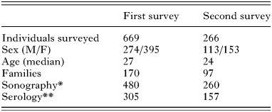
In total, 669 and 266 volunteers responded in the first and second survey respectively (Table 1). Data obtained from questionnaires in the first survey showed on the basis of family groups (n=170) that: 29% of the families practiced home slaughter; 51% consumed home-grown vegetables; 58% drank rural water and 81% of the families were dog owners, and all reported that dogs received Praziquantel, administered on a monthly basis by the Uruguayan Hydatid Disease Fund (“Comisión Honoraria de lucha contra La Hidatidosis”).
Table 1. Description of the study population (The number of individuals and families participating in both surveys is shown. The number of individuals screened by abdominal ultrasound (*) and serology (**) are indicated. In total, 58 sequential serum samples were obtained from voluntary donors participating in both surveys.)
A family history of CE was reported in 48% of the families (in the last 10 years), while 18% of the cases had occurred recently (in the last 5 years). All adult participants had a considerable degree of awareness concerning hydatid disease, and dog owners were acquainted with proper feeding procedures. The use of ‘water holes’ was no longer reported in the second survey; instead, collective deep wells had been constructed. No significant changes were reported with the other risk factors on the basis of 97 families surveyed.
Echographical prevalence
Groups of 480 and 260 individuals were screened by ultrasound imaging in the first and second survey respectively (Table 1). Ecographical prevalence (newly detected images) as determined in sequential surveys was 0·8% (4 individuals) and 1·1% (3 individuals) for either survey respectively. Individuals with presumptive cyst images belonged to different families and were studied clinically at the Regional Hospital.
Serological study
A study population of 305 and 157 serum samples was obtained in the first and second survey respectively. Results obtained from preliminary screening using the crude antigens HCFAg and PSAg, showed that 27 individuals exhibited significant levels of specific antibodies against one or both antigens in the absence of presumptive cyst images. Most seropositive individuals belonged to different families but 3 families exhibited 2 cases each. Questionnaire data from this group of individuals defined as the ‘ultrasound normal group’ was compared with that of the ‘seronegative group’ in order to correlate risk factors with the serological data. This study group comprised 21 women and 6 men of median age 32 and 29 respectively. Statistical analysis of data revealed a significant positive correlation between serological reactivity and certain risk factors: presence of family antecedent (P=0·005) and recent antecedents (P=0·0001) as well as the use of rural water (P<0·0001).
Further serological analysis of the ‘ultrasound normal group’ was performed through detection of antibodies specific to purified AgB, a more specific antigenic component expressed in E. granulosus (Maddison et al. 1989; González et al. 1996). Nine of the 27 sera exhibited reactivity against AgB (sera nos 1–8 and no. 27, Table 2). Reactivity was analysed simultaneously against antigens of T. hydatigena and T. canis. No significant correlations were found (AgB vs TxAg P=0·41; AgB vs ThAg P=0·16; TxAg vs ThAg, P=0·1; Chi-square test).
Table 2. Serological profile of the ‘ultrasound normal group’ (Results are indicated as +/− according to the cut-off value defined for each ELISA. All sera of the ‘ultrasound normal group’ (nos 1–27) were positive against Echinococcus granulosus crude antigens (EgAg) in the first survey. Sera nos 1–20 were also included in the second survey.)
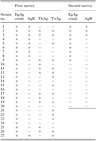
Sequential analysis of sera obtained from 20 out of 27 individuals of the ‘ultrasound normal group’, was made during the second survey (sera nos 1–20, see Table 2). Results showed that 9 sera (nos 1–9, Table 2) had remained reactive against the crude antigens while the rest had become negative.
All but 1 of the sera, having exhibited reactivity against AgB in the first survey, were included in the sequential analysis; 3 out of 8 sera analysed (nos 1–3, Table 2), had remained reactive against AgB while the other 5 sera were found to have become negative.
Image follow-up of this group showed that 1 individual (no. 4, Table 2) exhibited multiple calcified spots in the liver and spleen, in the second survey. These images were confirmed by CAT. No presumptive images were detected in the rest of the individuals.
DISCUSSION
Cystic echinococcosis is an important endemic disease among rural communities in Uruguay. Data obtained in this work confirm that CE still remains a public health hazard in the Department of Tacuarembó, as a local mean prevalence of 1% was found in two surveys carried out in 1998 and 2000 in the region of Caraguatá.
In asymptomatic carriers, liver cysts were found to grow very slowly – approximately 0·5 cm/year in diameter (Frider, Larrieu & Odriozola, 1999), thus single sonographic data values can not be used to evaluate the results of educational schemes in the short term.
The use of serology to assess exposure to parasite antigens was evaluated; thus constituting an indirect yet objective approach that may contribute to future assessment of the impact of control schemes. Serological data obtained from people without presumptive cyst images was studied in relation to risk factors. A group of individuals exhibiting no changes in abdominal sonography examination showed serological reactivity against E. granulosus antigens in accordance with results obtained in other rural communities of Uruguay (Bonifacino et al. 1991; Cohen et al. 1998) and other distant hyperendemic regions (Shambesh et al. 1999; Wang et al. 2001). A higher serological prevalence was obtained for this region (8·8%) when compared with the results of previous surveys in Uruguay (Bonifacino et al. 1991; Cohen et al. 1998) that could be attributed to the use of crude PSAg in addition to HCFAg. Crude protoscolex antigen was also used, since the expression of common antigens has been observed in oncospheres and larval stages of E. granulosus (Zhang et al. 2003) as well as with other helminths (Garcia-Allan et al. 1996).
The incidence of each risk factor was compared between the ‘ultrasound normal group’ (n=27) and the rest of people in the first survey. Individual analysis showed a statistically significant correlation (P<0·005) between the detection of antibodies specific to crude antigens, family antecedents, and the use of rural waters. Such relationship is relevant since the use of rural water had previously been correlated with CE prevalence in studies involving larger populations in other rural villages of Uruguay (Carmona et al. 1998). After 2 years, the absence of water holes and the consequent increase in the number of collective wells represent the most valuable improvement in sanitary conditions registered in the second survey. Follow-up analysis of the serological reactivity of the ‘ultrasound normal group’ was made, and each individual re-examined after 2 years, in an attempt to determine a predictive value of serological reactivity.
In the second survey, none of the individuals of the ‘ultrasound normal group’ had presumptive cyst images, except one individual exhibiting multiple calcified points in the liver and spleen, as both detected by sonography and confirmed by CAT Concomitant seroconvertion of specific IgG against AgB in this individual poses an interesting query, because AgB is only known to exist in hydatid fluid derived from fertile cysts, protoscoleces and germinative layer (Lightowlers & Gottstein, 1995) while no reactivity was observed against the other antigens tested. The presence of degenerated parasites in this individual might be suggested in a first speculative approach, as abortive lesions have been reported in patients with alveolar hydatidosis (Bresson-Hadni et al. 1994; Gottstein et al. 1996; Bartholomot et al. 2002). In addition, one possible role for antibodies in immunoresistance against E. granulosus was suggested by Rogan et al. (1992), though no updated evidence has thus far been made available.
Potential developing cysts could not be discounted in the rest of the individuals of this ‘ultrasound normal group’, because of the very slow growth rate of hydatid cysts and sonography sensitivity, so that preventive follow-up of these individuals is currently underway.
Antigens from other commonly occurring helminths in the region were taken in to account to study the significance of the reactivity observed against crude antigens.
T. hydatigena is a commonly occurring helminth parasite in Uruguayan dogs (Cabrera et al. 1996), with a higher probiotic potential (100-fold higher than E. granulosus) and a 4-fold higher number of eggs in the faeces of the co-infected dog (Gemmel & Roberts, 1995). These characteristics indicate that human exposure to oncospheres of T. hydatigena is putatively more frequent than exposure to E. granulosus.
Preliminary results showed a significant difference in the levels of specific IgG against T. hydatigena between communities living in Montevideo and those living in the rural area under the scope of this study (A. Hernández, personal communication); the level of antibodies against this non-pathogenic helminth may influence the definition of the cut-off and the design of diagnostic assays aimed for people living in rural areas.
Toxocara canis, is a commonly occurring helminth in both rural and urban areas in the region (Radman et al. 2000) and cross-reaction has been reported with E. granulosus (Bonifacino et al. 1991; Virginio et al. 2003).
Cross-reaction with T. solium and other species of Echinococcus is a major problem in CE immunodiagnosis (Rickard & Lightowlers, 1986; Kharebov, Nahmias & El-On, 1997), though in the case of Uruguay, infections with these parasites have not been reported. Cross-reacting antigens were taken into account in an attempt to discriminate between specific and non-specific response. The lack of a significant correlation suggested that cross-reaction alone does not account for the serological response observed, though a higher number of sera should be tested to confirm this idea.
Of the people that were seropositive (putatively exposed), approximately 60% became seronegative after two years. This profile of transient antibodies has been previously reported by García et al. (2001) with people living in endemic areas for Taenia solium; it has been suggested by the authors that people can develop only a transient serological antibody response after being exposed to parasites, without development of the disease or having self-cured infections. As antigen B was postulated to be genus specific (Maddison et al. 1989) it can be speculated that similar phenomena can occur with hydatid disease.
In conclusion, results of this work indicate that serology might be taken into account as an objective marker of environmental exposure of rural habitants. Further analysis of immunological parameters of individuals of the ‘ultrasound normal group’ is underway in our laboratory in an effort to contribute to the knowledge concerning natural and acquired resistance against cystic echinococcosis.
The authors thank the participation of people of rural communities for their invaluable cooperation. We are also particularly grateful to rural medical and nursing staff for assistance and support in the communities surveyed, Dr F. Alonso, Dr M. Vilan, D. Olivera, M. Correa, and M. Ferreira. We also thank the Instituto de Higiene for invaluable collaboration. This work was supported by PEDECIBA (Programa de desarrollo de las ciencias basiscas, PNUD; CSIC (Comisión Sectorial de Investigación Cientifica) and the Hospital Regional de Tacuarembó (Programa de Desarrollo de Atención Primaria en Salud, MSP-IMT-GTZ). The English version of the manuscript was revised by E. Speranza.
References
REFERENCES
BARBIERI, M.,
STERLA, S.,
BATTISTONI, J. &
NIETO, A. (
1993).
High performance latex reagent for hydatid serology using an Echinococcus granulosus lipoprotein antigen fraction purified from cyst fluid in one step.
International Journal for Parasitology 23,
565–572.CrossRefGoogle Scholar
BARTHOLOMOT, G.,
VUITTON, D.,
HARRAGA, S.,
SHI DA, Z.,
GIRAUDOUX, P.,
BARNISH, G.,
WANG, Y.,
MacPHERSON, C. &
CRAIG, P. (
2002).
Combined ultrasound and serologic screening for hepatic alveolar echinococcosis in central China.
American Journal of Tropical Medicine and Hygiene 66,
23–29.CrossRefGoogle Scholar
BONIFACINO, R.,
MALGOR, R.,
BARBEITO, R.,
BALLESTE, R.,
RODRIGUEZ, M.,
BOTTO, C. &
KLUG, F. (
1991).
Seroprevalence of Echinococcus granulosus infection in a Uruguayan rural human population.
Transactions of the Royal Society of Tropical Medicine and Hygiene 85,
769–772.CrossRefGoogle Scholar
BRESSON-HADNI, S.,
PETITJEAN, O.,
MONNOT-JACQUARD, B.,
HEYD, B.,
KANTELIP, B.,
DESCHASEAUX, M.,
RACADOT, E. &
VUITTON, D. A. (
1994).
Cellular localization of interleukin-1β, interleukin-6 and tumor necrosis factor-α mRNA in a parasitic granulomatous disease of the liver, alveolar echinococcosis.
European Cytokine Network 5,
461–468.Google Scholar
CABRERA, P. A.,
PARIETTI, S.,
HARAN, G.,
BENAVIDES, U.,
LLOYD, S.,
PERERA, G.,
VALLEDOR, S.,
GEMMEL, M. A. &
BOTTO, C. (
1996).
Rates of reinfection with Echinococcus, Taenia hydatigena, Taenia ovis and other cestodes in a rural dog population in Uruguay.
International Journal for Parasitology 26,
79–83.CrossRefGoogle Scholar
CARMONA, C.,
PERDOMO, R.,
CARBO, A.,
ALVAREZ, C.,
MONTI, J.,
GRAUERT, R.,
STERN, D.,
PERERA, G.,
LLOYD, S.,
BAZINI, R.,
GEMMEL, M. &
YARZABAL, L. (
1998).
Risk factors associated with human cystic echinococcosis in Florida, Uruguay: results of a mass screening study using ultrasound and serology.
American Journal of Tropical Medicine and Hygiene 58,
599–605.CrossRefGoogle Scholar
COHEN, H.,
PAOLILLO, E.,
BONIFACINO, E.,
BOTTA, B.,
PARADA, L.,
CABRERA, P.,
SNOWDEN, K.,
GASSER, R.,
TESSIER, E.,
DIBARBOURE, L.,
WEN, H.,
ALLAN, J.,
SOTO DE ALFARO, H.,
ROGAN, M. &
CRAIG, P. (
1998).
Human cystic echinococcosis in a Uruguayan community: a sonographic, serologic, and epidemiologic study.
American Journal of Tropical Medicine and Hygiene 59,
620–627.CrossRefGoogle Scholar
CRAIG, P. S.,
ROGAN, M. T. &
ALLAN, J. C. (
1996).
Detection, screening and community epidemiology of taeniid cestode zoonoses: cystic echinococcosis, alveolar echinococcosis and neurocysticercosis.
Advances in Parasitology 38,
196–250.CrossRefGoogle Scholar
FRIDER, B.,
LARRIEU, E. &
ODRIOZOLA, M. (
1999).
Long-term outcome of asymptomatic liver hydatidosis.
Journal of Hepatology 30,
228–231.CrossRefGoogle Scholar
GARCÍA, H.,
GONZALEZ, A.,
GILMAN, R.,
PALACIOS, L.,
JIMÉNEZ, I.,
RODRÍGUEZ, S.,
VARASTEGUI, M.,
WILKINS, P.,
TSANG, V. &
THE CISTICERCOSIS WORKING GROUP IN PERU (
2001).
Short report: Transient antibody response in Taenia solium infection in field conditions – A mayor contributor to high seroprevalence.
American Journal of Tropical Medicine and Hygiene 65,
31–33.CrossRefGoogle Scholar
GARCÍA-ALLAN, C.,
MARTÍNEZ, N.,
FLISSER, A.,
ALUJA, A.,
ALLAN, J. C. &
CRAIG, P. S. (
1996).
Immunocharacterization of Taenia solium oncosphere and metacestode antigens.
Journal of Helminthology 70,
271–280.CrossRefGoogle Scholar
GEMMELL, M. A. &
ROBERTS, M. G. (
1995).
Modelling E. granulosus life cycles. In
Echinococcus and Hydatid disease (
ed. Thompson, R. C. A. & Lymbery, A. J.), pp.
333–351.
CAB International,
Wallingford,
UK.
GONZÁLEZ, G.,
NIETO, A.,
FERNÁNDEZ, C.,
ORN, A.,
WERNSTEDT, C. &
HELLMAN, U. (
1996).
Two different 8 kDa monomers are involved in the oligomeric organization of the native Echinococcus granulosus antigen B.
Parasite Immunology 18,
587–596.CrossRefGoogle Scholar
GOTTSTEIN, B.,
BETTENS, F.,
PARKINSON, A. &
WILSON, F. (
1996).
Immunological parameters associated with susceptibility or resistance to alveolar hydatid diseas in Yupiks/Inupiats.
Arctic Medical Research 55,
14–19.Google Scholar
KHAREBOV, A.,
NAHMIAS, J. &
EL-ON, J. (
1997).
Cellular and humoral immune responses of hydatidosis patients to Echinococcus granulosus purified antigens.
American Journal of Tropical Medicine and Hygiene 57,
619–625.CrossRefGoogle Scholar
LIGHTOWLERS, M. W. &
GOTTSTEIN, B. (
1995).
Echinococcosis/hydatidosis: antigens, immunological and molecular diagnosis. In
Echinococcus and Hydatid Disease (
ed. Thompson, R. C. A. & Lymbery, A. J.), pp.
355–393.
CAB International,
Wallingford,
UK.
MADDISON, S. R.,
SLEMENDA, S.,
SCHANTZ, P. M.,
FRIED, J. A.,
WILSON, M. &
TSANG, V. C. (
1989).
A specific diagnostic antigen of Echinococcus granulosus with an apparent molecular weight of 8 kDa.
American Journal of Tropical Medicine and Hygiene 40,
377–383.CrossRefGoogle Scholar
MÍGUEZ, M.,
BAZ, A. &
NIETO, A. (
1996).
Carbohydrates on the surface of Echinococcus granulosus protoscoleces are immunodominant in mice.
Parasite Immunology 18,
559–569.CrossRefGoogle Scholar
PERDOMO, R.,
PARADA, R.,
ALVAREZ, C.,
CATTIVELLI, D.,
GENINAZZI, H.,
BARRAGUE, A. D.,
FERREIRA, C.,
RIVERO, E.,
MONTI, J. &
PARADA, J. (
1990).
Estudio epidemiologico de hidatidosis. Detección precoz por ultrasonido en zonas de alto riesgo.
Revisión Medica Uruguaya 6,
34–47.Google Scholar
RADMAN, N. E.,
ARCHELLI, S. M.,
FONROUGE, R. D.,
DEL V GUARDIS, M. &
LINZITTO, O. R. (
2000).
Human toxocariosis: its seroprevalence in the City of La Plata.
Memorias del Instituto Oswaldo Cruz 95,
281–285.CrossRefGoogle Scholar
RICKARD, M. D. &
LIGHTOWLERS, M. W. (
1986).
Immunodiagnosis of hydatid disease. In
The Biology of Echinococcus and Hydatid Disease (
ed. Thompson, R. C. A.), pp.
217–249.
George Allen & Unwin,
London,
UK.
ROGAN, M. T.,
CRAIG, P.,
ZEHYLE, E.,
MASINDE, G.,
WEN, H. &
ZHOU, P. (
1992).
In vitro killing of taeniid oncospheres, mediated by human sera from hydatid endemic areas.
Acta Tropica 51,
291–296.CrossRefGoogle Scholar
SHAMBESH, M. A.,
CRAIG, P. S.,
MacPHERSON, C. N. L.,
ROGAN, M. L.,
GUSBI, A. M. &
ECHTUISH, E. F. (
1999).
An extensive ultrasound and serologic study to investigate the prevalence of human cystic echinococcosis in northern Libya.
American Journal of Tropical Medicine and Hygiene 60,
462–468.CrossRefGoogle Scholar
VIRGINIO, V.,
HERNÁNDEZ, A.,
ROTT, M.,
MONTEIRO, A.,
ZANDONA, A.,
NIETO, A.,
ZAHA, A. &
FERREIRA, H. B. (
2003).
A set of recombinant antigens from Echinococcus granulosus with potential for use in the immunodiagnosis of human cystic hydatid disease.
Clinical and Experimental Immunology 132,
309–315.CrossRefGoogle Scholar
WANG, Y.,
ROGAN, M.,
VUITTON, D.,
WEN, H.,
BARTHOLOMOT, B.,
McPHERSON, C.,
ZOU, P.,
DING, Z.,
ZHOU, H.,
ZHANG, F.,
LUO, J.,
XIONG, H.,
FU, Y.,
McVIE, A.,
GIRAUDOUX, P.,
YANG, W. &
CRAIG, P. (
2001).
Cystic echinococcosis in semi-nomadic pastoral communities in north-west China.
Transactions of the Royal Society of Tropical Medicine and Hygiene 95,
153–158.CrossRefGoogle Scholar
ZHANG, W.,
LI, J.,
YOU, H.,
ZHANG, Z.,
TURSON, G.,
LOUKAS, A. &
McMANUS, D. P. (
2003).
Short report: Echinococcus granulosus from Xinjiang, PR China; cDNAs encoding the EG95 vaccine antigen are expressed in different life cycle stages and are conserved in the oncosphere.
American Journal of Tropical Medicine and Hygiene 68,
40–43.Google Scholar
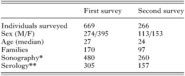
Table 1. Description of the study population
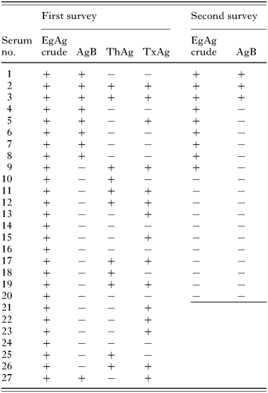
Table 2. Serological profile of the ‘ultrasound normal group’
 You have
Access
- 21
- Cited by