Introduction
Human American and African trypanosomiases are major tropical diseases, infecting millions of people every year, and yet are widely neglected worldwide (Kennedy, Reference Kennedy2013; Bern, Reference Bern2015). The haemoflagellate protozoan Trypanosoma cruzi causes the American trypanosomiasis or Chagas disease, which is transmitted primarily through contact with feces of haematophagous triatomine insects (Chagas, Reference Chagas1909; Santana et al., Reference Santana, Souza, Lima, Araújo, Justiniano, Dantas and Melo2012). The highest disease incidence is in Latin America (Pan American Health Organization, 2006; Pan American Health Organization, 2014). Transmission of parasites also occurs through non-vector pathways such as blood transfusion (Moraes-Souza and Ferreira-Silva, Reference Moraes-Souza and Ferreira-Silva2011), organ transplantation (Márquez et al., Reference Márquez, Crespo, Mir, Pérez-Sáez, Quintana, Barbosa and Pascual2013), laboratory accidents (Dias, Reference Dias2006) and by ingestion of contaminated food (Shikanai-Yasuda and Carvalho, Reference Shikanai-Yasuda and Carvalho2012; Domingues et al., Reference Domingues, Hardoim, Souza, Cardoso, Mendes, Previtalli-Silva, Abreu-Silva, Pelajo-Machado, da Costa and Calabrese2015).
During its life cycle, T. cruzi is usually found in three different infective forms: epimastigotes, trypomastigotes and amastigotes (Chagas, Reference Chagas1909; Tyler and Engman, Reference Tyler and Engman2001). As an epimastigote, the parasite replicates inside the insect vector stomach, and later in the intestine, thus becoming a trypomastigote. The latter infects the host animal and moves through the bloodstream to infect different tissues. Trypomastigotes then change into amastigotes and begin to multiply (Elias et al., Reference Elias, Marques-Porto, Freymüller and Schenkman2001; Tyler and Engman, Reference Tyler and Engman2001). Eventually, some parasites return to their trypomastigote form to move through the bloodstream and infect new organs, which is when a haematophagous insect can feed on contaminated blood and repeat the infectious process (Chagas, Reference Chagas1909; Tyler and Engman, Reference Tyler and Engman2001; Minning et al., Reference Minning, Weatherly, Atwood, Orlando and Tarleton2009).
The signs and symptoms caused by Chagas disease have two successive stages: acute and chronic (Rassi et al., Reference Rassi, Rassi and Marin-Neto2010). The acute phase usually last from 2 to 4 weeks. The main symptoms are redness, oedema and fever (Coura and Viñas, Reference Coura and Viñas2010; Rassi et al., Reference Rassi, Rassi and Marin-Neto2010). After this phase, the parasite number in the bloodstream usually decreases and the symptoms disappear, causing a latency state that lasts indefinitely (Prata, Reference Prata2001). In some cases, the disease develops, originates the chronic phase, characterized by tissue damage in parasite multiplication. Heart, brain, oesophagus and colon are the most affected tissues showing necrosis and physiological impairments (Coura and Dias, Reference Coura and Dias2009; Rassi et al., Reference Rassi, Rassi and Marin-Neto2010). Chagas disease is the leading cause of non-ischaemic cardiomyopathy worldwide (Feldman and McNamara, Reference Feldman and McNamara2000) due to cardiac chambers enlargement, congestive heart failure, dysrhythmias and thromboembolism (Marin-Neto et al., Reference Marin-Neto, Cunha-Neto, Maciel and Simões2007; Bern, Reference Bern2015).
In addition to humans, T. cruzi parasitosis has been identified in a wide variety of domestic and wild mammals, such as bats, anteaters, marsupials, carnivorous, rodents, rabbits, primates, pigs and caprines (Barretto, Reference Barretto1964; Deane, Reference Deane1964; Añez et al., Reference Añez, Crisante and Soriano2009; Herrera, Reference Herrera2010). Dogs are their main domestic reservoir (Montenegro et al., Reference Montenegro, Jiménez, Dias and Zeledón2002), whereas bats were recently shown to be an important wild reservoir, with a hypothesis that states that T. cruzi has a long history of co-evolution shared with bats (Stevens et al., Reference Stevens, Noyes, Dover and Gibson1999; Hamilton et al., Reference Hamilton, Teixeira and Stevens2012). Other groups of vertebrates are refractory to T. cruzi due to antigenic incompatibility, lack of recognition and/or cell signalling or inadequate levels of systemic temperature (Herrera, Reference Herrera2010). Experimental studies show that infected hosts with different T. cruzi strains show different parasite levels and immune responses according to parasite–host evolutionary interactions and to a wide variety of parasite genotypes (Roellig et al., Reference Roellig, McMillan, Ellis, Vandeberg, Champagne and Yabsley2010; Duz et al., Reference Duz, Vieira, Roatt, Aguiar-Soares, Cardoso, Oliveira, Reis, Tafuri, Veloso, Reis and Carneiro2014).
An effective way to evaluate the health of an individual infected with T. cruzi is by using haematological parameters analysis (Beldomenico et al., Reference Beldomenico, Telfer, Gebert, Lukomski, Bennett and Begon2008; Stockham and Scott, Reference Stockham and Scott2013) through morphological and quantitative analysis. In addition, haemoglobin concentration can diagnose anaemia produced by blood loss, haematopoiesis suppression or bone marrow pathologies (Van Hove et al., Reference Van Hove, Schisano and Brace2000). White blood cells or leucocytes are the performers of the immune response, thus intervening in the defence of the body against foreign substances or infectious agents (Teske, Reference Teske, Weiss and Wardrop2011). Alterations in the number of leucocytes usually reflect virus, bacteria or protozoa infections, and allergic reactions to medications, bone marrow damage, cancer or autoimmune disorders (Stockham and Scott, Reference Stockham and Scott2013). Among them, neutrophils are the main immune cells that fight the pathogens at infection site, where they recognize and eliminate the invading microorganisms through cytotoxic mechanisms (Mayadas et al., Reference Mayadas, Cullere and Lowell2014). Lymphocytes are the effectors of innate and acquired immunity, and they proliferate in response to antigenic stimuli. Therefore, circulating levels of lymphocytes may be used as indicators of the immunological response (Beldomenico et al., Reference Beldomenico, Telfer, Gebert, Lukomski, Bennett and Begon2008; Dotiwala et al., Reference Dotiwala, Mulik, Polidoro, Ansara, Burleigh, Walch, Gazzinelli and Lieberman2016). Monocytes eliminate invading microorganisms or cellular debris by phagocytosis. The number of peripheral monocytes is usually increased during subacute and chronic inflammatory responses caused by bacterial and protozoa infections (Weiss and Souza, Reference Weiss, Souza, Weiss and Wardrop2011). Eosinophils are multifunctional effector cells with proinflammatory and destructive abilities implicated in tissue homeostasis, modulation of the adaptive immune response and innate immunity to certain microorganisms (Kita, Reference Kita2011), whereas basophils release chemical mediators that alter the vascular structure and recruit other inflammatory cells to the site of infection (Obata et al., Reference Obata, Mukai, Tsujimura, Ishiwata, Kawano, Minegishi and Karasuyama2007). Therefore, total leucocyte counting is important to the immunological status as their numerical and morphological variation may reflect nutritional deficiencies, functional bone marrow changes and works as an important marker for parasitic infections (Stockham and Scott, Reference Stockham and Scott2013).
Studies with experimental animal models provide the empirical basis for determining the major haematological parameters usually analysed in parasitic infections (Cardoso and Brener, Reference Cardoso and Brener1980; Pung et al., Reference Pung, Hulsebos and Kuhn1988). However, as preclinical studies usually involve small, heterogeneous experiments, their findings sometimes cannot contribute directly for improvements on human health (Hooijmans and Ritskes-Hoitinga, Reference Hooijmans and Ritskes-Hoitinga2013; Van Luijk et al., Reference Van Luijk, Bakker, Rovers, Ritskes-Hoitinga, de Vries and Leenaars2014). A systematic review is intended to show preclinical evidence in an objective manner, unlike widely used narrative reviews (Uman, Reference Uman2011). To our knowledge, this is the first study to provide new, reliable evidence on the major haematological alterations during the development of T. cruzi infection in different species. These findings may increase the knowledge of Chagas disease signs and have a potential value to understand the relevance of the host–parasite interactions, in order to identify a pattern of haematological alteration and possible species-specific differences (Olifiers et al., Reference Olifiers, Jansen, Herrera, De Cassia Bianchi, D'Andrea, De Miranda Mourão and Gompper2015). We also aimed at investigating the relevance of these alterations on the infection diagnosis during the initial asymptomatic phase, as well as determining the main T. cruzi strains, type of infections, inoculum and inoculation routes used in studies on American trypanosomiasis, and the methodological bias found in the selected studies.
Material and methods
The systematic review was adhered to the PRISMA guidelines (Kilkenny et al., Reference Kilkenny, Browne, Cuthill, Emerson and Altman2010) (Preferred Reporting Items for Systematic Reviews and Meta-Analysis), including search strategy, selection criteria, extraction and data analysis.
Literature research
We completed a comprehensive bibliographic search using the electronic database PubMed (https://www.ncbi.nlm.nih.gov/pubmed) and Scopus (https://www.scopus.com/home.uri) on 31 August 2016 at 8:00 p.m. The keywords for filters for three criteria were Chagas disease, haematology and animal model (Supplementary file S1). The hierarchical distribution of the MeSH terms was the strategy used to develop the filter on the PubMed platform. We applied a standardized filter in the Scopus platform for animal studies and the same PubMed search strategy was adapted and used. We only included experimental studies published in English, Portuguese and Spanish. Views, comments, notes and unpublished studies were not included.
We selected only studies that met all of the eligibility criteria listed below:
(1) Studies on mammals experimentally or naturally infected with T. cruzi;
(2) Studies with at least one control group infected with T. cruzi and not receiving any type of treatment (e.g. drug treatment);
(3) Experimental subjects with no functional alterations due to lesions or other manipulations, and non-genetically modified;
(4) Studies in which at least two of the major haematological parameters of erythrocytes (total erythrocyte counting, haematocrit and haemoglobin dosage) and at least two leucocytes (total and differential leucocyte counting) were measured.
Extraction and data management
Selection by abstract: two independent reviewers (E.V. and D.L.J.C.) selected eligible studies based on title and abstract analysis. In the case of disagreements, another group of reviewers (R.V.G., M.B.F., M.M.S. and R.D.N.) decided whether the study fulfilled the inclusion and exclusion criteria.
To discard the subjectivity in the data collection and selection process, the information was independently extracted by four reviewers (R.V.G., M.B.F., M.M.S. and R.D.N.) and analysed separately. Data from each study were extracted and tabulated using standardized information such as publication characteristics (title, author, year and country); experimental model (animal species, sex, age, weight, number of animals and experimental groups); infection characteristics (T. cruzi strain, inoculation route, inoculum quantity, time of evolution of parasitaemia) and haematological parameters (haemoglobin, haematocrit, total erythrocyte counting, leucocytes, lymphocytes, neutrophils, monocytes, eosinophils and basophils). We asked the authors, by email, to provide the papers in cases that they were not easily available. Data were subsequently compared and conflicting information were identified and corrected after discussion among the reviewers.
Bias analysis
The quality of the articles was analysed according to the criteria described in the research ‘Animal Research Reporting of in Vivo Experiments’ (ARRIVE). These criteria are based on brief descriptions of the essential characteristics of all studies using animal models such as theoretical and methodological basis, research objective, improvement of analytical methods, statistical design, sample calculations and result measures (Kilkenny et al., Reference Kilkenny, Browne, Cuthill, Emerson and Altman2010). Considering that this systematic review aimed at evaluating important aspects of the publications referenced, we developed a table summarizing all investigated aspects as well as their relevance, in addition to describing positive and negative aspects of the retrieved studies.
Results
Included studies
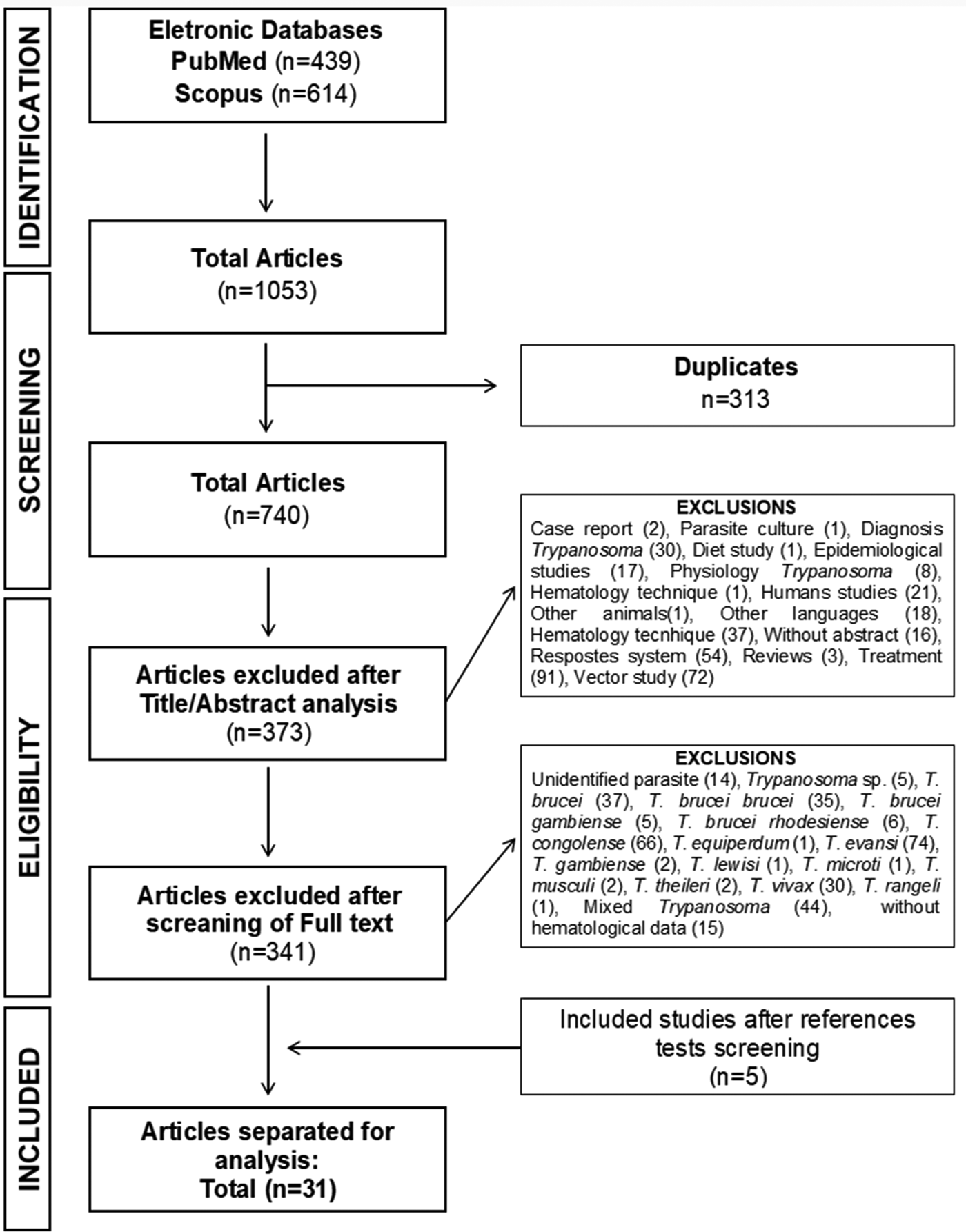
The PRISMA diagram illustrates the selection process of the studies (Fig. 1). The initial research resulted in 1053 studies; 313 were duplicates. After reading the title and abstract, we excluded 373 studies with subjects not related to the chosen topic. From these, 72 studies were related to Trypanosoma vectors, 59 were excluded for not addressing haematological aspects, 18 were not in English, Portuguese or Spanish and three consisted of non-original (revision) research articles. After this initial selection, the remaining 367 articles were then completely read and 341 articles were excluded for using other Trypanosoma species (312), other types of pathogens (14) and without haematological data (15). Therefore, 26 studies were included in this systematic review. The bibliographical references of the 26 selected articles were analysed, and five studies were added in accordance with the inclusion criteria, which brought us to a total of 31 studies (Fig. 1).
PRISMA diagram. Different phases of selection of studies for conducting qualitative and quantitative analyses. Flow diagram of the systematic review literature search results. Based on ‘Preferred Reporting Items for Systematic Reviews and Meta-Analyses: The PRISMA Statement’. http://www.prisma-statement.org. From: Moher D, Liberati A, Tetzlaff J, Altman DG, The PRISMA Group (2009).

Qualitative and quantitative analysis
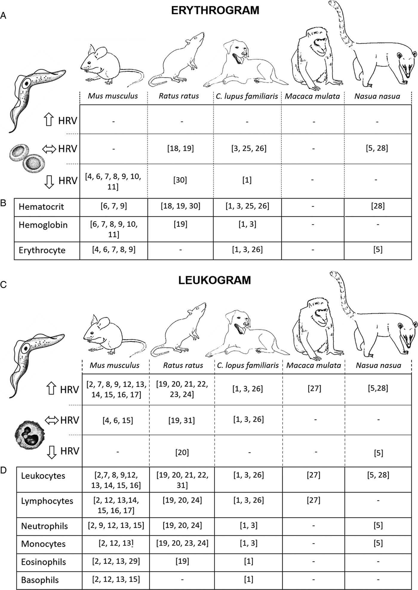
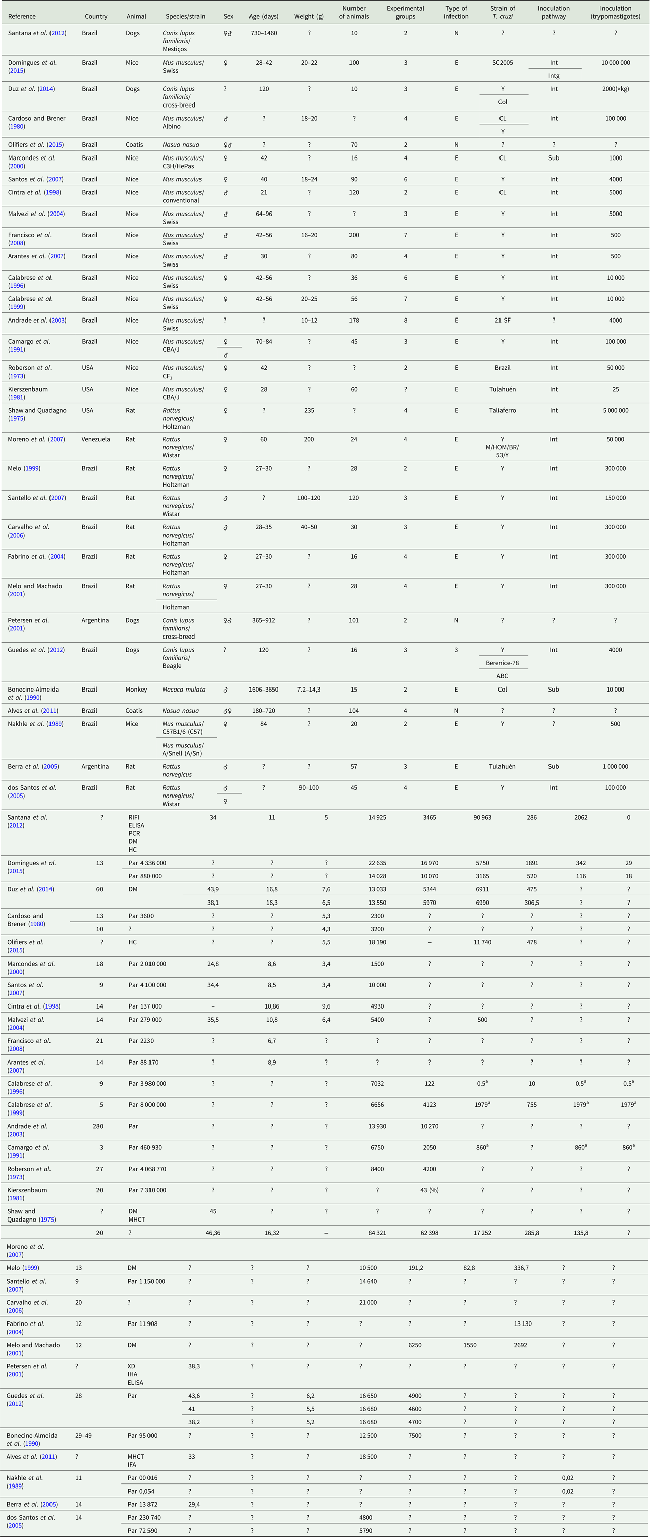
The selected studies were performed in four different countries, most of them in Brazil (n = 25) followed by the USA (n = 3). The most used animals were mice (48%), followed by rats (29%). The most used T. cruzi strains were Y (54%) followed by CL (10%), although 12.9% of the studies do not specify this in their articles. Tests to confirm infection (parasitological characterization) were not described in 6.5% of the studies; the route of inoculation was not provided in 19.35% and the inoculum concentration is absent in 12.9% of them (Table 1). The results for the different haematological parameters varied according to the animal species. For mice, there was a decrease in erythrocyte values (Cardoso and Brener, Reference Cardoso and Brener1980; Cintra et al., Reference Cintra, Silva, Silva, Silva, Afonso, Nicoli, Bambirra and Vieira1998; Marcondes et al., Reference Marcondes, Borelli, Yoshida and Russo2000; Malvezi et al., Reference Malvezi, Cecchini, de Souza, Tadokoro, Rizzo and Pinge-Filho2004; Santos et al., Reference Santos, Levy, Toldo, Azevedo and Júnior2007), haematocrit (Marcondes et al., Reference Marcondes, Borelli, Yoshida and Russo2000; Malvezi et al., Reference Malvezi, Cecchini, de Souza, Tadokoro, Rizzo and Pinge-Filho2004; Santos et al., Reference Santos, Levy, Toldo, Azevedo and Júnior2007) and haemoglobin (Cintra et al., Reference Cintra, Silva, Silva, Silva, Afonso, Nicoli, Bambirra and Vieira1998; Marcondes et al., Reference Marcondes, Borelli, Yoshida and Russo2000; Malvezi et al., Reference Malvezi, Cecchini, de Souza, Tadokoro, Rizzo and Pinge-Filho2004; Arantes et al., Reference Arantes, Pedrosa, Martins, Veloso, de Lana, Bahia, Tafuri and Carneiro2007; Santos et al., Reference Santos, Levy, Toldo, Azevedo and Júnior2007; Francisco et al., Reference Francisco, de Abreu Vieira, Arantes, Pedrosa, Martins, Silva, Velosa, de Lana, Bahia, Tafuri and Carneiro2008) and a significant increase in leucocyte levels (Roberson et al., Reference Roberson, Hanson and Chapman1973; Camargo et al., Reference Camargo, Araujo, Sakurada, Stach-machado, Rancel and Silva1991; Calabrese et al., Reference Calabrese, Lagrange and Da Costa1996; Cintra et al., Reference Cintra, Silva, Silva, Silva, Afonso, Nicoli, Bambirra and Vieira1998; Calabrese et al., Reference Calabrese, Paradela, do Valle, Tedesco, Silva, Mortara and da Costa1999; Andrade et al., Reference Andrade, Mesquita, Jambeiro, Santos and Portella2003; Malvezi et al., Reference Malvezi, Cecchini, de Souza, Tadokoro, Rizzo and Pinge-Filho2004; Santos et al., Reference Santos, Levy, Toldo, Azevedo and Júnior2007; Domingues et al., Reference Domingues, Hardoim, Souza, Cardoso, Mendes, Previtalli-Silva, Abreu-Silva, Pelajo-Machado, da Costa and Calabrese2015) mainly in the lymphocyte proportion (Roberson et al., Reference Roberson, Hanson and Chapman1973; Kierszenbaum, Reference Kierszenbaum1981; Calabrese et al., Reference Calabrese, Paradela, do Valle, Tedesco, Silva, Mortara and da Costa1999; Andrade et al., Reference Andrade, Mesquita, Jambeiro, Santos and Portella2003; Domingues et al., Reference Domingues, Hardoim, Souza, Cardoso, Mendes, Previtalli-Silva, Abreu-Silva, Pelajo-Machado, da Costa and Calabrese2015), neutrophils (Camargo et al., Reference Camargo, Araujo, Sakurada, Stach-machado, Rancel and Silva1991; Calabrese et al., Reference Calabrese, Paradela, do Valle, Tedesco, Silva, Mortara and da Costa1999; Domingues et al., Reference Domingues, Hardoim, Souza, Cardoso, Mendes, Previtalli-Silva, Abreu-Silva, Pelajo-Machado, da Costa and Calabrese2015), monocytes (Calabrese et al., Reference Calabrese, Paradela, do Valle, Tedesco, Silva, Mortara and da Costa1999; Domingues et al., Reference Domingues, Hardoim, Souza, Cardoso, Mendes, Previtalli-Silva, Abreu-Silva, Pelajo-Machado, da Costa and Calabrese2015) and eosinophils (Camargo et al., Reference Camargo, Araujo, Sakurada, Stach-machado, Rancel and Silva1991; Calabrese et al., Reference Calabrese, Paradela, do Valle, Tedesco, Silva, Mortara and da Costa1999; Domingues et al., Reference Domingues, Hardoim, Souza, Cardoso, Mendes, Previtalli-Silva, Abreu-Silva, Pelajo-Machado, da Costa and Calabrese2015) when compared with the standard values for these species (Supplementary file S2). In infected rats, the haematocrit (Shaw and Quadagno, Reference Shaw and Quadagno1975; Malvezi et al., Reference Malvezi, Cecchini, de Souza, Tadokoro, Rizzo and Pinge-Filho2004) and haemoglobin (Moreno et al., Reference Moreno, Araujo, Alarcon, Lugo, Moreno and Borges2007) values remained within the normal range described for these species. However, the number of leucocytes (Melo, Reference Melo1999; Carvalho et al., Reference Carvalho, Camargos, Almeida, Peluzio, Alvarez-Leite, Chiari and Reis2006; Moreno et al., Reference Moreno, Araujo, Alarcon, Lugo, Moreno and Borges2007; Santello et al., Reference Santello, Frare, Santos, Toldo, Kawasse, Zucoloto and Prado2007) had a significant increase when compared with the reference values, with special emphasis on monocytes (Melo, Reference Melo1999; Melo and Machado, Reference Melo and Machado2001; Fabrino et al., Reference Fabrino, Leon, Parreira, Genestra, Almeida and Rossana2004; Moreno et al., Reference Moreno, Araujo, Alarcon, Lugo, Moreno and Borges2007) (Table 1 and Supplementary file S2). When dogs were used as model, erythrocyte, haematocrit and haemoglobin levels remained within normal reference values after T. cruzi infection (Petersen et al., Reference Petersen, Gürtler, Cecere, Rubel, Lauricella, Hansen and Carlomagno2001; Guedes et al., Reference Guedes, Veloso, Mineo, Santiago-Silva, Crepalde, Caldas and Bahia2012; Duz et al., Reference Duz, Vieira, Roatt, Aguiar-Soares, Cardoso, Oliveira, Reis, Tafuri, Veloso, Reis and Carneiro2014). Similarly, leucocytes remained in the normal range for dogs, with increases only in the proportions of lymphocytes and eosinophils (Guedes et al., Reference Guedes, Veloso, Mineo, Santiago-Silva, Crepalde, Caldas and Bahia2012; Duz et al., Reference Duz, Vieira, Roatt, Aguiar-Soares, Cardoso, Oliveira, Reis, Tafuri, Veloso, Reis and Carneiro2014). In our selection, we found a study with monkeys (Bonecine-Almeida et al., Reference Bonecine-Almeida, Galvão-Castro, Pessoa, Piramez and Laranja1990) and two with coatis (Alves et al., Reference Alves, Olifiers, Bianchi, Duarte, Cotias, D'Andrea and Jansen2011; Olifiers et al., Reference Olifiers, Jansen, Herrera, De Cassia Bianchi, D'Andrea, De Miranda Mourão and Gompper2015). The study with monkeys showed a significant increase in the number of leucocytes, especially lymphocytes, with values above the reference reported for primates (Table 2). In coatis, the authors reported an increase in the amount of leucocytes (Alves et al., Reference Alves, Olifiers, Bianchi, Duarte, Cotias, D'Andrea and Jansen2011; Olifiers et al., Reference Olifiers, Jansen, Herrera, De Cassia Bianchi, D'Andrea, De Miranda Mourão and Gompper2015) with slightly elevated values for neutrophils (Olifiers et al., Reference Olifiers, Jansen, Herrera, De Cassia Bianchi, D'Andrea, De Miranda Mourão and Gompper2015) and lower values for monocytes (Olifiers et al., Reference Olifiers, Jansen, Herrera, De Cassia Bianchi, D'Andrea, De Miranda Mourão and Gompper2015) (Table 1 and Supplementary file S2). In general, 50% of the studies measuring haematological parameters associated with erythrocytes showed erythropenia (Fig. 2a and b), and 68.2% of the studies that estimated leucocyte values showed leucocytosis. In 25.8% of the studies, the parasitaemia level was not shown. Figure 2c and d schematically shows all the results of haematological parameters analysed in the different non-human mammals infected with T. cruzi mentioned above.
Schematic representation of studies in different species showing fluctuations in the haematological reference values (HRV), according to total erythrogram (a), erythrogram-specific parameters (b), total leukogram (c), leukogram-specific parameters (d) in non-human mammals infected with Trypanosoma cruzi.

Qualitative descriptors of the studies of the haematological parameters in non-human mammals infected with Trypanosoma cruzi

a Total number of polymorphonuclear leucocytes (neutrophils, eosinophils and basophils). ♂, Male; ♀, female;?, uninformed; N, natural; E, experimental; Sub, subcutaneous; Int, intraperitoneal; Intg, intragastric; RIFI, indirect immunofluorescence test; ELISA, enzyme-linked immuno sorbent assay; PCR, polymerase chain reaction; DM, direct microscopy; HC, haemoculture; Par, parasitaemia; MHCT, micro centrifuge technique; XD, xenodiagnosis; IHA, indirect haemagglutination test.
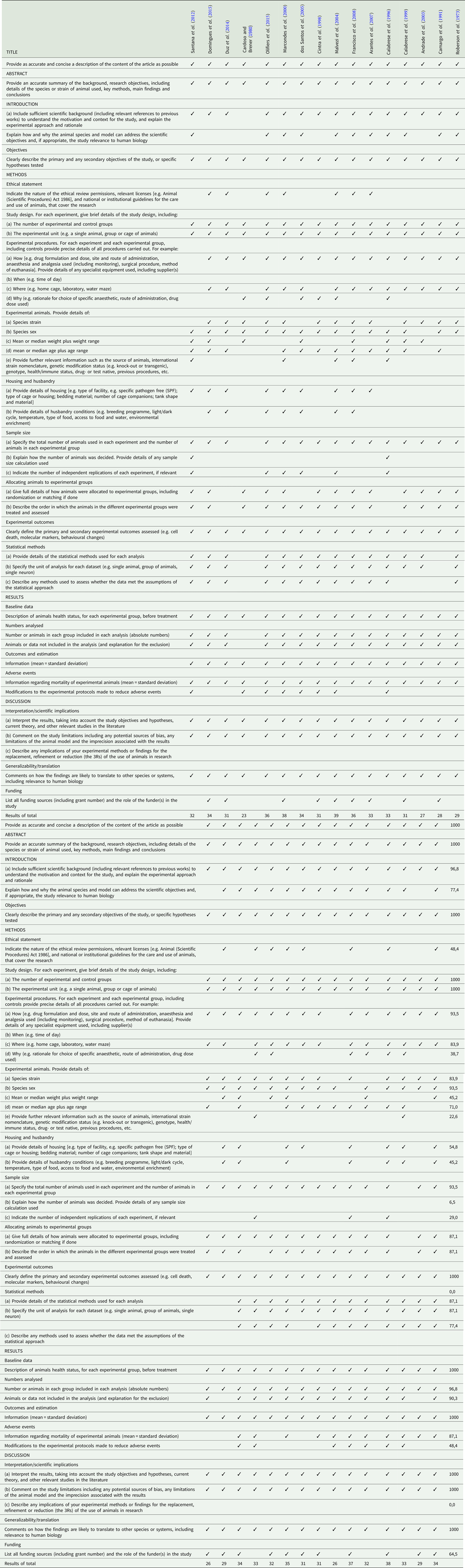
Biases analyses (ARRIVE) of the haematological parameters in non-human mammals infected with T rypanosoma cruzi

Bias analysis
All studies showed a title coherent with the content and abstracts containing literature review, objectives, methods, main results and final remarks. Ethics committee approval was reported in 48.3%. Experimental design is shown in 87% (n = 27); however, none of them reported measures to minimize bias. Regarding the experimental animal, all studies reported the species name. Sex, weight and age were reported in 93.5% (n = 29), 45.1% (n = 14) and 70.9% (n = 22) of the studies, respectively. The detailed description of the statistical analysis was reported in 87.0% (n = 27). Only 15 studies reported modifications to the experimental protocol by adverse events. No study reported on the decision about the number of animals used (Table 2).
Discussion
In this study, we conducted a systematic review to identify the main haematological parameters analysed in non-human mammals infected with T. cruzi and evaluated the possible relevance of these analyses to study the development of Chagas disease. Despite the great heterogeneity of the studies, in general, the presence of T. cruzi in the organisms promoted no changes or a decrease in blood erythrocytes and an increase in leucocytes, which may represent anaemia and severe infection. Studies in humans have shown that some patients diagnosed in the acute phase developed the indeterminate chronic form of Chagas disease, which is asymptomatic and does not require treatment, as it may remain stable for 20 or more years (Maya et al., Reference Maya, Orellana, Ferreira, Kemmerling, López-Muñoz and Morello2010; Rassi et al., Reference Rassi, Rassi and Marin-Neto2010). An early diagnose, however, which might be facilitated by alterations in the haematological patterns, may prevent the further development of the disease. In contrast, a delayed diagnose, confirmed only in the chronic phase, may cause irreversible lesions in several organs and systems (Rassi et al., Reference Rassi, Rassi and Marin-Neto2010; Martins-Melo et al., Reference Martins-Melo, Ramos, Alencar and Heukelbach2012).
The detection of parasites in the peripheral blood, as well as serological tests, direct microscopy, blood culture, polymerase chain reaction and immunological tests (Gascón et al., Reference Gascón, Albajar, Cañas, Flores, Herrera, Lafuente, Luciardi, Moncayo, Molina, Muñoz and Puente2008) are tools with definitive evidence of T. cruzi infection. In addition, these analyses may indicate the parasite strain infectivity, the tissue parasitism degree and the immune response efficiency against the parasite (de Castro et al., Reference de Castro, Araújo-Jorge, Rivera, Junqueira, de Araújo-Jorge and de Castro2000). Our study showed that 6.5% of the studies neglected this information.
For many years, different studies have attempted to associate the haematological parameters to the parasitological profile and the histopathological changes of Chagas disease (Guedes et al., Reference Guedes, Veloso, Mineo, Santiago-Silva, Crepalde, Caldas and Bahia2012; Ferraz et al., Reference Ferraz, Bilotti, Aleixo, Herrero, do Nascimento and de Araújo2014). Although haematological data do not show a strong association with parasitaemia, parasite load or organ damage, it may provide additional information to assess the impact of Chagas disease on the host organism as a whole (Moreno et al., Reference Moreno, Araujo, Alarcon, Lugo, Moreno and Borges2007; Lopes et al., Reference Lopes, Ramos, Gomez-Hernandez, Ferreira and Rezende-Oliveira2015; Santana et al., Reference Santana, Gaspar, Rossi, Nascentes, de Oliveira, da Costa and Rodrigues2015). Haematological analysis is a simple, affordable test and may play a role as a complementary tool to evaluate the Chagas disease evolution and the effectiveness of the aetiological treatment of T. cruzi infection (Kaushansky and Shattil, Reference Kaushansky and Shattil2007; Stockham and Scott, Reference Stockham and Scott2013). On the other hand, the relevance and applicability of these parameters to characterize Chagas disease require a more comprehensive analysis of the spectrum of haematological changes induced by the infection (Malvezi et al., Reference Malvezi, Cecchini, de Souza, Tadokoro, Rizzo and Pinge-Filho2004; Guedes et al., Reference Guedes, Veloso, Mineo, Santiago-Silva, Crepalde, Caldas and Bahia2012), especially considering the variability of haematological characteristics in different host species.
Carlos Chagas, in his pioneering study, was the first to describe the development of anaemia in patients diagnosed with trypanosomiasis (Chagas, Reference Chagas1909). Trypanosoma cruzi infection is also associated with severe haematological abnormalities that may contribute to mortality (Martins-Melo et al., Reference Martins-Melo, Ramos, Alencar and Heukelbach2012). Studies in inoculated rats with different T. cruzi strains showed anaemia, thrombocytopenia (Cardoso and Brener, Reference Cardoso and Brener1980; Marcondes et al., Reference Marcondes, Borelli, Yoshida and Russo2000) and neutropenia, followed by neutrophilia and eosinopenia within the second and the fifth weeks of infection (Repka et al., Reference Repka, Rangel, Atta, Gavino and Piedrabuena1985). However, the mechanisms underlying these haematological changes are not fully understood (Marcondes et al., Reference Marcondes, Borelli, Yoshida and Russo2000; Malvezi et al., Reference Malvezi, Cecchini, de Souza, Tadokoro, Rizzo and Pinge-Filho2004), but bone marrow suppression in haematopoiesis may be involved (Marcondes et al., Reference Marcondes, Borelli, Yoshida and Russo2000).
In this review, 50% of the studies in different species reported anaemia. Changes in haemoglobin and percentage of total erythrocytes are common in diseases caused by bacteria or protozoa (Stockham and Scott, Reference Stockham and Scott2013). Similar results from studies not included here were found in murine (Repka et al., Reference Repka, Rangel, Atta, Gavino and Piedrabuena1985; Thomsen et al., Reference Thomsen, Pisa, Bro-Jørgensen and Kiessling1986), coatis (Silva et al., Reference Silva, Victório, Ramirez, Dávila, Trajano and Jansen1999; Herrera et al., Reference Herrera, Aquino, Menezes, Marques, Moraes, Werther and Machado2001, Reference Herrera, Dávila, Norek, Abreu, Souza, D'Andrea and Jansen2004) and dogs (Klein and Camacho, Reference Klein and Camacho1997; Rashid et al., Reference Rashid, Rasheed and Hussain2008) infected with Trypanosoma, which also showed a decrease in haematocrit and high anaemia incidence. Anaemia in bacterial, protozoan and viral infections may occur due to erythrocyte-reduced lifespan or destruction by autoantibodies, decreased bone marrow activity and decreased erythrocyte precursors due to T. cruzi cytotoxicity (Marcondes et al., Reference Marcondes, Borelli, Yoshida and Russo2000; Kook et al., Reference Kook, Zeng, Guibin, Kirby, Young and Maciejewski2001), or by cytokines secreted by leucocytes or macrophages as a consequence of the inflammatory response of the organism (Marcondes et al., Reference Marcondes, Borelli, Yoshida and Russo2000; Tatakihara et al., Reference Tatakihara, Malvezi, Panis, Cecchini, Zanluqui, Yamauchi and Martins-Pinge2015). Studies in rodents reveal that T. cruzi infection can induce the secretion of a haemolysin capable of inducing the destruction of erythrocytes in many species of animals, including humans (Andrews and Whitlow, Reference Andrews and Whitlow1989; Mendonça et al., Reference Mendonça, da Rocha, de Braz Moraes, LaRocque-de-Freitas, Logullo, Morrot, Nunes, Freire-de-Lima and Decote-Ricardo2017).
Another haematological alteration shared by most studies we analysed was the decreased number of leucocytes. During parasite infection, molecules produced by the host immune system and molecules produced by T. cruzi induce the haematopoietic system to increase the production of defence cells (Tribulatti et al., Reference Tribulatti, Mucci, Van Rooijen, Leguizamón and Campetella2005), thus demonstrating a positive correlation between parasitaemia level and leucocyte counting. However, this leucocytosis may vary according to the animal model genetics and the parasite strain used (Domingues et al., Reference Domingues, Hardoim, Souza, Cardoso, Mendes, Previtalli-Silva, Abreu-Silva, Pelajo-Machado, da Costa and Calabrese2015).
Regarding the distribution of different types of leucocytes in all evaluated animal models, a more significant increase occurred in the number of lymphocytes and monocytes, although in dogs only eosinophils were increased. Similar studies have shown the characterization of several isolates of T. cruzi and all of them showed the same tendency in the proportion of altered leucocytes (de Bonilla et al., Reference de Bonilla, Ewald and Negrette1973; Rivera et al., Reference Rivera, Moreno, González and de Yarbuh2000). These changes might be associated with different variations in haematological profiles during parasitaemia, which might be a species-specific characteristic and may also vary with age and health status. Although the role of lymphocytes in parasite control is still not well known, a possible participation involves CD4 T lymphocytes in the production of cytokines such as interferon-γ (IFN-γ), which positively regulates the release of nitric oxide by macrophages, thus controlling the parasite replication through the synthesis of immunoglobulin isotopes that fight against the immunogen (Brener and Gazzinelli, Reference Brener and Gazzinelli1997; Zhu et al., Reference Zhu, Yamane and Paul2009; Green et al., Reference Green, DiFazio and Flynn2013). In fact, IFN-γ directs the differentiation of CD4 T lymphocytes into Th1 lymphocytes, active to dendritic cells, macrophages, natural killer lymphocytes, interleukin-12, amplifying the destruction of microorganisms (Schoenborn and Wilson, Reference Schoenborn and Wilson2007; Artis and Spits, Reference Artis and Spits2015).
These cells of the monocytic lineage are directly involved in the parasite control during the first stages of acute infection with T. cruzi (Fabrino et al., Reference Fabrino, Leon, Parreira, Genestra, Almeida and Rossana2004). In addition, a high migration of monocyte-derived macrophages has been observed in infected tissues during the acute phase and is believed to act as an inflammatory mediator (Melo and Machado, Reference Melo and Machado2001; Melo et al., Reference Melo, Fabrino, D’Ávila, Teixeira and Ferreira2003a). These inflammatory macrophages, triggered by the Chagasic infection, show a consistent increase in their organelle system and nuclear components, which usually distinguish them from other resident macrophages (Melo and Machado, Reference Melo and Machado2001; Melo et al., Reference Melo, D’Ávila, Fabrino, Almeida and Bozza2003b). Another significant feature is the increased formation of cytoplasmic lipid bodies, intracellular domains involved in the inflammatory mediator production (Tribulatti et al., Reference Tribulatti, Mucci, Van Rooijen, Leguizamón and Campetella2005).
Regarding the role of eosinophils during Chagas disease, there is evidence indicating an antiparasitic effect (Guedes et al., Reference Guedes, Veloso, Afonso, Caliari, Carneiro, Diniz, Marques-da-Silva, Caldas, Do Valle Matta, Souza, Lana, Chiari, Galvão and Bahia2009). In the presence of antibodies and components of the complement system, eosinophils may release lipid mediators, such as leukotriene C4 (Shaw et al., Reference Shaw, Cromwell and Kay1984; Shi, Reference Shi2004), which is an activating factor of platelets, liposins and granular cationic proteins. All these mediators can cause dysfunction and destruction of other cells membranes, facilitating its task as parasite eliminators (Shi, Reference Shi2004; Acharya and Ackerman, Reference Acharya and Ackerman2014).
Taken together, the results gathered in this systematic review reveal a high level of scientific evidence and a great contribution on the global estimate of the importance of haematological analyses to Chagas disease. In this context, both blood parasitaemia and haematocrit analysis were found to be important for the diagnoses and the level of infection evaluation. In addition, the analysis of haematological parameters also seems important to evaluate the physiological condition of the infected individual. The selection process was based on widely accepted and recommended practices for systematic reviews. Publication bias is a relevant issue highlighted in our work. Therefore, we used the ARRIVE Guide, which allowed us testing the degree of reliability of the studies individually and later collectively. After this analysis, we realized that the aspects related to the experiments organization were neglected, including lack of randomization and double-blind studies. These points raise additional concerns that may compromise the results in some studies.
These factors highlight the need to enhance experimental designs and current guidelines in reporting animal experiments to ensure an adequate level of scientific evidence. Finally, the methodologies and evaluation parameters are extremely heterogeneous with different measures being reported in all the studies. Interestingly, most studies did not report whether their results could be used to other species and systems, showing a lack of relevance to human biology. Considering the experimental model used in most studies and the social relevance of T. cruzi infection for the world population, the transfer of the results and the applicability to the disease treatment in humans are essential to address the neglecting aspects over the last decades.
As a conclusion, this systematic review shows that there is an association between haematological parameters and T. cruzi infection. Regarding the different mammalian species analysed, we conclude that most of them showed the same pattern of haematological changes following T. cruzi infection: decreased or normal erythrogram and increased leukogram values, except for the laboratory mouse Mus musculus, which showed a consistent decrease in erythrogram parameters in 100% of analysed studies. This species is an important research tool for modelling human disease progression and development (Vandamme, Reference Vandamme2014); therefore, changes in their haematological parameters might be valuable for future clinical research. In addition, we observed that haematological parameters might direct the diagnosis of Chagas disease in the initial phase, which might improve the quality of life. Regarding the lack of important methodological information, compromising the replicability of the studies, we hope that our critical analysis help future studies to reduce methodological bias, thus improving data reliability.
Supplementary material
The supplementary material for this article can be found at https://doi.org/10.1017/S0031182018001294.
Acknowledgments
The authors would like to thank the ‘Conselho Nacional de Desenvolvimento Científico e Tecnológico’ (CNPq) and ‘Fundação de Amparo à Pesquisa do Estado de Minas Gerais’ (FAPEMIG).
Financial support
This research received no specific grant from any funding agency, commercial or not-for-profit sectors.
Conflict of interest
None.
Ethical standards
Not applicable.