Introduction
The vast majority of laryngeal carcinomas are of the squamous cell type.Reference Gianoli, Butcher and Martin1 Other malignant variants are rare, particularly primary neuroendocrine laryngeal tumours, with approximately 500 cases having been reported worldwide. Combined primary squamous and small cell carcinoma of the larynx is even more rarely encountered, with only 14 cases of this so-called composite tumour published to date.Reference Gianoli, Butcher and Martin1–Reference Yucel, Sokmensuer, Gedikoglu and Ayas12 In all these cases, squamous cell carcinoma was found in synchrony with small cell carcinoma.
We report what we believe to be the first documented case of combined atypical carcinoid and squamous cell carcinoma of the larynx.
Case report
A 55-year-old woman presented to the ENT out-patients department with a five-month history of hoarseness. She reported a 25-pack-year smoking history, with no other significant co-morbidity.
Flexible nasoendoscopy revealed a tumour involving the petiole area of the larynx.
The patient proceeded to microlaryngoscopy, during which biopsies were obtained from the anterior commissure.
Four weeks later, the patient underwent trans-oral laser resection of the tumour, which was converted to a debulking procedure intra-operatively due to an unacceptable risk of vocal fold damage. Further biopsies were obtained and sent for analysis.
On review of both biopsy specimens, histologically distinct tumour morphologies were apparent. A diagnosis of combined atypical carcinoid and squamous cell carcinoma of the larynx was subsequently made.
Chest radiography and computed tomography of the chest and abdomen demonstrated prominent level 1b lymph nodes, which were deemed clinically insignificant, and a discrete thyroid mass. Fine needle aspiration of this mass demonstrated colloid, macrophages and follicular epithelium, in keeping with features of a colloid nodule. A focal abnormality in segment five of the liver was also identified, which had the appearance of a simple hepatic cyst.
The patient subsequently received external beam radiotherapy (55 Gy in 20 fractions of 2.75 Gy over four weeks). The larynx and adjacent level 3 and lower level 2 lymph nodes were included in the treated volume.
At one-year follow up, the patient remained free of recurrence.
Pathological findings
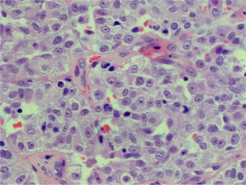
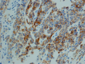
The primary biopsy specimen consisted of multiple mucosal fragments. In each, there was extensive growth of malignant tumour, comprising round to spindle-shaped cells arranged in broad sheets and trabeculae (Figure 1). Cells were of varying sizes, with many exhibiting prominent nucleoli and several mitotic figures (four mitoses per 10 high power fields; Figure 2). Small areas of necrosis were noted, and cells showed positivity for cytokeratin and CD56 and scattered positivity for synaptophysin (Figure 3). The tumour was negative for carcinoembryonic antigen and calcitonin. The results of histological analysis were consistent with an atypical carcinoid.
Photomicrograph of first biopsy specimen showing atypical carcinoid, with strands and nests of intermediate-sized, atypical cells. Normal overlying laryngeal squamous epithelium is also present (H&E; ×400).

Photomicrograph of atypical carcinoid at higher magnification, showing irregular nuclei and several mitoses (H&E; ×600).

Photomicrograph showing atypical carcinoid with positive immunostaining for CD56 (×400).

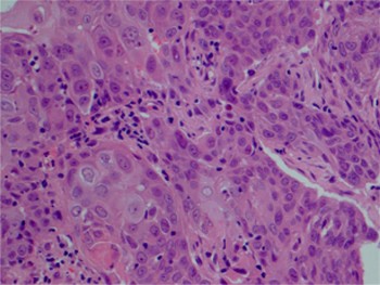
The second biopsy specimen, taken four weeks later, failed to show any of these features, but instead exhibited those of a conventional, moderately well differentiated squamous cell carcinoma. This tumour was entirely negative for CD56, synaptophysin and chromogranin A (Figure 4).
Photomicrograph of second biopsy specimen showing moderately well differentiated squamous cell carcinoma with interspersed lymphocytes. Tumour cells were negative for CD56 (H&E; ×400).

Discussion
The incidence of laryngeal cancer is relatively low compared with that of carcinomas of all sites, comprising 2 to 5 per cent of cancers worldwide.Reference Fu, Wenig, Abemayor and Wenig13 Approximately 99 per cent of malignant laryngeal tumours are squamous cell in origin, arising from the stratified squamous epithelium.Reference Fu, Wenig, Abemayor and Wenig13 Islands, tongues and clusters of invasive, atypical cells within underlying stroma are characteristic. Areas of keratosis, dysplasia and carcinoma in situ are often evident adjacent to the primary invasive lesion. Cells typically demonstrate squamous differentiation, including keratinisation, keratin pearl formation and intercellular bridging.
Neuroendocrine neoplasms represent a rare, heterogeneous subset of laryngeal malignancies, and are classified into distinct groups. These include typical carcinoid (carcinoid, grade 1), atypical carcinoid (malignant carcinoid, grade 2), small cell carcinoma, (small cell neuroendocrine carcinoma, grade 3), combined small cell (composite carcinoma) and the paraganglia-derived paraganglioma.Reference Barnes, Barnes, Eveson, Reichert and Sidransky14 The present case represented a composite tumour consisting of squamous cell carcinoma and atypical carcinoid. All 14 previously described cases of laryngeal composite tumour have consisted of combined squamous cell and small cell carcinoma.Reference Gianoli, Butcher and Martin1–Reference Yucel, Sokmensuer, Gedikoglu and Ayas12 To our knowledge, our case is the first to document a composite tumour including an atypical carcinoid.
Histological analysis of the patient's neuroendocrine carcinoma demonstrated cells that were both large and pleomorphic, with too many mitoses to suggest typical carcinoid. Furthermore, the histological features did not resemble the closely packed arrangement of small cells, with inconspicuous cytoplasm and absent nucleoli, characteristic of small cell neuroendocrine carcinoma. Necrosis was also a minor feature, contrary to the appearance of small cell carcinoma, in which it is marked. Extensive clinical investigation failed to reveal evidence of concomitant primary neoplasms, particularly in the lungs and thyroid, thus excluding metastatic disease as a differential diagnosis.
Primary atypical carcinoid is the most frequent non-squamous cell carcinoma of the larynx.Reference Woodruff and Senie15 More than 90 per cent originate in the supraglottic larynx, and there is a predilection for the arytenoids, the aryepiglottic folds and the laryngeal aspect of the epiglottis. The peak incidence is in the sixth and seventh decades of life. Primary atypical carcinoid is more common in men, and the majority of those afflicted have a history of heavy smoking. The tumour has a propensity to metastasise, and so emphasis is placed on the importance of staging procedures to detect both primary and secondary disease.Reference Ferlito, Friedmann and Goldman16
Surgery is the primary treatment for primary atypical carcinoid tumours; an elective, modified or radical neck dissection may be indicated. Tumours are rarely responsive to chemotherapy.Reference Ferlito, Friedmann and Goldman16 Radiotherapy has also been shown to be ineffective in pre-operative, post-operative and primary settings.Reference Snyderman, Johnson and Barnes17 Overall survival rates of 48 per cent at five years and 30 per cent at 10 years have been reported.Reference Woodruff and Senie15
Interestingly, laryngeal atypical carcinoids typically stain positive for cytokeratin and for neuroendocrine markers such as synaptophysin, chromogranin and CD56. Positive staining for calcitonin and carcinoembryonic antigen is also seen in 75–80 per cent of cases.Reference Barnes, Barnes, Eveson, Reichert and Sidransky14 However, we failed to demonstrate this after repeated staining of our patient's tissue samples. Staining for S-100 also proved negative.
• Laryngeal composite carcinoma represents a rare subset of primary laryngeal neoplasms
• The majority of composite tumours consist of combined squamous and small cell carcinoma
• We report a laryngeal composite tumour comprising squamous cell carcinoma combined with atypical carcinoid
• Treatment of primary neoplasms consisting of more than one histological type is tailored to the most biologically aggressive tumour
• Accurate diagnosis of the histological nature of laryngeal composite tumours is imperative to ensure optimal therapy
Small cell laryngeal carcinoma is rare. Approximately 160 cases have been reported in the last 30 years, the majority of which involved the supraglottic region.Reference Gnepp18 The tumour mainly occurs in male smokers, with a peak incidence in the sixth, seventh and eighth decades of life. Concomitant para-neoplastic syndromes have been occasionally documented, including Eaton–Lambert syndrome, Cushing syndrome and Schwartz–Barrter syndrome.Reference Medina, Moran and Geopfert10, Reference Bishop, Osamura and Tsutsumi19, Reference Trotoux, Glickmanas, Sterkers, Trousset and Pinel20 Small cell laryngeal carcinoma is histologically identical to small cell carcinoma of the lung, and exhibits similarly aggressive biological characteristics. Treatment consists of chemotherapy and radiotherapy. The prognosis is relatively poor, with two and five year survival rates of only 16 and 5 per cent, respectively.Reference Gnepp18
When considering two individual but synchronous neoplasms, it is logical that therapy should be dictated by the most aggressive biological subtype. In the context of laryngeal composite tumours, treatment has historically been tailored to small cell carcinoma. The current report highlights the possibility of variable neuroendocrine tumours being present in vivo with squamous cell carcinoma of the larynx. Our patient received treatment as for squamous cell carcinoma, comprising surgical excision followed by curative radiotherapy. The fact that she remained free of disease at one year follow up supports our diagnosis of synchronous atypical carcinoid rather than small cell carcinoma, which would be much more likely to recur in this time period following radiotherapy alone.