Implications
The need to investigate more sensitive and reliable indicators of animal health and welfare is of primary importance, not only to research, but also with a view to possible applications in the field. Heat shock proteins (HSPs) have become increasingly important because of their correlation to some pathological and poor welfare conditions. HSP70 expression has never been investigated in pig liver, and this study proposes a quantification analysis by Western blot. Moreover, this method detected significant differences between pigs belonging to the same original group and reared in intensive/indoor v. extensive/outdoor conditions.
Introduction
Several factors in modern pig production, such as limited freedom of movement, the lack of stimuli, aggressive temperament, noise, low lighting levels, atmospheric ammonia, and the rapid growth rate, may be stressful for pigs (D'Eath et al., Reference D'Eath, Turner, Kurt, Evans, Thölking, Looft, Wimmers, Murani, Klont, Foury, Ison, Lawrence and Mormède2010; O'Connor et al., Reference O'Connor, Parker, McLeman, Demmers, Lowe, Cui, Davey, Owen, Wathes and Abeyesinghe2010; Parker et al., Reference Parker, O'Connor, McLeman, Demmers, Lowe, Owen, Davey, Wathes and Abeyesinghe2010). As part of the multidisciplinary approach of scientific research on Animal Welfare, major efforts have been made in the investigation of new, reliable and sensitive physiological parameters: acute phase proteins (Murata et al., Reference Murata, Shimada and Yoshioka2004), serum antioxidants (Brambilla et al., Reference Brambilla, Civitareale, Ballerini, Fiori, Amadori, Archetti, Regini and Betti2002) and stress hormones (Kaneko, Reference Kaneko2008) are well-known examples. These variables respond to stressful situations and are more likely to indicate short-term rather than long-term stress. Immune response has also been the object of interest, but its analysis failed to detect the presence of social stress in sows, unlike salivary cortisol (Couret et al., Reference Couret, Otten, Puppe, Prunier and Merlot2009).
At the cellular levels, stress response includes the synthesis of special stress proteins, heat shock proteins (HSPs), which represent a class of highly conserved cellular proteins (Morimoto et al., Reference Morimoto, Tissieres and Georgopoulos1990; Feder and Hofmann, Reference Feder and Hofmann1999).
HSPs are classified mainly based on their molecular weight (kDa). The major HSPs include HSP110, HSP100, HSP90, HSP70, HSP60, HSP40 and the small HSP family (normally 20 to 25 kDa). The human HSP70 family has an unknown number and, although some have been described, the information is inconsistent and incomplete (Brocchieri et al., Reference Brocchieri, Conway de Macario and Macario2008). The four major members are: the stress-inducible HSP70 (72 kDa), the constitutively expressed HSC70 (73 kDa), the glucose-regulated protein 78 (GRP78 or Bip, 78 kDa), which is mainly located in the endoplasmic reticulum, and HSP75 (75 kDa), also known as mortalin and mtHSP70 that is mainly located in the mitochondrial matrix (Bausero et al., Reference Bausero, Gastpar, Multhoff and Asea2005). In mammals, the constitutive HSP family members (HSC70) play an important chaperoning role in unstressed cells, whereas the expression of the inducible (HSP70) forms is known to be activated by acute stressor insults (Morimoto et al., Reference Morimoto, Tissieres and Georgopoulos1990) and pathological and environmental factors (Kiang and Tsokos, Reference Kiang and Tsokos1998), including psychological stress (Fukudo et al., Reference Fukudo, Abe, Hongo, Utsumi and Itoyama1997). HSPs, especially HSP70, can interact with denatured or misfolded proteins and assist in their recovery from stressful events, either by repairing (protein refolding) or degrading them (Ciocca et al., Reference Ciocca, Osterreich, Chamness, McGuire and Fuqua1993; Hartman and Gething, Reference Hartman and Gething1996; Voellmy, Reference Voellmy1996).
A significant decrease in the levels of HSP70, one of the most important HSPs, was reported in the liver of acutely stressed male rats, whereas only a weak decrease in the levels of HSP70 was demonstrated in chronic stress models (Filipović et al., Reference Filipović, Gavrilović, Dronjak, Demajo and Radojcić2008). Moreover, the overexpression of HSP70 in the stomach of male rats seemed to protect against gastric ulcers, through its cytoprotective effects on the gastric mucosa, by increasing mucosal blood flow (Shichijo et al., Reference Shichijo, Ihara, Matsuu, Ito, Okomura and Sekine2003).
Few data are available about expression of HSPs in pig tissues and their role in response to acute and chronic stress conditions. Expression of HSPs in the heart of transported pigs was investigated by Bao et al. (Reference Bao, Sultan, Nowak and Hartung2008). In swine myocardium, HSP70 levels decreased in response to a 6-h journey (Bao et al., Reference Bao, Sultan, Nowak and Hartung2008), whereas in swine stomach HSP70 levels increased significantly after 1, 2 and 4 h of transportation. Moreover, Marruchella et al. (Reference Marruchella, Di Leonardo, Di Guardo, Romanucci, Marà, Tiscar, Mosca and Della Salda2004) observed that expression of HSP70 was higher in pre-ulcerative mucosa of the swine gastric pars oesophagea than in normal mucosa. HSP70 expression was also investigated in serum, colon and small intestine of pigs (Sepponen and Pösö, Reference Sepponen and Pösö2006), showing a negative correlation with the carcass and live weight of pigs. Authors speculated that pigs with a higher concentration of inducible HSP70 were more stressed and did not grow as well as those less stressed and with a lower concentration of inducible HSP70 (Sepponen and Pösö, Reference Sepponen and Pösö2006). These rather conflicting results need further investigation, particularly regarding heavy pigs (at least 9 months of age and with a slaughter live weight of 160 kg), on which very few data are available. In particular, the role of HSP70 expression after a chronic stress, attributable to overall husbandry conditions (European Food Safety Authority (EFSA), 2007), has to be further investigated. The purpose of this study was to determine whether HSP70 was expressed in porcine liver, and to assess the level of expression in two groups of pigs reared in the conventional and extensive system, respectively, by Western blot (WB) and immunohistochemistry (IHC).
Material and methods
Animals, facilities and management
Thirty-six crossbred commercial heavy pigs (Landrace ×Large White) were divided into two groups: Group A (eight castrated males and eight females) was equally distributed into two single-sex partly slatted indoor pens. Group B (11 castrated males and nine females) was kept in one single open area of about 6000 m2. Animals of both groups were fed twice a day with the same liquid diet consisting of three parts of water and one part of commercial dry feed containing wheat, corn and soybean meal (87.5% of dry matter).
In Group A, the space allowance was 1.02 m2/pig during the entire fattening cycle; the individual space allowance at the feeding trough was 0.4 m/pig. The pens consisted of an indoor area with a solid floor and an outdoor defecation area with a slatted floor. Drinking water was available ad libitum through a nipple (one per pen). As environmental enrichment, each pen contained two metal chains. In Group B, the outdoor hill area was partially grassy and partially wooded (with the presence of oak and beech). It contained four covered dry resting areas, a feeding trough and two tanks with fresh water always available. This particular environment is adopted in the Veneto Region within the Piccole Produzioni Locali, a livestock production dedicated to traditional specialities of cured cuts such as Soppressa and Culatello (De Rui et al., Reference De Rui, Buffon, De Lucchi, Vio, Favretti and Cereser2010).
The animals of both groups were kept under the described conditions from 11 weeks of age, after weaning into an indoor conventional pig unit (average BW = 25 ± 4 kg), to 41 weeks of age for the Group A (males’ BW = 170.2 ±8.8 kg; females’ BW = 170.5 ± 9.1 kg) and 48 weeks of age for the Group B (males’ BW = 171.8 ± 12.8 kg; females’ BW = 172.4 ± 12.8 kg). Both groups were slaughtered after a transport period of 1 h. The animals were transported between about 0600 and 0700 h in a commercial trailer pulled by a van. Pigs were stunned with low voltage tongs.
Sampling at the abattoir
At the abattoir all the animals were inspected by the official veterinary services for assessing their health status, in accordance to the Good Health guidelines of the Welfare Quality® (2009) Protocol. Liver samples (about 1 g) were collected from all the animals and immediately frozen in liquid nitrogen and stored at −80°C until analysis. Moreover, a further liver sample for each animal was collected and immediately fixed in 10% neutral buffered formalin at room temperature (RT).
WB
Tissue samples were carried out at the level of the left lateral lobe of the liver. Each sample was weighted, thawed out and homogenized with Ultra-Turrax (IKA, Staufen, Germany) in five volumes of 0.125 M Tris-HCl (pH 6.8) buffer containing protease inhibitor cocktail (Sigma-Aldrich, St. Louis, MO, USA) according to the manufacturer's protocol. Homogenized samples were centrifuged at 13 000 × g at 4°C for 15 min and the suspension was stored at −80°C until analysis.
Protein concentration for the liver was measured by the Coomassie Brilliant Blue method using bovine serum albumin as standard in accordance with Bradford (Reference Bradford1976). A total of 100 μg of protein extract were added to an equal volume of Ssample Buffer Laemmli 2X (Sigma-Aldrich) and boiled for 5 min. Ten micrograms of liver homogenate was separated using 10% SDS-polyacrylamide mini-gel (100 × 100 mm) electrophoresis under reducing conditions according to Laemmli (Reference Laemmli1970). The transfer was carried out at 250 mA for 2 h at 4°C using a 25 mM Tris-base, 192 mM glycine and 20% methanol as electrode solution in a mini trans-blot electrophoretic transfer cell (GE Healthcare, Chalfont St. Giles, UK). Nitrocellulose was treated with blocking solution (3% skim milk, 0.5% Tween-20 in Tris-buffered saline pH 7.6) overnight at 4°C, to prevent non-specific binding, and then incubated with HSP70 primary mouse monoclonal antiserum (Stressgen Biotechnologies, San Diego, CA, USA) in 3% of blocking solution for 60 min. Membranes were next incubated with HRP-labelled (Horseradish peroxidase) goat anti-mouse IgG diluted 1 : 8000 (Bio-Rad, Hercules, CA, USA) for 60 min. All membranes were visualized using Chemiluminescent HRP substrate (Millipore, Billerica, MA, USA).
Band intensity was normalized from one gel to another using an internal standard on each gel and loading the same amount of liver homogenate. Densitometric analysis was performed using an Image Scanner (Amersham Pharmacia Biotech AB, Uppsala, Sweden), and the data were quantified by the ImageMaster Total Lab 2.0 Software (Amersham Pharmacia Biotech AB), bordering the complete band and determining the area and the intensity of the band of interest.
IHC
Formalin fixed samples were routinely processed and embedded in paraffin blocks. IHC was carried out by an automated immunostainer (Autostainer link 48; Dako, Milano, Italy). Four-micrograms sections were deparaffinized in xylene, rehydrated in graded ethanol and rinsed in distilled water. Heat-induced antigen retrieval was performed in 10 mM citrate buffer (pH = 6.0) at 97°C for 15 min. Endogenous peroxidases were neutralized by incubating the sections with the EnVision FLEX Peroxidase-Blocking Reagent (SM801, Dako Denmark A/S, Glostrup, Denmark), for 10 min at RT; the sections were incubated with HSP70 primary mouse monoclonal antiserum (Stressgen Biotechnologies) overnight at +4°C. The EnVision FLEX/HRP (Dako) and the EnVision FLEX Substrate Buffer EnVision FLEX DAB+ were used as a detection system and chromogen, respectively. The sections were then counterstained with the EnVision FLEX Hematoxylin (Dako). The specificity of the immunostaining was verified by incubating the sections with a mouse pre-immune serum and phosphate-buffered saline instead of the specific primary antibody.
Statistical analysis
HSP70 distribution was evaluated using a GLM. The HSP70 was the dependent variable, whereas sex, group and the interaction were the independent variables (fixed effects). Given the unequal sample sizes for the groups, least squared means (LS-means) were preferred to standard means. The contrast analysis was used to analyse the interaction between sex and group, and to calculate the statistical significance for each combination of the two variables. The analysis of residuals was adopted for checking the model. The results were expressed as LS-means of the HSP70 variable and root mean squared error of the model. All data analyses were performed using SAS 9.1 version.
Results
Slaughtered animals were in good body condition, were declared in good health status and were then delivered for human consumption. No lame animals were detected when being unloaded from the lorry. The carcasses did not present any evidence of pneumonia, pleurisy, pericarditis, skin lesions, tail biting or ear biting in both groups. The livers of all the animals did not show evidence of either gross lesions or abnormalities in shape, size and consistency. The extensive group resulted in a higher variability of final live weight and a longer fattening period.
WB
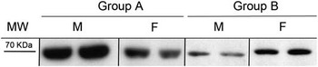
The antibody used in the present study specifically recognized HSP70 in heavy pigs, determining the presence of a band of about 70 kDa in the liver of both males and females (Figure 1). The entire membrane is shown in Supplementary Figure S1. Results of GLM are given in Table 1. The interaction between sex and group was statistically significant (P = 0.001). When stratified by sex, Group A showed significantly higher HSP70 expression values compared with Group B for both male and female subjects (P < 0.001). Stratifying by group, males showed significantly higher HSP70 values than females in Group A (P < 0.001), whereas no statistical differences were observed between sexes for Group B (P = 0.653).
Representative Western blot analysis of liver homogenates in intensively and extensively kept heavy pigs. Lanes represent heat shock protein 70 (about 70 kDa). MW = molecular weights in kDa; Group A = intensive/indoor confinement system; Group B = extensive/outdoor confinement system; M = males; F = females.

Effect of sex and two different confinement systems (Group A: intensive/indoor; Group B: extensive/outdoor) on the expression of HSP70 in swine liver, investigated by Western blot

HSP70 = heat shock protein 70; RMSE = root mean squared error; LS-mean = least squared mean.
Results of the GLM analysis are given in the original scale of pixels as adjusted means (LS-mean) and RMSE of the model. The number of animals is reported (No.).
Exponent letters A, B and C refer to P < 0.001.
IHC
HSP70 cytoplasmic immunoreactivity was seen in a granular pattern in the liver of pigs from both groups, and was characterized by a multifocal distribution, mainly observed in the centrilobular and perilobular hepatocytes (Supplementary Figures S2 and S3).
Discussion
Nowadays, several studies are focused on the welfare of farm animals. A major objective is to find reliable and easily measurable biomarkers related to rearing and transportation. In this framework, the identification of biomarkers collectable at the abattoir, instead of in vivo, can be considered a feasible solution to investigate stress physiology in livestock without raising ethical issues related to animal experimentation and unnecessary sufferings.
Important differences between production systems can be established on the basis of animal-based measures of the good feeding and housing principles of the Welfare Quality® Protocol. Recently, Temple et al. (Reference Temple, Courboulay, Manteca, Velarde and Dalmau2012) demonstrated that pigs reared in the conventional system presented the lowest prevalence of poor body condition, whereas pigs reared in extensive systems were associated with a decreased prevalence of bursitis and pig dirtiness. Some physiological parameters have also been proved to be influenced by the rearing system. For example, comparing the effects of different rearing systems for growing pigs on stress indicators determined at slaughter, Foury et al. (Reference Foury, Lebret, Chevillon, Vautier, Terlouw and Mormède2011) detected the lowest levels of plasma creatine kinase and urinary catecholamines in animals reared outdoors.
Although cortisol is considered an important ‘stress hormone’ in swine, because excess cortisol is secreted in response to physical or psychological stress (Kaneko, Reference Kaneko2008), several authors demonstrated that, in pigs, its concentration in saliva (de Jong et al., Reference de Jong, Prelle, van de Burgwal, Lambooij, Korte and Blokhuis2000) and urine (Foury et al., Reference Foury, Lebret, Chevillon, Vautier, Terlouw and Mormède2011; Lebret et al., Reference Lebret, Prunier, Bonhomme, Foury, Mormède and Dourmad2011) was not affected by the rearing system.
Interestingly, although many studies have evaluated HSP response to acute and chronic stress conditions (heat, hypoxia, infections; Ciocca et al., Reference Ciocca, Osterreich, Chamness, McGuire and Fuqua1993; Hartman and Gething, Reference Hartman and Gething1996; Voellmy, Reference Voellmy1996), the possible role of HSP in animal response to rearing conditions has been poorly investigated. Particularly in pigs, no study has been conducted to evaluate the effect of the rearing system on HSP expression.
HSP70 is the most widely studied of all HSPs and has numerous important chaperoning functions. In the unstressed cell, there is a constitutive production of these proteins that are required in various aspects of cellular homeostasis, whereas the inducible (HSP70) form is expressed at detectable levels after stressor insults (Morimoto et al., Reference Morimoto, Tissieres and Georgopoulos1990). HSP70 is known to assist the folding of nascent polypeptide chains, to act as a molecular chaperone and to mediate the repair and degradation of altered or denatured proteins.
In this study, the anti-HSP70 antibody recognized a band of about 70 kDa in the liver of both males and females by WB. When stratified by sex, the intensively kept group showed significantly higher HSP70 values than the extensively kept group for both males and females; moreover, stratifying by group, males showed significantly higher values than females in the intensively kept group, whereas no statistical differences were observed between sexes for the extensively kept group.
These results could indicate that pigs reared in intensive systems are more susceptible to stress than those reared outdoors. Moreover, the significantly higher expression of HSP70 in males of the intensively kept group than in females may suggest that males are physiologically more sensitive to stress factors resulting from intensive husbandry systems, such as high density, limited free movement, lack of stimuli and rapid growth rate. This hypothesis seems supported also by the prevalence of gastric ulcer detected at slaughter, that was reported to be significantly higher in males than in females from the intensive rearing system (Di Martino et al., Reference Di Martino, Scollo, Capello, Stefani, Schiavon, Rampin, Marangon, Gottardo and Bonfanti2011).
When the same antibody, recognizing only the inducible HSP70 isoform, was used by IHC, a multifocal amount of protein expression was observed in the cytoplasm of the hepatocytes, particularly in the central vein region and in the perilobular region. In mammalian cells, there are two nucleo-cytoplasmic HSP70 isoforms (a constitutive isoform and a stress-inducible one) with highly homologous amino acid sequences, but with different patterns of expression and as yet unclear differences in cellular functions. In a previous study on rat liver, the exposure of the animal to stress was followed by rapid HSP70 nuclear accumulation, which at first resulted from pre-existing HSC70 shifting from the cytoplasm to the nucleus, and eventually from the induction of the inducible isoform synthesis (Cvoro et al., Reference Cvoro, Korać and Matić2004). The maximal level of HSP70 nuclear accumulation coincided with the maximal extent of inducible isoform induction and was achieved 5 h after the stress. At the same time, the cytoplasmic accumulation of the protein was noticed for the first time. These findings suggest that, during stress and at the beginning of the recovery period, HSP70 is needed more in the nuclei than in the cytoplasm (Cvoro et al., Reference Cvoro, Korać and Matić2004). These data confirm the previously established concept of nuclear localization of HSP70 during stress and its cytoplasmic storage during recovery (Velazquez and Lindquist, Reference Velazquez and Lindquist1984; Welch and Feramisco, Reference Welch and Feramisco1984). In our study, HSP70 was recognized only in the cytoplasm of hepatocytes, in the liver of pigs from both groups. This finding could indicate that liver is involved in chronic stress response in pigs, and HSP70 expression could be a useful stress biomarker. Anyway, the lack of nuclear positivity seems to indicate that the rearing conditions are not stressful enough to cause HSP70 nuclear accumulation in liver. Our results agree with those obtained in a previous study that investigated the expression and localization of four HSPs (HSP70, HSP86, HSP90 and HSP27) in the heart tissue of pigs transported for 6 h (Bao et al., Reference Bao, Sultan, Nowak and Hartung2008). Immunostaining detected the consistent presence of all HSPs in pig myocardial cells both during transport and under normal housing conditions. IHC analysis revealed predominance of HSP70 (significantly the highest levels) and HSP27 in the cytoplasm of myocardial cells (Bao et al., Reference Bao, Sultan, Nowak and Hartung2008).
In conclusion, this study has, for the first time, investigated the expression of HSP70 by liver cells in pigs reared with two different methods, indicating a possible role of HSPs in stress response to husbandry conditions.
Acknowledgments
This work was funded by Regione del Veneto ‘Direzione Prevenzione U.C. Sanità Animale e Igiene Alimentare’ (DGRV 3594/10). The authors are grateful to Dr Francesco Chisini Granzotto (Ulss 7 of Pieve di Soligo) and Dr Fiorenzo Signor (Ulss 3 of Bassano) for their support during the activities at the abattoir.