Isolated ventricular septal defects have generally been thought to have great long-term prognosis – either after surgical closure or if the defect is deemed small and not requiring surgery.Reference Gabriel, Heger and Innerhofer 1 Lately, however, patients with either surgically closed or small, unrepaired ventricular septal defects have been found with cardiac abnormalities in their adult years,Reference Gabriels, De Backer and Pasquet 2 – Reference Menting, Cuypers and Opic 6 illustrating that long-term outcomes are not without complications as previously thought. At our institution, we have recently investigated patients in their twenties, born with simple, isolated ventricular septal defects that were either successfully closed in childhood or remained unrepaired. Both patient groups exhibited 20% lower peak exercise capacity compared with healthy controls.Reference Heiberg, Laustsen, Petersen and Hjortdal 7 , Reference Maagaard, Heiberg and Hjortdal 8 Importantly, the mechanisms behind the impaired exercise can not only be explained by preceding surgery but must be linked to the defect itself.
In a previous study, an abnormal pattern of force–frequency relationship in the right ventricle was demonstrated in young adults with operated ventricular septal defects, which could be directly correlated with the lower peak exercise capacity.Reference Heiberg, Schmidt, Redington and Hjortdal 9 The underlying mechanism for the disrupted contractility could be a rise in right ventricle afterload during exercise, as demonstrated by Moller et al.Reference Moller, Brun and Fredriksen 10 In a normal active life, the exercise-induced right ventricular afterload could have an effect on the morphology of the right ventricle over many years. It is important to clarify whether a possible abnormal morphology exists in adults with ventricular septal defects as it may progress with time and potentially result in cardiac abnormalities, already reported by others,Reference Neumayer, Stone and Somerville 3 , Reference Karonis, Scognamiglio and Babu-Narayan 4 , Reference Menting, Cuypers and Opic 6 , Reference Gabriels, Van De Bruaene and Helsen 11 or even cardiac failure. Whether small, unrepaired ventricular septal defects reveal the same pattern is not yet known, as their force–frequency relationship has not been investigated. Still, they exhibit decreased peak exercise capacity similar to that of operated ventricular septal defects. In a study from 2015,Reference Heiberg, Ringgaard, Schmidt, Redington and Hjortdal 12 a degree of structural changes of the right ventricle was suggested when investigating adults with surgically corrected ventricular septal defects with cardiac MRI. This study was recently followed up by an editorial,Reference Mertens 13 stressing the need for additional imaging studies that could further clarify these findings in this growing patient cohort. So far, it is unknown whether ventricular volumes are also affected in adults with ventricular septal defects, as this long-term outcome has not yet been investigated. Therefore, the aim of this prospective, long-term follow-up study was to examine biventricular volumes by MRI in adults with either surgically corrected or small, unrepaired ventricular septal defects.
Materials and methods
Ethics
This study protocol complies with the ethical standards of The Regional Committee on Biomedical Research Ethics of the Central Denmark Region (charts: 1-10-72-74-14, 1-16-02-12-14), The Danish Data Protection Agency (chart: 2007-58-0010), and with the Helsinki Declaration of 1975, revised in 2008. Written informed consent was obtained from all patients at the beginning of the study in consistency with Danish law.
Study population
The study population was made up of patients with operated ventricular septal defects, patients with unrepaired ventricular septal defects, and two groups of healthy controls with similar age and gender distribution as their respective patient group. The inclusion criteria for the patients were age between 18 and 40 years and a congenital ventricular septal defect that had either been surgically closed through a right atrial approach at our institution between 1990 and 1995 or left open and unrepaired. Exclusion criteria for the patients included spontaneous closure of the defect, surgical closure by ventriculotomy, coexistence of other congenital cardiac defects than ventricular septal defect, associated syndromes, for example, Down’s syndrome, documented arrhythmia other than right bundle branch block, and cardiac or pulmonary disease including any valve pathology, pregnancy, and missing patient chart.
All operated patients had undergone echocardiographic examinations 2 years previously as part of another studyReference Heiberg, Schmidt, Redington and Hjortdal 9 ruling out other cardiac defects. All patients with unrepaired ventricular septal defects underwent clinical examination and echocardiography in order to exclude spontaneous defect closure since last echocardiography. The echocardiographic data obtained on the unrepaired patients and their controls were included in this study in order to give further details on their current shunt size. Patients and controls were included in random order, and all were between the age of 18 and 40 years. All patients have previously undergone an upright bicycle test between November 2011 and May 2016, where reduced oxygen uptake was demonstrated. Methods and results from the exercise tests are reported previously.Reference Heiberg, Laustsen, Petersen and Hjortdal 7 , Reference Maagaard, Heiberg and Hjortdal 8 , Reference Asschenfeldt, Heiberg, Ringgaard, Maagaard, Redington and Hjortdal 14
Echocardiography
The patients with unrepaired ventricular septal defects and their control group underwent transthoracic echocardiography at rest using a GE Vivid 7 (GE Healthcare, Horten, Norway) with a 2.5-MHz probe. The unrepaired ventricular septal defect was visualised in either the parasternal long- or short-axis view or the apical four-chamber view, from which the diameter of the defect was measured on at least two different images. The area was calculated from the estimated diameter, using the mean radius2×π. The velocity through diastole and systole, respectively, was measured with continuous wave and subsequently traced on a frozen parasternal long-axis view of the unrepaired defect containing three consecutive heart cycles. Integrating the mean velocity through the defect area over time, the volume through the shunt was estimated and then indexed to body surface area. Additional echocardiographic resting values included posterior wall index and inner diameter of left ventricle at end-diastole and end-systole, as well as interventricular septal index at end-diastole and end-systole, tricuspid annular plane systolic excursion, tricuspid regurgitation, and pulmonary regurgitation.
MRI
MRI was performed using a 1.5-T Philips Achieva dStream whole-body scanner (Philips, Amsterdam, The Netherlands) equipped with 40 mT/m gradients with a slew rate of 180 mT/m per ms and software release R517 using an 18-cm surface coil and a spine coil array. Breathing frequency was measured with an air-filled belt fastened around the patient’s waist, and cardiac rhythm was monitored with integrated four-lead electrocardiography. First, standard scout images of the heart and surroundings were obtained in the transaxial, coronal, and sagittal direction. Thereafter, an interactive MRI platform with single-shot planning was used to locate the four-chamber view using clinical MRI guidelines. This single-shot position was used to produce five cine images during three breath-holds of the four-chamber view. For the left ventricle, short-axis stacks were planned from the four-chamber cine scan, with slices being placed parallel to the mitral valve in end-diastole. Using a kT-blast sequence, 28–30 slices were acquired during two breath-holds lasting approximately 24 seconds, sampling 16 heart phases with a slice thickness of 4 mm, a field of view of 320×304 mm, and a matrix of 164×162. Short-axis scans of the right ventricle were acquired in a similar manner, with slices parallel to the tricuspid valve in end-diastole. The short-axis scans covered from the apex of the ventricle and past the atrioventricular valve orifice.
Flow measurement for the calculation of stroke index was acquired using phase-contrast scans. The scans were produced using the aforementioned single-shot planning, identifying the left ventricular outflow tract. At the upper limit of the aortic bulb, an orthogonal plane was placed, approximately 2 cm above the aortic valve, thereby obtaining a transverse image of the ascending aorta. In a similar manner, the right ventricular outflow tract was located just above the pulmonary valve but proximal to the bifurcation, resulting in a transversal view of the pulmonary trunk. Following this, flow measurements were obtained using a free-breathing, electrocardiography-triggered phase-contrast sequence. The field of view was 300×300 mm and pixel size was 2.3×3.1 mm. A total of 50 cardiac phases were covered and the scan time was 2:30 minutes at a heart rate of 60 beats/min. The velocity sensitivity was 150 cm/seconds. The order of the scanning was randomised for the entire group of patients, ensuring that equal amounts of scans were performed starting with the aorta, then the pulmonary trunk or starting with the pulmonary trunk followed by the aorta. For three patients with unrepaired ventricular septal defects and three matched controls, the electrocardiography-triggered phase-contrast sequence could not be acquired because of unreliable electrocardiography signal. Instead, real-time scans – segmented gradient-echo phase-contrast echo planar imaging – were chosen, consisting of 170 consecutive, phase-contrast flow acquisitions without electrocardiography triggering, each lasting 81 ms with a frame rate of 12.2 frames per second, field of view of 320×305 mm, and pixel size of 3.64×4.49 mm. Velocity sensitivity was set to 150 cm/second.
Data analysis
An independent observer blinded to group allocation performed offline MRI analyses of ventricular volumes and flow through the ascending aorta and pulmonary trunk. Ventricular volume measurements were performed using the Medviso Segment Software. The outline of the endocardial contours was traced manually throughout all slices, with the papillary muscles and trabeculation being included as part of the ventricular volume for better reproducibility.Reference Sievers, Kirchberg, Bakan, Franken and Trappe 15 , Reference Winter, Bernink and Groenink 16 Volumetric analyses were carried out automatically by the applied software. Flow measurements in the ascending aorta and pulmonary trunk were performed using the in-house produced software Siswin. The vessel contours of the ascending aorta and pulmonary trunk were traced manually using a four-point adjustable, elliptical ROI tool. Heart rate and cardiac output were calculated as mean values from each sequence. Mean cardiac index from the pulmonary trunk and aorta was defined as the net amount of blood per time per body surface area reaching the systemic circulation or the pulmonary arteries, respectively. Therefore, the mean cardiac index was defined as the mean blood flow across the scanning plane, which is a result of the mean forward flow subtracted from the mean retrograde flow.Reference Hundley, Li and Lange 17 Stroke index from left and right ventricles was calculated from cardiac index and heart rates. For the unrepaired ventricular septal defects, the size of the shunt was calculated as the flow ratio between the pulmonary and systemic circulation. The size of the operated ventricular septal defects was previously measured with echocardiography elsewhere.Reference Heiberg, Schmidt, Redington and Hjortdal 9
Intra-observer variability of the primary observer was assessed using right ventricular end-diastolic and end-systolic volume index scans of 12 randomly chosen patients. The scans were analysed twice with a time interval of more than a month between the first and second analyses. For interobserver variability, 15 MRI scans of right ventricular end-diastolic and end-systolic volume index were randomly selected and evaluated by two experienced observers.
Correlation analyses were applied for both patient groups on their current end-diastolic volume indices and their previously achieved peak oxygen uptake during bicycle exercise.Reference Heiberg, Laustsen, Petersen and Hjortdal 7 , Reference Maagaard, Heiberg and Hjortdal 8 For the surgically repaired patients, further correlations were examined concerning their operative age, type of defect, and pre-operative size of defect with the biventricular end-diastolic volume indices. For the unrepaired patients, analyses were made between their current size of the shunt and type of defect with their biventricular end-diastolic volume indices.
Endpoints
Our primary end point was right ventricular end-diastolic volume in operated and unoperated patients, whereas secondary endpoints were left ventricular end-diastolic volumes and biventricular stroke volume indices.
Statistical methods
Continuous, normally distributed data were reported as means with standard deviations and otherwise as medians with 95% confidence intervals. The unpaired Student t-test was used for normally distributed data. p-values <0.05 were considered statistically significant. Inter- and intra-observer agreements were assessed with intraclass correlation coefficient using a two-way mixed model for the differences between measurements.Reference Shrout and Fleiss 18 Correlation analyses were calculated using Pearson’s sample correlation r. Statistical analyses were performed using StataIC 11.2 (StataCorp LP, College Station, Texas, United States of America).
Results
Study population
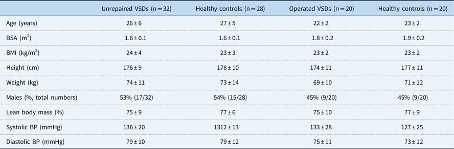
The patients were included from May 2014 to May 2016 at Aarhus University Hospital, Denmark. Patient groups and their respective healthy control groups displayed similar demographics and clinical characteristics as presented in Table 1. Comparative statistics were performed and no differences were found in demographic or clinical parameters between the groups. No gender- or ethnicity-based differences were present. None of the patients dropped out of the study.
Demographics and clinical characteristics of patients with unrepaired ventricular septal defects (VSDs), patients with operated VSDs, and their respective control groups.

BMI=body mass index; BP=blood pressure; BSA=body surface area
Data presented as mean±standard deviation or percentages total numbers
The anatomical positions of the ventricular septal defects were, for the operated patients, 14 perimembranous defects and six muscular defects. Their current median flow ratio between pulmonary trunk and ascending aorta was 1.0 (95% confidence interval 0.9 to 1.1). The unrepaired ventricular septal defects were made up of 23 perimembranous defects and nine muscular defects. Their median flow ratio between pulmonary trunk and ascending aorta was 1.2 (95% confidence interval 1.2 to 1.3). The operated patients underwent surgical closure at a mean age of 2.1±1.4 years and the size of the ventricular septal defect was 7.9±3.2 mm as judged by echocardiography. Five operated patients had their ventricular septal defects closed with direct suture, and the rest were closed by patch. In two operated patients, a trivial, residual shunt had been noted previously following childhood surgery. However, their current flow ratio between pulmonary trunk and ascending aorta was 1.0 according to our MRI flow measurements, and ventricular volumes did not differ from the rest of the operated patients. Therefore, they remained in the group of surgically repaired ventricular septal defects. No patients demonstrated significant valvular regurgitation. All patients had normal biventricular ejection fractions and none of them were found with peak tricuspid regurgitation velocities ≥2.8 m/second, suggesting normal estimated pulmonary pressures at rest.
Echocardiography
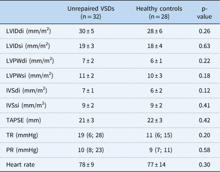
In 26 patients with an unrepaired ventricular septal defect, it was possible to estimate a diastolic and systolic shunting using echocardiography. The mean shunt, indexed to body surface area, during diastole was found to be 3.1±2 ml/m2 and during systole 16.2±10 ml/m2. The remaining echocardiographic values of the patients and their healthy controls are presented in Table 2.
Echocardiographic parameters of patients with unrepaired ventricular septal defects (VSDs) and their group of healthy controls.

IVSdi and IVSsi=interventricular septum index at end-diastole and end-systole; LVIDdi and LVIDsi=left ventricular inner diameter index at end-diastole and end-systole; LVPWdi and LVPWsi=left ventricular posterior wall index at end-diastole and end-systole; PR=pulmonary regurgitation; TAPSE=tricuspid annular place systolic ejection; TR=tricuspid regurgitation
Data presented as mean±standard deviation or as medians with (95% confidence intervals)
MRI
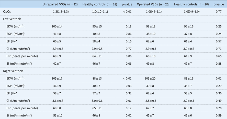
Biventricular end-diastolic, cardiac, and stroke volume indices are displayed in Table 3. Among the 20 operated patients, a larger right ventricular end-diastolic volume index was demonstrated, 103±20 ml/m2, when compared with their healthy control group, 88±16 ml/m2, p=0.01. Similarly, for the unrepaired patients, a higher right ventricular end-diastolic volume index was observed, 105±17 ml/m2, compared with their controls, 88±13 ml/m2, p<0.01. Left ventricular end-diastolic volume index was comparable between both patient groups and their respective healthy controls. Comparative analyses were made between the two patient groups, but no differences were found regarding end-diastolic indices of the right (p=0.66) or left ventricle (p=0.64).
Flow and volume measurements in patients with unrepaired ventricular septal defects (VSDs), patients with operated VSDs, and their respective control groups.

Ci=cardiac index; EDVi=end-diastolic volume index; EF=ejection fraction; ESVi=end-systolic volume index; HR=heart rate; QpQs=flow ratio between pulmonary trunk and ascending aorta; Si=stroke index
Data presented as mean with±standard deviation or median with 95% confidence interval
* Results on 25 unrepaired ventricular septal defect patients and 23 healthy controls
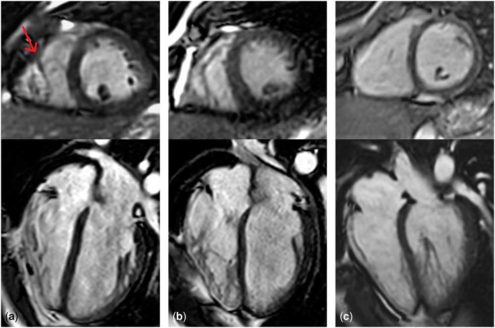
Right ventricular stroke index was larger in unrepaired patients, 53±12 ml/minute/m2, compared with controls, 46±8 ml/minute/m2, p=0.02, whereas no difference was found between operated patients and their controls. Examples of right ventricular morphology from MRI scans for patients with operated ventricular septal defects, patients with unrepaired ventricular septal defects, and their respective control groups are displayed in Figure 1a–c. The scans show coarse trabeculation of the right ventricle in patients. Their right ventricle generally demonstrated a thickened moderator band with increasingly trabeculated myocardial tissue towards the apical part of the ventricle.
( a ) Coarsely trabeculated right ventricle with a considerable moderator band (arrow) in a male patient, aged 20 years, operated for a ventricular septal defect in childhood. ( b ) Male patient, aged 25 years, with a small, unrepaired ventricular septal defect, showing extensive trabeculation of the right ventricle. ( c ) Healthy male, aged 24 years, with visually normal trabeculation of the right ventricle.

Inter- and intra-observer correlations
The observer variability for the right ventricular end-diastolic volume measurements demonstrated good agreements between inter- and intra-observer measurements, with an intraclass correlation coefficient of 0.93 for the intra-observer and an intraclass correlation coefficient of 0.76 for the interobserver. For the right ventricular end-systolic volume measurements, the intraclass correlation coefficient was 0.75 for interobserver variability and 0.86 for intra-observer variability
Correlation analyses
For the surgically corrected patients, a positive correlation was found between peak exercise capacity and end-diastolic volume in the right ventricle (r=0.52, p=0.02), but not the left ventricle (r=0.38, p=0.10). No correlations were found between biventricular end-diastolic volume indices and operative age or pre-operative size of the defect. For the patients with unrepaired defects, a positive correlation was found between peak exercise capacity and end-diastolic volume in the right ventricle (r=0.51, p=0.003) and the left ventricle (r=0.63, p<0.001). The size of the shunt and the biventricular end-diastolic volume indices revealed no relationship.
Discussion
Adults with either surgically closed or unrepaired, small ventricular septal defects have altered right ventricular morphology with larger end-diastolic volumes and more pronounced trabeculation compared with healthy controls. These findings may be linked to the previously found abnormal exercise outcomes.Reference Heiberg, Laustsen, Petersen and Hjortdal 7 – Reference Heiberg, Schmidt, Redington and Hjortdal 9 , Reference Heiberg, Ringgaard, Schmidt, Redington and Hjortdal 12 , Reference Maagaard, Heiberg, Asschenfeldt, Ringgaard and Hjortdal 19 We propose two possible mechanisms for the larger right ventricular volumes and distinct trabeculation: first, pulmonary hyperperfusion may permanently change the pulmonary vascular bed and lead to increased right ventricle afterload or second, the shunt itself may cause a remodelling of the right ventricular structure, a remodelling that does not normalise after surgical closure.
A congenital ventricular septal defect results in hyperperfusion of the pulmonary vascular bed – either in large quantities for a short time period as in the patients who underwent surgical closure or in small quantities for a long period as in the patients with unrepaired defects. This hyperperfusion may cause alterations in the pulmonary vascular endothelium, increasing vascular resistance, thereby resulting in higher pulmonary artery pressure. This theory was previously suggested, as Möller et alReference Moller, Brun and Fredriksen 10 demonstrated higher right ventricular systolic pressure during exercise in adults with septal defects. The pulmonary vasculature of closed ventricular septal defects was moreover found to react abnormally when observing a lack of pulmonary vasodilatation during physical activity under administration of sildenafil.Reference Brun, Moller, Fredriksen, Thaulow, Pripp and Holmstrom 20 In a new study from our group, the pulmonary trunk was further investigated with MRI in adults with small, unrepaired ventricular septal defects and a larger retrograde flow along with greater diameter was discoveredReference Maagaard, Heiberg, Asschenfeldt, Ringgaard and Hjortdal 19 – perhaps demonstrating the very early, subclinical signs of abnormal pulmonary vascular resistance. In another study from our institution, it was also demonstrated that patients with surgically closed ventricular septal defects exhibited abnormal contractility at rest and, even more so, during increasing exercise.Reference Heiberg, Schmidt, Redington and Hjortdal 9 Abnormal contractility and a larger pulmonary retrograde flow may both reflect increased right ventricular afterload as the consequence of increased pulmonary vascular resistance in patients with ventricular septal defects.
Another explanation for our findings could be that the higher volume burden on the right ventricle from the ventricular septal defect results in a remodelling of the ventricular structure and consequently a larger diastolic volume. This potential remodelling in relation to a ventricular septal defect has previously been investigated and demonstrated in a porcine study from 2010.Reference Monreal, Youtz and Phillips 21 Their findings revealed subclinical remodelling with abnormal contraction relaxation in the myocytes implying right ventricular diastolic dysfunction. Furthermore, they also found an upregulation of the mitochondrial proteins indicating a heightened right ventricular work. A heightened workload may eventually result in larger myocardial mass, which has already been demonstrated in adults with surgically closed ventricular septal defects.Reference Heiberg, Ringgaard, Schmidt, Redington and Hjortdal 12 In addition, a higher mass may be a reflection of our findings of a markedly trabeculated myocardium seen in both surgically closed and unrepaired ventricular septal defects (Fig 1a and b).
The ventricular dimensions measured in our study were mostly within normal values, as judged by a systematic review from 2015.Reference Kawel-Boehm, Maceira and Valsangiacomo-Buechel 22 Nevertheless, these same patient groups have previously been found with impaired outcomes on a number of different clinical tests. A consequence of a larger right ventricular volume is perhaps a change in right ventricular function as seen by lower fractional area change in surgically closed ventricular septal defects.Reference Menting, Cuypers and Opic 6 , Reference Gabriels, Van De Bruaene and Helsen 11 , Reference Heiberg, Ringgaard, Schmidt, Redington and Hjortdal 12 This could potentially indicate an altered ejection fraction and a degree of right ventricular dysfunction. The larger right ventricle is possibly just one of the very early signs of a stressed ventricular component in relation to a ventricular septal defect in young adults, progressing with advancing age. Potentially, the larger volume can be seen as a compensatory step to the right on the Frank–Starling curve as a way to maintain stroke volume in the face of higher right ventricular afterload. Possibly, this is also reflected in the positive correlation found between the size of the right ventricle and the exercise capacity in both patient groups. Taking previous findings into consideration, this would imply that at rest no clinical signs can be readily demonstrated. During exercise, however, a number of parameters are affected as a reflection of the ventricle not being able to honour the increasingly higher demand arising with physical activity. In two older studies, this has perhaps already been demonstrated: Otterstad et alReference Otterstad, Simonsen and Erikssen 23 found that patients with unrepaired, small ventricular septal defects had subnormal increases in ventricular cardiac outputs during exercise, but with normal resting haemodynamics. In addition, Jablonsky et alReference Jablonsky, Hilton and Liu 24 saw abnormal, flat responses to exercise when looking at ventricular ejection fractions in both surgically closed and small, unrepaired ventricular septal defects.
A third possible option that could explain the findings of a larger right ventricular end-diastolic volume index in the patients with unrepaired defects could be that the defect causes diastolic shunting that could cause a larger volume in the right ventricle before systole. Judging from the echocardiographic data on the unrepaired patients, the diastolic shunting takes up around 16% of the total amount of blood being shunted through the open defect. Whether this could play a part in the larger volumes found in the right ventricle can only be speculated upon. Nevertheless, our findings do underline that the majority of blood is primarily being shunted during systole in these small defects.
Finally, the finding of a larger right ventricular end-diastolic volume index was a bit surprising as the presence of a ventricular septal defect is more often associated with left ventricle enlargement and hypertrophy. In particular, in the patients with an unrepaired defect and a current shunt ratio of 1.2, it would be expected that the concomitantly measured volume of the left ventricle would be larger in end-diastole than that of the controls. With a shunt ratio of 1.2, the difference in the total cardiac index from the left ventricle would correspond to 20%, whereas the difference in ventricular end-diastolic volume would more accurately be around 12% with an ejection fraction of 60%. Looking at the results of the left ventricular end-diastolic volume index of the unrepaired patients, the difference compared with that of the controls is around 5%. A possible explanation for this difference not being closer to 12% could lie in the diastolic shunting. This, however, only comprises 3.1±2 ml/m2, still leaving roughly 5 ml/m2 to be accounted for. A possible reason could obviously be ascribed to the methods applied for estimating end-diastolic volumes, or perhaps the patients were examined too young for any possible dilatation of the left ventricle to manifest. The remaining echocardiographic parameters did not reveal any hints of a modified left ventricle as no differences were found in left ventricular diameters or wall dimensions, suggesting that no dilatation or hypertrophy is present in this patient group.
Study limitations
Our study has some limitations. First, it is well known that quantification of the right ventricle is complex and reproducibility therefore can be low, although MRI is considered the gold standard technique for the assessment of ventricular volumes. In choosing to include papillary muscles and trabeculation as part of the right ventricle volume, the reproducibility increasedReference Sievers, Kirchberg, Bakan, Franken and Trappe 15 , Reference Winter, Bernink and Groenink 16 with satisfactory inter- and intra-observer agreements. Second, the end-systolic volumes in the unrepaired patients and their controls were reflected in a subgroup, as it was not possible to retrieve the results in seven patients and five controls. It should also be stressed that the definition of the endocardial borders was especially difficult to delineate in end-systole in the more heavily trabeculated right ventricles of the patients. Finally, the patients with corrected ventricular septal defects underwent surgical closure at a higher age than would be anticipated in a contemporary cohort. However, our results still reflect a large cohort of current adults living with the long-term consequences of a congenital ventricular septal defect.
Conclusion
With this study, we have demonstrated that young adults with either a surgically closed or a small, unrepaired ventricular septal defect reveal a larger right ventricular end-diastolic volume. Our findings, along with those previously demonstrated in this growing patient population, underline the importance of continuing follow-ups of adults as they may not prove to be completely cured.
Acknowledgements
The authors warmly acknowledge research secretary Jette Breiner for her appreciated contribution and medical student Christine Bauer-Jensen for her assistance in the data analysis of the phase-contrast MRI.
Financial Support
This study was supported by Aarhus University, Lægeforeningens Forskningsfond, and Snedkermester Sophus Jacobsen & Hustru Astrid Jacobsen’s Fund.
Conflict of Interest
None.
Ethical Standards
The authors assert that all procedures contributing to this work comply with the ethical standards of the Regional Committee on Biomedical Research Ethics of Central Denmark Region and with the Helsinki Declaration of 1975, as revised in 2008, and the study has been approved by the institutional committees.