Introduction
Fasciola gigantica, a causative agent of tropical fasciolosis in domestic ruminants in Asia and Africa, inflicts an annual economic loss of over US$3 billion to the livestock sector (Spithill et al., Reference Spithill, Smooker and Copeman1999). Infection with liver flukes is also recognized as an emerging human health problem in several countries of the world (Mas-Coma et al., Reference Mas-Coma, Bargues and Valero2005). Prevalence of F. gigantica infection in some ruminants ranges from 80 to 100% in several countries of west Africa and Asia (Sharma et al., Reference Sharma, Dhar and Raina1989; Sanyal, Reference Sanyal2001; Keyyu et al., Reference Keyyu, Kassuku, Msalilwa, Monrad and Kyvsgaard2006). The infection is treated by regular use of anthelmintics but vaccination is the long-term strategy for the intervention of this scourge. Fasciola proteins that have been evaluated as potential vaccine candidates include glutathione-S-transferase, cathepsin-L protease and fatty acid binding proteins (FABP) (Wijffels et al., Reference Wijffels, Salvatore, Dosen, Waddington, Wilson, Thompson, Campbell, Sexton, Wicker, Bowen, Friedel and Spithill1994; Dalton et al., Reference Dalton, McGonigle, Rolph and Andrews1996; Morrison et al., Reference Morrison, Colin, Sexton, Bowen, Wicker, Friedel and Spithill1996; Tendler et al., Reference Tendler, Brito, Vilar, Serra-Freire, Diogo, Almeida, Delbem, Silva, Savino, Garratt, Katz and Simpson1996; Estuningsih et al., Reference Estuningsih, Smooker, Wiedosari, Widjajanti, Vaiano, Partoutomo and Spithill1997), of which FABP is a prominent vaccine candidate and has shown promise in inducing protection against fasciolosis and schistosomosis in various vaccine trials (Tendler et al., Reference Tendler, Brito, Vilar, Serra-Freire, Diogo, Almeida, Delbem, Silva, Savino, Garratt, Katz and Simpson1996; Estuningsih et al., Reference Estuningsih, Smooker, Wiedosari, Widjajanti, Vaiano, Partoutomo and Spithill1997; Ramajo et al., Reference Ramajo, Oleaga, Casanueva, Hillyer and Muro2001; Nambi et al., Reference Nambi, Yadav, Raina, Sriveny and Saini2005). Trematode parasites lack a de novo pathway of lipid metabolism, utilizing host fatty acids and sterols for their lipid metabolism, and appear to use FABPs as transport molecules (Meyer et al., Reference Meyer, Meyer and Bueding1970; Ockner, Reference Ockner1990). Therefore, vaccination with FABP would disrupt this vital link in the survival of the parasite in its natural host.
A few reports are available on the vaccination potential of Fasciola antigens, when delivered to the host as DNA constructs (Smooker et al., Reference Smooker, Steeper, Drew, Strugnell and Spithill1999, Reference Smooker, Kennedy, Steeper, Christopoulos and Spithill2001; Kofta et al., Reference Kofta, Mieszczanek, Plucienniczak and Wedrychowicz2000, Wedrychowicz et al., Reference Wedrychowicz, Lamparska, Kesik, Kotomski, Mieszczanek, Jedlina-Panasiuk and Plucienniczak2003; Espino et al., Reference Espino, Osuna, Gil and Hillyer2005; Kennedy et al., Reference Kennedy, Spithill, Tennent, Wood and Piedrafita2006), but most of the work on DNA vaccination in helminths is restricted to Schistosoma infection (Shi et al., Reference Shi, Zhang, Ye, Lin, Cai, Shen, Bickle and Taylor2001; Zhu et al., Reference Zhu, Ren, Da'dara, Harn, Xu, Si, Yu, Liang, Ye, Yin, He, Xu, Cao and Hua2004; Dai et al., Reference Dai, Wang, Yu, Xu, Peng, He, Liu, Zhou and Liu2007; Tang et al., Reference Tang, Zhou, Chen, Luo, Wang, Chen, Huang, Zeng and Yi2007). Various scientific reports have demonstrated the effectiveness of DNA vaccines in providing potent immune responses against viruses, bacteria and parasites (Shi et al., Reference Shi, Zhang, Ye, Lin, Cai, Shen, Bickle and Taylor2001; Smooker et al., Reference Smooker, Rainczuk, Kennedy and Spithill2004: Garzon et al., Reference Garzon, Berraondo, Crettaz, Ochoa, Vera, Lasarte, Vales, Van Rooijen, Ruiz, Prieto, Zulueta and Gonzalez-Aseguinolaza2005; Zhou et al., Reference Zhou, Liu, Yu, Zha, Zhang, Chen, Wang, Jin, Wu, Chen, Shan, Chen, Liu, Kong and Shen2007) but the limitation to DNA vaccination lies in the delivery of DNA and its stability in the target cells. As such, several non-viral DNA delivery vehicles have been developed for enhancing the cellular uptake of DNA in gene therapy and in an attempt to enhance the potency of DNA vaccines.
Polyethylenimine (PEI), a cationic polymer with varying molecular sizes and structures (branched 800 kDa, 50 kDa and 25 kDa, and linear form with average molecular weight of 22 kDa, and so on) is considered to be a versatile gene delivery vehicle for mammalian cells. But so far, use of cationic polymers for gene delivery has been restricted to viral and bacterial DNA targeting and in gene therapy in cancer biology. No reports are available on the use of a cationic polymer in a DNA vaccination strategy against parasites such as Fasciola or Schistosoma. The present communication reports on the adjuvant effect of PEI, when administered with plasmid DNA encoding F. gigantica fatty acid binding protein in the muscles of mice and rabbits.
Materials and methods
Experimental animals
One hundred, 6-week-old, randomly bred Swiss albino mice (20-25 g body weight) and 62 New Zealand white rabbits of either sex (weighing 300–400 g) were obtained from the Laboratory Animal Resource Section, Indian Veterinary Research Institute (IVRI), Izatnagar and reared in the Small Animal House of the Division of Parasitology, IVRI, Izatnagar. Institute animal ethics committee guidelines on the use of experimental animals were followed for conducting experiments on these laboratory animals.
Cloning of F. gigantica fatty acid binding protein gene
Fasciola gigantica fatty acid binding protein gene was cloned in a mammalian expression vector pVR1020, with a cytomegalovirus early promoter/enhancer sequence and tissue plasminogen activator signal sequence (Vical Incorporated, San Diego, California, USA). The 399 bp gene was amplified by polymerase chain reaction, using primers with a BamH1 restriction site in each primer (forward primer 5′-A CCG GAT CCG CTG ACT TTG TGG GTT CG-3′ and reverse primer 5′-A CCG GAT CCT CAC GCT TTG AGC AGA GT-3′), and cloned immediately downstream and in frame with the signal sequence of the expression vector, for secretion of the expressed protein. Plasmid DNA was propagated in Escherichia coli strain DH5α and large-scale production of endotoxin-free plasmid DNA was achieved by using the Endofree Plasmid Maxi kit (Qiagen Incorporated, California, USA).
Polyethylenimine complexation of the plasmid DNA
Polyethylenimine, 25 kDa branched cationic polymer (Sigma-Aldrich, St Louis, Missouri, USA), was used for gene delivery in mice and rabbit muscle cells. The complexation of the PEI with DNA was done at amine nitrogen over DNA phosphate (N/P) ratios of 4, 6 and 8, respectively. PEI was used as a 1 m monomer aqueous stock solution (43 mg of commercial stock diluted in 1 ml water) at the N/P ratio of 4, 6 and 8, respectively, using the formula:

(1 μg of DNA contains 3 nmoles of anionic phosphate).
The appropriate quantity of PEI was mixed, dropwise with the DNA, at 5% final concentration of glucose, vortexed and incubated for 15 min at room temperature (RT) before immunizing the animals.
In vitro DNase degradation assay
Aliquots of naked DNA and PEI–DNA complexed at N/P = 4 were treated with DNase I (1 μg μl− 1) at 37°C for 30 min, to study the protection conferred to the DNA against DNase degradation in vitro by its complexation to PEI. Residual DNase activity in the PEI complexed DNA aliquot was inhibited by adding 5 μl of 0.5 m EDTA to the reaction mixture. The PEI–DNA complex was then dissociated by treating this reaction mixture with 5–10 μl of 5000 U ml− 1 heparin for 10 min. Stability of the PEI–DNA complex to DNase degradation was checked by agarose gel electrophoresis.
In vitro transfection
In vitro transfection of the pVR1020–FABP recombinant plasmid was carried out in a Madin Darby Canine Kidney (MDCK) cell line, using lipofectamine transfection reagent (Invitrogen, Carlsbad, California, USA) as per the manufacturer's recommendations. The cells were also transfected with PEI–DNA complex (N/P = 4) to study its transfection efficiency. Various concentrations of PEI alone were added to the cells for cytotoxicity assay. The cells were grown overnight (O/N) in 10 cm2 culture tubes in RPMI-1640 medium to 90–95% confluence and transfected with the plasmid DNA–lipofectamine complex for 3–4 h in serum-free medium, followed by culture in complete growth medium with 10% fetal calf serum for the next 36 h at 37°C. The cell monolayer was washed (2 × ) with phosphate-buffered saline (PBS), pH 7.2, and fixed with chilled, 80% acetone (acetone in water) for 20 min at room temperature. Acetone was decanted and the cell monolayer air dried for the next 15 min. The free sites between the cell monolayer were blocked with 2% bovine serum albumin (BSA) for 1 h at 37°C and subsequently washed with PBS (3 × ). Rabbit anti-FABP antibodies were diluted at 1:100 in PBS − 1% BSA and incubated with the cell monolayer for 1 h at 37°C. Subsequent to the washing step, the cell monolayer was treated with goat anti-rabbit–fluorescein isothiocyanate (FITC)-conjugated secondary antibody (Sigma, St Louis, Missouri, USA), at 1:100 dilution in PBS − 1% BSA at 37°C for 1 h in the dark. The cell monolayer was washed with PBS (5 × ) and layered with 0.5 ml PBS–glycerol (50%). Cells were examined for fluorescence under a fluorescence microscope.
DNA immunization
Mice
The amounts of DNA received per mouse were: Group I, 10 μg; Group II, 25 μg; Group III, 50 μg; and Group IV, 50 μg. Groups I, II and III received DNA complexed to PEI at N/P = 4 and Group IV received naked DNA. Groups V and VI served as PEI and healthy controls, respectively.
In another experiment, three groups of mice were administered 50 μg DNA complexed to PEI at N/P ratios of 4, 6 and 8, respectively (Group I, Group II and Group III), and Group IV received 50 μg of naked DNA. Each group in both experiments comprised ten mice.
Rabbits
Two immunization experiments were conducted in rabbits. In the first experiment, Group I rabbits received 100 μg DNA complexed with PEI at N/P = 8; Group II, 100 μg naked DNA; and Group III served as infected controls. In the second experiment, Group I rabbits were immunized with 25 μg DNA complexed with PEI at N/P = 4; Group II received 25 μg naked DNA; and Group III rabbits served as infected controls. Metacercarial challenge of the immunized and non-immunized control rabbits was carried out 1 month after the last booster in the immunized groups, at 100 metacercariae per rabbit. The metacercariae were harvested from field-infected Lymnaea auricularia and used for oral infection of rabbits within 15–20 days of their harvesting. Each group in both experiments comprised ten rabbits.
DNA immunization of the mice and rabbits was done through an intramuscular route in the thigh muscles at 3-week intervals, with each animal receiving four immunizing doses. Blood from the immunized mice was sampled from the tail vein at weekly intervals, pre-and post-immunization, for probing the antibody response. Rabbits were bled intracardially for the periodic collection of sera.
Cytotoxicity study of polyethylenimine in mouse muscles
Polyethylenimine cytotoxicity was evaluated in mouse muscles by injecting PEI at a dose of 12.5 μg (corresponding to the amount used in 50 μg DNA complex at an N/P ratio of 4) in 25 μl of 5% glucose in the thigh muscles. Also, PEI–DNA complexes of 25 μg DNA were injected at N/P ratios of 4, 6 and 8, respectively, in a volume of 40 μl of 5% glucose in each thigh muscle of other groups of mice. Muscle tissue receiving either polymer or polymer–DNA complex were isolated at 7 days, fixed in phosphate-buffered formalin (10%), washed and embedded in paraffin. Tissue sections, 5 μm in thickness, were cut and stained with haematoxylin and eosin for histological examination. Mice receiving intramuscular injection of 40 μl of 5% glucose served as control animals.
Enzyme linked immunosorbent assay
The 96-well flat-bottomed polystyrene microtitre plates (Greiner, Bio-Ore GmbH, Germany) were coated with 2 μg ml− 1 recombinant FABP in carbonate-bicarbonate buffer, pH 9.6, at 4°C O/N. The plates were washed (3 × ) with PBS, pH 7.2, containing 0.05% Tween-20 (PBS-T) for 15 min. A 300 μl volume of 3% skimmed milk in PBS was then dispensed into each well and incubated for 2 h at 37°C to block the free sites, followed by PBS-T washing. Serum samples were diluted 1:100 in 1% skimmed milk–PBS and incubated for 60 min at 37°C. Following a wash with PBS-T, species-specific secondary antibody conjugated with horse radish peroxidase (HRP) (diluted in 1% skimmed milk–PBS, 1:5000–1:10,000) was incubated per well at 37°C for 1 h. The plates were washed with PBS-T, and orthophenylene diamine (OPD) substrate in citrate phosphate buffer, pH 5.0, was dispensed into each well. The reaction was stopped with 50 μl of 3N HCl and absorbance read at 492 nm on an ELISA reader (Bio-Rad, Hercules, California, USA). The volumes of antigen, test serum and secondary antibody used in the assay were 100 μl/well, respectively.
Immunoglobulin isotype response of the mouse to DNA immunization was detected by using goat anti-mouse IgG, IgG1, IgG2a and IgG2b antibodies coupled to HRP (Santa Cruz, Biotechnology Incorporated, California, USA), at appropriate dilutions.
Statistical analysis
Statistical analysis was done by one-way ANOVA using SPSS 10.0 software (SPSS Inc., Chicago, Illinois, USA).
Results
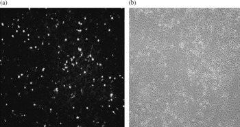
Cloning of the 399 bp FABP gene in the correct reading frame with the vector signal sequence was confirmed by sequencing of the gene. In vitro transfection of the MDCK cell line with recombinant plasmid DNA showed efficient transfection and expression of the fatty acid binding protein by immunofluorescence assay. Distinct fluorescence in the cell monolayer transfected with the DNA–FABP plasmid, with no fluorescence in the cells transfected with non-recombinant plasmid, confirmed the expression of the protein in the MDCK cells (fig. 1). However, when the cells were treated with varying concentrations of PEI, they died and were detached from the monolayer. Also, transfection efficiency with PEI complexed DNA (N/P = 4) was lower than that shown by DNA–lipofectamine reagent (data not shown).
Immunofluorescence assay showing in vitro transfection of MDCK cells with pVR1020–FABP plasmid: (a) with fluorescence in the recombinant vector-transfected cells; (b) cells transfected with a non-recombinant plasmid.

Stability of the PEI–DNA complex to DNase degradation
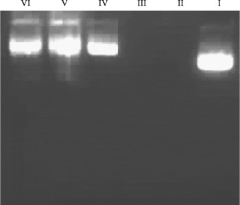
The protective effect of the polymer on its condensation of the DNA against DNase degradation was confirmed by in vitro DNase I treatment of the PEI–DNA complex, followed by its heparin treatment. Naked DNA was completely degraded on incubation with DNase, as detected by agarose gel electrophoresis. However, condensation of DNA with PEI protected it from DNase degradation, though a certain degree of nicking occurred after the treatment. There was retardation of the electrophoretic mobility of the PEI–DNA complex, with quenching of fluorescent signal on ethidium bromide staining (fig. 2). These results confirmed the complexation of the cationic polymer with the DNA, leading to its protection from DNase degradation.
DNase degradation assay for confirmation of DNA–PEI complex formation. Lanes: I, control DNA; II, plasmid DNA treated with DNase I at 37°C for 30 min; III, plasmid DNA–PEI complex (ethidium bromide quenching); IV, plasmid DNA–PEI complex showing dissociation of DNA from PEI with heparin treatment; V and VI, plasmid DNA–PEI complex showing intact DNA after DNase I treatment, followed by dissociation of the complex with heparin.

Humoral response in mice
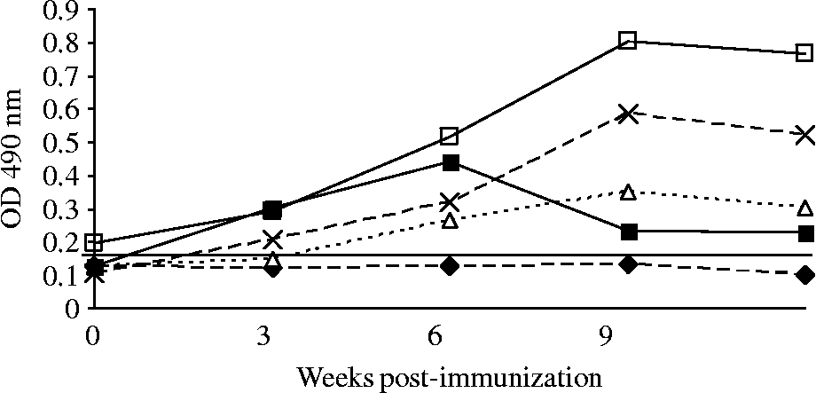
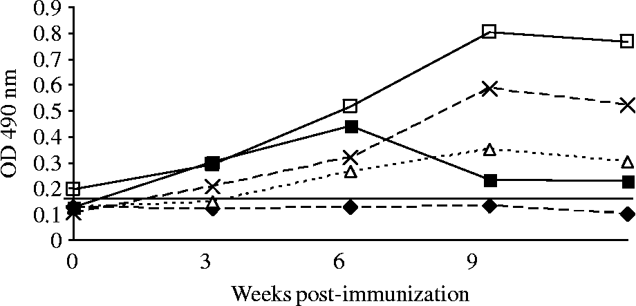
The antibody (IgG) response was probed in all the groups of mice receiving 50 μg naked DNA and PEI-complexed DNA at 10, 25 and 50 μg concentration, respectively, at weekly intervals post-immunization. Mice receiving 50 μg naked DNA showed an increase in the antibody titre after the first booster, which increased gradually at the third booster and maintained a plateau until the termination of the experiment. The isotype response of these mice to DNA immunization indicated a mixed type Th1 and Th2 response, showing elicitation of IgG1, IgG2a and IgG2b immunoglobulins. These isotypes increased from the first booster and maintained a plateau until the termination of the experiment. The IgG2a response was dominant over IgG1 and IgG2b (fig. 3).
ELISA showing the mean antibody isotype response in mice receiving 50 μg naked DNA for four immunizations given at 3-week intervals. □, IgG; × , IgG2a; △, IgG1; ■, IgG2b; ♦, healthy control.
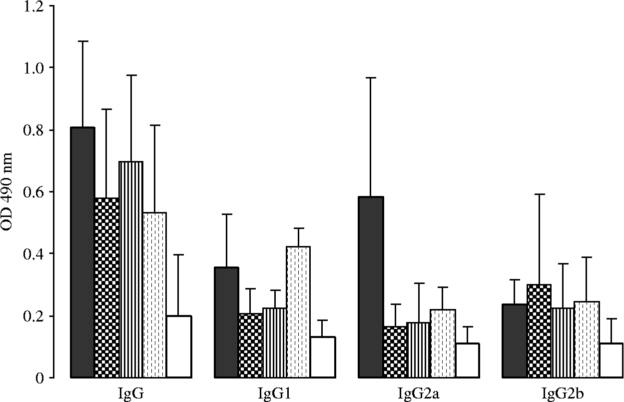
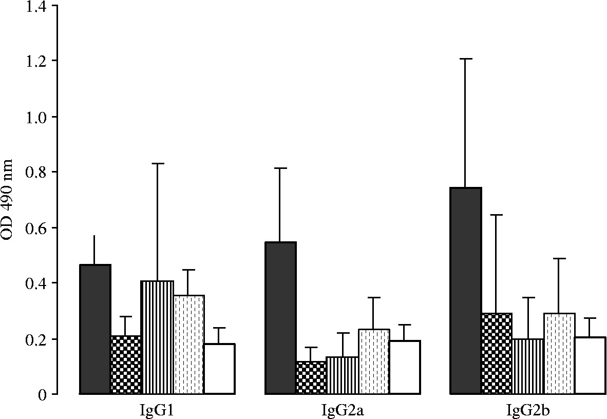
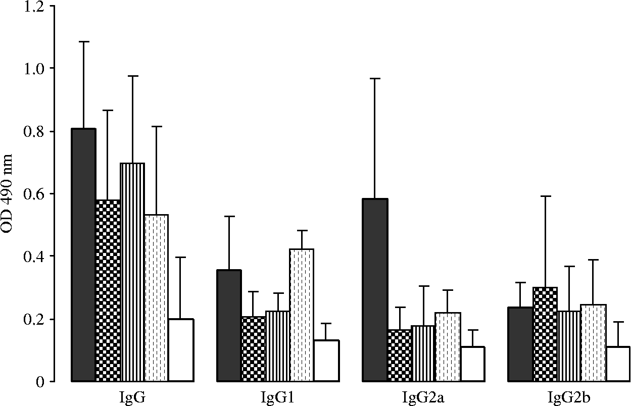
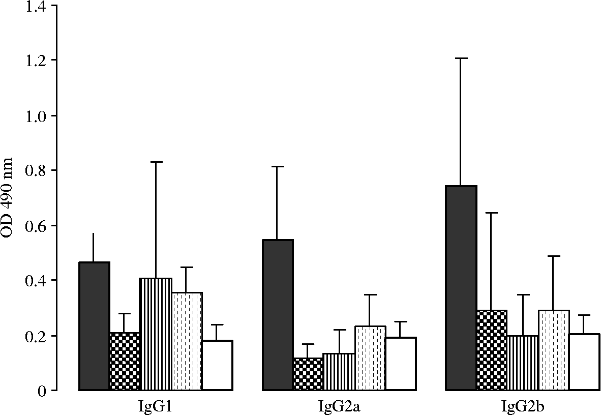
Mice receiving 10, 25 and 50 μg DNA complexed with PEI showed a lower response of IgG antibodies from first immunization to third booster, when compared with the naked DNA group (fig. 4). With each immunization dose, there was no perceptible increase in the titres of IgG and other isotypes, with the exception of one or two mice in each PEI group which showed an antibody response comparable to the naked DNA group. No significant difference in the antibody titres was observed between the naked DNA group and other PEI-treated groups, except for IgG2a, where the difference was significant at P < 0.05. Likewise, IgG1, IgG2a and IgG2b immunoglobulins did not show any significant increase in their titre with each immunization dose at N/P ratios of 4, 6 and 8, respectively (fig. 5). The difference in the antibody titres was significant between naked DNA and other groups with relation to IgG2a and IgG2b, but not with respect to IgG1 (P < 0.05).
Mean immunoglobulin isotype response of mice immunized with different concentrations of DNA complexed to PEI at a N/P ratio of 4. ■, 50 μg naked DNA;  , 10 μg DNA complexed to PEI;
 , 25 μg DNA complexed to PEI;
 , 50 μg DNA complexed to PEI; and □, healthy control (antibody titre probed after the third booster).
Mean immunoglobulin isotype response in mice immunized with 50 μg naked DNA or constant concentration of DNA complexed with PEI but at different N/P ratios. ■, naked DNA; , N/P ratio 4; , N/P ratio 6; , N/P ratio 8; and □, healthy control (antibody titre probed after the third booster).
In both the groups of mice receiving either naked DNA or PEI-condensed DNA, 50–60% mice were responders to immunization, as determined by their antibody response. The control group of mice, receiving PEI only, did not evoke any antibodies to the polymer.
Antibody response and fluke recovery in rabbits
The OD values of the antibody (IgG) ranged between 0.211 and 0.990 in the group receiving 100 μg naked DNA, and in the group administered with DNA–PEI (N/P = 8) it ranged from 0.240 to 0.427, showing that the antibody response was not enhanced in these animals, in spite of using PEI as adjuvant. Only 4/10 and 3/10 rabbits in the naked DNA and PEI groups, respectively, responded to immunization by producing anti-FABP antibodies, whereas after metacercarial challenge 50% and 30% of the rabbits in the naked DNA and DNA–PEI groups, respectively, evoked anti-FABP antibodies with a titre of 0.447–0.705. The antibody response of rabbits of the second experimental groups, administered with 25 μg DNA–PEI (N/P = 4) or naked DNA, was not probed, but instead the protection level with respect to a reduction in fluke burden was studied in these groups.
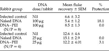
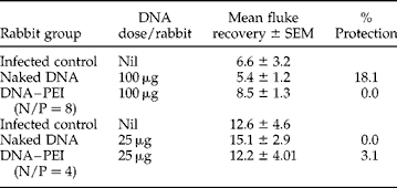
Immunization of rabbits with 100 μg DNA/rabbit showed a mean fluke recovery of 5.4 in the naked DNA group, 8.5 in DNA–PEI (N/P = 8) group and 6.6 in the infected control group. The naked DNA group showed 18.1% protection, which was statistically insignificant (P>0.7), while the DNA–PEI group showed 0.0% protection. Rabbits administered with 25 μg DNA/rabbit, had a mean fluke recovery of 15.1 in the naked DNA group, 12.2 in the DNA–PEI group and 12.6 in the infected control group, respectively. The naked DNA group showed 0.0% and the DNA–PEI group 3.1% protection, which was statistically insignificant (P>0.05) (table 1).
Fluke recovery in rabbits immunized with 100 μg DNA–PEI complex, 25 μg DNA–PEI complex and naked DNA, respectively.
% Protection = 1 − (mean number of flukes in vaccinated rabbits/mean number of flukes in challenged control rabbits) × 100.
There were no signs of retardation of parasite growth or reduction of liver pathological lesions in response to naked or PEI-complexed DNA immunization, when compared with the infected control group.
Muscle cytotoxicity
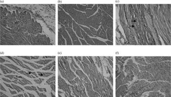
PEI-administered mice showed moderate infiltration with polymorphs, mononuclear cells and fibroblasts in the dermis, while adjoining muscles had necrotic debris between the fibres and centrally placed chain of nuclei, indicating regeneration. In the DNA-injected mice, there was only a mild fibrocellular reaction within the dermis, while muscle fascicles remained unaffected. Mice inoculated with PEI-condensed DNA had muscle fascicles showing a varying degree of infiltration and muscle necrosis, which was inversely related to the N/P ratio, with higher level of cytotoxicity at N/P = 4 than at N/P ratios of 6 and 8 (fig. 6).
Thigh muscle sections of mice, stained with haematoxylin and eosin (magnification × 20). Muscle fibres injected with (a) DNA and (b) glucose, show apparently normal architecture, whereas the 12 μg PEI-injected site (c) shows necrosis and regeneration of muscle fibres (arrows). In the lower panel, muscle injected with PEI–DNA at N/P = 4 (d) shows mild necrosis of muscle fibres (arrows), while at N/P = 6 (e) and N/P = 8 (f) no significant changes are observed.

Discussion
Several studies aimed at developing a viable vaccine against fasciolosis have raised a realistic possibility of immunological control of this parasite, though at present there is no vaccine available for the control of infection. A number of Fasciola proteins, including fatty acid binding protein, cathepsin-L, glutathione-S-transferase, myosin, paramyosin and fluke haemoglobin, are under evaluation for developing a viable vaccine against fasciolosis.
The present investigation was aimed at studying the adjuvant effect of polyethylenimine administered with plasmid DNA encoding F. gigantica FABP in mice and rabbits, and the kinetics of the immune responses generated thereof. Mice immunized with the naked DNA–FABP construct evoked an FABP-specific humoral immune response. This response was a mixed-type Th1/Th2, with both IgG1 and IgG2 antibodies being elicited and IgG2a being predominant over IgG1 and IgG2b. These results are in agreement with those of Smooker et al. (Reference Smooker, Kennedy, Steeper, Christopoulos and Spithill2001), where F. gigantica FABP DNA induced an IgG1-dominant response with significant IgE, IgG2a and IgG2b responses (indicating a mixed Th1/Th2 response), while immunization with F. hepatica cathepsin-L5 DNA induced a Th2 response in mice. A mixed Th1/Th2 type of immune response to vaccination with F. hepatica saposin-like protein-2 in BALB/c mice, with the production of IgG1, IgG2, IgE and high levels of interleukin-10 (IL-10) and interferon-γ (IFN-γ), was found (Espino et al., Reference Espino, Osuna, Gil and Hillyer2005). Clonorchis sinensis FABP encoding plasmid DNA induced predominantly IgG2 antibodies, with production of IFN-γ but no IL-4 in Sprague–Dawley rats, indicating a typical Th1-dominated immune response with this protein (Lee et al., Reference Lee, Kim, Sohn, Lee and Yong2006). Kofta et al. (Reference Kofta, Mieszczanek, Plucienniczak and Wedrychowicz2000) reported 100% and 74% protection in male and female rats, respectively, on DNA immunization with F. hepatica cysteine proteinase. None of these vaccination studies attempted any strategic use of gene delivery vehicles or ligand-directed targeting of the parasite gene to the host tissue. Strategic targeting of a DNA vaccine encoding F. hepatica pro-cathepsin B antigen to the immune cells in sheep, by combining the use of CpG motifs with cytotoxic lymphocyte antigen 4 (CTLA-4) mediated targeting, with the aim of eliciting both antibody and cellular responses, was reported for the first time by Kennedy et al. (Reference Kennedy, Spithill, Tennent, Wood and Piedrafita2006).
Polyethylenimine, a cationic polymer, is known to be an efficient carrier of DNA, whose transfection efficiency lies in extensive endosome buffering with its consequent swelling and rupture, thus providing the DNA–PEI particles an escape mechanism in the cell (Boussif et al., Reference Boussif, Lezoualc'h, Zanta, Mergny, Scherman, Demeneix and Behr1995). But in the present experimental study, PEI-complexed DNA administration in the mouse muscle cells did not seem to have a positive effect on DNA transfection, as it failed to enhance the antibody response, when compared with mice receiving naked DNA. Mice receiving 10, 25 and 50 μg DNA complexed to PEI, respectively, at a N/P ratio of 4, did not evoke any perceptibly higher humoral response. Different N/P ratios of 4, 6 and 8 again had no positive effect on the antibody response in these mice. Similar results in rabbits with DNA–PEI polyplex administration indicated no enhanced adjuvanticity of the PEI in both these animal species. This is in contrast to several other reports on DNA vaccination using PEI as a versatile adjuvant (Boussif et al., Reference Boussif, Lezoualc'h, Zanta, Mergny, Scherman, Demeneix and Behr1995; Oh et al., Reference Oh, Park, Kang, Ko, Kim and Kim2003, Garzon et al., Reference Garzon, Berraondo, Crettaz, Ochoa, Vera, Lasarte, Vales, Van Rooijen, Ruiz, Prieto, Zulueta and Gonzalez-Aseguinolaza2005). Systemically delivered PEI–DNA complexes resulted in the transduction of lung, spleen, liver, heart, brain and kidney, with no histological change observed in these organs (Oh et al., Reference Oh, Kim, Yoon, Kim, Yang and Kim2001). Garzon et al. (Reference Garzon, Berraondo, Crettaz, Ochoa, Vera, Lasarte, Vales, Van Rooijen, Ruiz, Prieto, Zulueta and Gonzalez-Aseguinolaza2005) demonstrated that a single immunizing dose of human immunodeficiency virus (HIV) gp-120 complexed with linear PEI was able to induce a strong cellular immune response capable of providing both systemic and mucosal protection in mice against vaccinia virus challenge. Enhanced adjuvanticity of murine IL-2 plasmid DNA when administered with PEI in mice intramuscularly was reported by Oh et al. (Reference Oh, Park, Kang, Ko, Kim and Kim2003). PEI has been used for in vivo gene delivery in a variety of cells, since it has high transfection efficiency due to its proton sponge effect over a broad pH range.
However, cytotoxic effects associated with PEI-condensed DNA administration have been reported. Wang et al. (Reference Wang, Zhang, Mao and Leong2002) reported an inflammatory response and severe necrosis in all the muscle samples receiving PEI injection at a dose of 2.5 μg. Local toxicity after intramuscular injection of PEI in mice, with lower transfection of muscle cells with DNA–PEI complex versus naked DNA have been observed (Anderson et al., Reference Anderson, Peng, Akinc, Hossain, Kohn, Padera, Langer and Sawicki2004; Chang et al., Reference Chang, Choi, Kim, Yockman, Christensen, Kim and Kim2007). Toxic effects of DNA–PEI in liver at higher concentrations of DNA in intravenous immunization resulted in the suppression of immune response (Garzon et al., Reference Garzon, Berraondo, Crettaz, Ochoa, Vera, Lasarte, Vales, Van Rooijen, Ruiz, Prieto, Zulueta and Gonzalez-Aseguinolaza2005). In the present study also, administration of PEI alone (12.5 μg) in mice thigh muscles caused significant muscle cytotoxicity, but condensation of DNA with PEI had a lesser toxic effect on the muscle cells, which was inversely related to N/P ratio. Although side-effects associated with PEI–DNA complex administration have been reported, the evidence is still contradictory (Chollet et al., Reference Chollet, Favrot, Hurbin and Coll2002; Kang et al., Reference Kang, Lim, Seo, Jeon, Lee and Kim2008). This could be due to the use of PEI with different molecular weights, structures and chemical modifications, and different nitrogen/phosphate ratios (Wightman et al., Reference Wightman, Kircheis, Rossler, Carotta, Ruzicka, Kursa and Wagner2001; Kunath et al., Reference Kunath, Harpe, Fischer, Petersen, Bickel, Voigt and Kissel2003; Garzon et al., Reference Garzon, Berraondo, Crettaz, Ochoa, Vera, Lasarte, Vales, Van Rooijen, Ruiz, Prieto, Zulueta and Gonzalez-Aseguinolaza2005). Also, the presence of glycoaminoglycans (GAGs) in the extracellular matrix of skeletal muscle cells has been proposed to inhibit binding of the cationic polyplex to the cell membrane and to destabilize cationic polyplex formation, leading to reduction of muscle cell transfection (Chang et al., Reference Chang, Choi, Kim, Yockman, Christensen, Kim and Kim2007). Therefore, failure of polyethylenimine (branched, 25 kDa) in the present study to boost the effective immune response in both the animal species could be attributed to either cytotoxicity associated with this cationic polymer or to muscle cells being an unsuitable target for this polymer (Wang et al., Reference Wang, Zhang, Mao and Leong2002; Anderson et al., Reference Anderson, Peng, Akinc, Hossain, Kohn, Padera, Langer and Sawicki2004; Chang et al., Reference Chang, Choi, Kim, Yockman, Christensen, Kim and Kim2007).
Acknowledgements
The authors are thankful to the Director, Indian Veterinary Research Institute, Izatnagar, India, for providing the necessary facilities to conduct this research work.