Introduction
The parapharyngeal space is a complex anatomical region located between the mandibular ramus and the lateral pharynx and extending as an inverted pyramid from the skull base superiorly down to the hyoid bone inferiorly. A parapharyngeal space tumour is a comparatively rare disease which is difficult to diagnose and treat. Moreover, because important internal organs are located in the cervical vertebrae, such as the cervix, cranial nerves (IX to XII) and sympathetic nerves, extreme care is required during surgery.
This study examined clinical aspects of cases of parapharyngeal space tumour treated by our department.
Patients and methods
Twenty-four patients with parapharyngeal space tumours were treated at the Kurume University Hospital department of otolaryngology between 1990 and 2007. Patients comprised 13 males and 11 females ranging in age from 11 to 80 years (mean 51 years). The primary tumour site involved the parotid glands (n = 10), nerve origins (10; vagal nerve, hypoglossal nerve, sympathetic nerve and glossopharyngeal nerve) or one small salivary gland; the primary tumour site was uncertain in eight cases. Surgical excision was performed in 22 patients and radiation therapy was administered to two patients. One case in which the tumour barely extended into the parapharyngeal space was excluded. The subjective symptoms, location, pathology, organisation, diagnosis, surgical approach and coexisting morbidity were all examined.
Results
Subjective symptoms
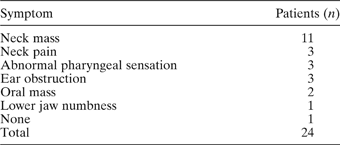
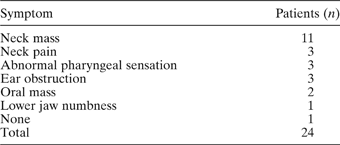
The patients' subjective symptoms were: neck mass (n = 11), neck pain (three), abnormal pharyngeal sensation (three), ear obstruction (three), oral mass (two) and lower jaw numbness (one) (Table I).
Subjective symptoms
Tumour location
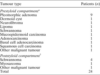
The parapharyngeal space tumours were found in the prestyloid compartment in 18 patients and in the poststyloid compartment in six. All of the pleomorphic adenomas were found in the prestyloid compartment. Four schwannomas and one myosarcoma were found in the poststyloid compartment (Table II).
Relation between compartment and tumour type

*n = 18; †n = 6.
Pathology
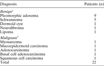
The histological diagnosis was generally obtained from examination of the surgically excised tumour (22/24). Seventy-seven per cent (17 of 22) of the parapharyngeal space tumours were benign and 23 per cent (five of 22) were malignant. Thirty-six per cent (eight of 22) were of salivary origin, 23 per cent (five of 22) were neurogenic and 41 per cent (nine of 22) were of miscellaneous origin. Table III presents the most commonly encountered histological diagnoses.
Histopathological diagnoses

*n = 17; †n = 5.
Surgical approaches
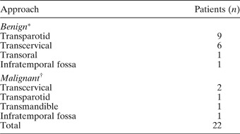
The benign parapharyngeal space tumours were surgically managed by the transparotid approach in 53 per cent of patients (nine of 17), transcervical removal in 35 per cent (six of 17), transoral removal in 6 per cent (one of 17) and infratemporal fossa approach in 6 per cent (one of 17). The malignant parapharyngeal space tumours were surgically managed by transcervical removal in 40 per cent of patients (two of five), transparotid approach in 20 per cent (one of five), transmandible approach in 20 per cent (one of five) and infratemporal fossa approach in 20 per cent (one of five) (Table IV).
Surgical approach

*n = 17; †n = 5.
Complications
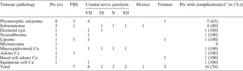
Post-operative complications were identified in 16 of the 22 patients and are listed in Table V. The most common complication, facial nerve paralysis, was identified in eight patients, seven of whom demonstrated first bite syndrome; all schwannoma complication resolved in all schwannoma patients within six months. However, post-operative nerve paralysis associated with schwannoma resection was permanent.
Complications

*Some patients experienced more than one complication. Pts = patients; FBS = first bite syndrome; Ca = carcinoma
Discussion
The parapharyngeal compartment is a potential space shaped like an inverted pyramid, with its base at the cranial base and its apex at the greater cornu of the hyoid bone. It is defined superiorly by the temporal bone, posteriorly by the vertebrae and prevertebral muscles enveloped by the prevertebral fascia, and laterally by the mandible, the posterior belly of the digastric muscle and the medial pterygoid muscle.Reference Carrau, Myers and Johnson1–Reference Takahashi3
The fascia from the styloid process to the tensor velipalatini divides the parapharyngeal space into anterolateral, prestyloid and poststyloid compartments. The prestyloid compartment is occupied by the parotid gland, fat and possibly lymph nodes. The poststyloid compartment contains the carotid sheath with the internal carotid artery, internal jugular vein, cranial nerves IX to XII, and lymph nodes. Tumours arising in these compartments may originate from any of the structures normally occupying each compartment.Reference Work and Hybels4, Reference Batsakis and Neige5
The clinical picture of parapharyngeal space tumours may be diverse and non-specific, and patients may present with a wide variety of symptoms and signs.Reference Carrau, Myers and Johnson1, Reference Tomita, Ogawa, Tagawa, Fujii and Nameki6–Reference Tsuji, Yagi, Minamino, Nagata, Yukawa and Minami8 In the current study, the most common symptom was a neck mass. All such masses presenting as bulging in the oropharyngeal area measured greater than 3 cm, an observation consistent with the findings of Som et al. Reference Som, Biller and Lawson9 Only one patient presented with an asymptomatic mass found incidentally during a routine physical examination.
The parapharyngeal compartment is divided anterolaterally by the styloid process and the fascia that adheres to it.Reference Som, Biller and Lawson9 Tumours in the prestyloid compartment are probably of parotid gland, whereas tumours in the poststyloid compartment are usually paragangliomas or of neurogenic origin. In the current study, all pleomorphic adenomas, and almost all malignant tumours, were found in the prestyloid compartment, while four of the five schwannomas were found in the poststyloid compartment. It is felt that surgery is automatically indicated for prestyloid parapharyngeal space tumours, because the surgical risk is lower than the risk of inaction.Reference Tomita, Ogawa, Tagawa, Fujii and Nameki6, Reference Sumi and Kishimoto10 This is especially true when the tumour is malignant.
In the current study, five (24 per cent) of 21 patients had malignant tumours, and four of these five patients had a poor prognosis. It is important to consider the existence of a malignant tumour, and it is necessary to diagnose such tumours at an early stage and to treat them as soon as possible.
The surgical approaches used were categorised into several different types.Reference Yoshihara11 A transcervical approach accesses the parapharyngeal space tumour through the neck and does not require a mandibulotomy. A transcervical-transparotid approach combines a neck and parotid dissection in order to allow identification of the facial nerve. An extended approach was employed in cases in which the digastric, styloid musculature, stylomandibular ligament and/or styloid process were removed. A transcervical-mandibulotomy dissection extended the cervical exposure with a mandibulotomy. The transcervical-transmastoid approach allowed the exposure to proceed superiorly from the neck by removing the mastoid and potentially exposing the middle cranial fossa. In this report, the surgical approaches used for benign tumours included transoral, transcervical and transcervical-transparotid approaches (excluding one case of recurrence). The surgical approaches used for malignant tumours included the transcervical-transparotid and transmandible approaches and infratemporal fossa dissection.
An important concern in parapharyngeal space tumour surgery is the potential for injury to cranial nerves VII, IX, X, XI and XII and to the sympathetic trunk, due to the proximity to or involvement of these nerves with the local disease. With the exception of direct invasion by malignancy or a neurogenic tumour, permanent cranial nerve injury is uncommon. In the current series, facial nerve paralysis was the most common post-operative complication, occurring in eight patients. Seven of these patients demonstrated first bite syndrome, which involves the development of pain in the parotid region after the first bite of each meal and can result from surgery of the parapharyngeal space. The cause is unclear but has been thought to involve a loss of sympathetic nerve function to the parotid, thus causing a denervation supersensitivity of the salivary gland myoepithelial cells.Reference Chiu, Cohen, Burningham, Anderson and Dabidson12, Reference Netterville, Jackson and Miller13 Surgeons who operate in the parapharyngeal space should therefore be aware of first bite syndrome. Patients should be counselled regarding this potential complication, and surgeons should consider first bite syndrome a potential consequence of injury or sacrifice of the sympathetic chain or its branches to the parotid gland.
Conclusion
Because of their complexity, parapharyngeal space tumour masses are challenging lesions to treat. Patient comorbidity and pre-operative functional deficits, as well as the tumour type, pathology, histology and anatomy, will influence management. The tumour location, i.e. prestyloid or poststyloid, does not in itself appear to influence the type of resection. Tumour vascularity, skull base involvement and the potential risk to the facial nerve are key factors. In consideration of the high incidence of post-operative complications, the surgical approach should therefore be carefully selected when treating parapharyngeal space tumours.