Introduction
The African catfish Clarias gariepinus Burchell 1822 is an economically important siluriforme species that contributes significantly to annual fish productions in many countries of the world, but most especially in Africa and many parts of Asia. It is cultured both intensively and extensively in Africa and Asia (Huisman & Richter, Reference Huisman and Richter1987), but most intensively in some countries in Europe (Galbusera et al., Reference Galbusera, Volckaert and Ollevier2000) and in many continents across the world (Galbusera et al., Reference Galbusera, Volckaert and Ollevier2000). This species belongs to the Clariidae family teleost catfish that is found mostly as freshwater fish, except for two families, Ariidae and Plotosidae, that have marine species (Teugels, Reference Teugels1996). C. gariepinus is also referred to as the sharptooth catfish because of their fine, pointed bands of teeth (Bovendeur et al. Reference Bovendeur, Eding and Henken1987; Skelton, Reference Skelton1993). They have the highest diversity in Africa and also populate North, Central and South America, Eurasia, Japan, Australasia, Asia Minor, South and Southeast Asia, (Ozouf-Costaz et al., Reference Ozouf-Costaz, Teugels and Legendre1990; Richter et al., Reference Richter, Eding, Verreth, Fleuren, Bromage and Roberts1995; Teugels, Reference Teugels1996; Agnèse & Teugels, Reference Agnèse and Teugels2001), but are mostly cultured in tropical and subtropical Africa and South-East Asia (Lenormand et al., Reference Lenormand, Slembrouck, Pouyaud, Subadgja, Legendre, Legendre and Pariselle1988; Hecht et al., Reference Hecht, Oellermann and Verheust1996). They are bottom dwellers but obligate air-breathers. They inhabit a variety of freshwater environments, including quiet waters such as lakes, ponds, and pools; are very prominent in flowing rivers, rapids, and around dams; and very adaptive to extreme environmental conditions. C. gariepinus can live in very poorly oxygenated and eco-degraded waters (Pienaar, Reference Pienaar1968). They can live at a pH range of 6.5–8.0; and at temperatures of 8–35°C with their optimal growth temperature being 28–30°C (Teugels, Reference Teugels1986).
At this time, C. gariepinus has become increasingly important globally, being a good source of protein as well as being easy to culture, plus having a high growth rate and resistance to diseases. Moreover, it can tolerate high stocking densities, a wide range of temperatures, low dissolved oxygen as well as high salinity levels, and most importantly it has low production input requirements and high returns (Teugels, Reference Teugels1986; Bovendeur et al., Reference Bovendeur, Eding and Henken1987; Goos & Richter, Reference Goos and Richter1995; Omitogun & Aluko, Reference Omitogun and Aluko2002; Mahmoud et al., Reference Mahmoud, Mekkawy and Sayed2009). With these attributes, C. gariepinus species are among other farmed species that contribute greatly to the total world fish supply (Bromage & Roberts, Reference Bromage and Roberts1995; FAO, 2001); however in most developing countries of Africa and Asia, production is not optimally reached to meet the rising demand of fish because of lack of seed and affordable good quality feeds especially in Nigeria where catfish aquaculture is gaining popularity among resource-poor as well as large-scale commercial farmers (Omitogun et al., Reference Omitogun, Ilori, Olaniyan, Amupitan, Oresanya, Aladele, Odofin and Katkov2012).
Aquaculture development is needed for sustainable food fish production in order to supplement capture fisheries. This situation is necessary to bridge the existing productivity gap between fish supply and demand in many parts of African countries. In view of this factor, a sustainable production of quality fish seed will provide a significant solution to challenges being faced by farmers. Moreover, adequate information on egg, embryonic and larval development is essential for successful seed production by employing genetic manipulation techniques. The present study will aid the understanding of their embryology and ontogenic development.
Some embryological studies on C. gariepinus (Legendre & Teugels, Reference Legendre and Teugels1991; Haylor & Mollah, Reference Haylor and Mollah1995) as well as other species, viz. Tilapia nilotica (Galman, Reference Galman1980), Rhombosolea tapirina and Ammotectis rotratus (Crawford, Reference Crawford1986), Oreochromis niloticus (Galman & Avtalion, Reference Galman and Avtalion1989), Heterobranchus longifilis (Legendre & Teugels, Reference Legendre and Teugels1991), zebrafish (Kimmel et al., Reference Kimmel, Ballard, Kimmel and Ullmann1995), and Heteropneustes fossilis (Puvaneswari et al., Reference Puvaneswari, Marimuthu, Karuppasamy and Haniffa2009; Korzelecka-Orkisz et al., Reference Korzelecka-Orkisz, Smaruj, Pawlos, Robakowski, Tanski, Szulc and Formicki2010) have been carried out. Further studies with focus on chronological organogenesis are significant to a full understanding of the early embryological and larval developments vis-à-vis incubation temperature, transitional stage, quasi-peristaltic and cardiac movement of the forming embryo, mechanical effect on the eggs, hatching moment and final hatching until complete yolk absorption. The chronological developmental stages of C. gariepinus described in this study will be indispensable in the genetic manipulation investigation to produce monosex and polyploid catfish for aquaculture.
Materials and methods
Fish sample and hypophysation
The study was carried out in June 2011 in the Wet and Biotechnology Laboratories of the Department of Animal Sciences, Obafemi Awolowo University, Ile-Ife, Nigeria. C. gariepinus breeders used were from stock held at the Wet Laboratory of the Department. Breeders were kept in standard conditions as described by Huisman & Richter (Reference Huisman and Richter1987). Sexually matured male breeders (weight of 1500 ± 100 g) were identified by their pinkish-coloured pointed genital papillae and the gravid females by free oozing out of eggs upon gentle pressure on their abdomen from the pinkish or reddish swollen vents (Puvaneswari et al., Reference Puvaneswari, Marimuthu, Karuppasamy and Haniffa2009). Artificial breeding was carried out using Ovaprim® (Syndel, Canada) as inducer and following standard protocols (Váradi et al., Reference Váradi, Benkó, Varga and Horváth1999). Fertilized eggs from three females and four males were then incubated in a closed circulating aquarium, and 10 sampled fertilized oocytes were incubated in a Petri dish at controlled temperature of 28.5 ± 0.5°C.
Microscopy and photomicrography
Embryonic development was observed, monitored, photographed and documented live with the aid of a light microscope (Olympus Trinocular Microscope XSZ-156T) with its digital camera (DCM 130, Res 1.3 M pixels) and a PC computer. Ten sampled fertilized oocytes in a Petri dish were mounted on the microscope. With these samples, the morphological developments, organogenesis, pigmentation and the time sequence of the different developmental stages were recorded. Upon yolk absorption, larvae were fed ad libitum with Artemia naupli shell-free feed, 54% Crude Protein (INVE®; Aquaculture Nutrition, Ogden, Utah, USA).
Results
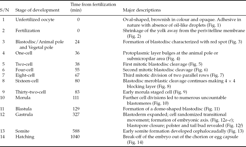
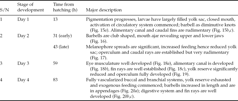
Matured unfertilized eggs were examined under the microscope and fertilized; early and post-embryonic developments were observed and photographs were taken from 0 min of fertilization through a cell stage to alevin and complete yolk absorption, fry swimming stages larvae and exogenous feeding level (Figs. 1–21). Hatchability rate was 85% at the controlled temperature of 28.5 ± 0.5°C. Tables 1 and 2 show the average timing of the embryonic and post-embryonic and larval developmental stages respectively. This study is the first time that early, post-embryonic and larval development of C. gariepinus was captured with full sequential photomicrographs and timing of transition revealed.
Embryonic developmental stages in Clarias gariepinus with respect to timing at 28.5 ± 0.5°C

Post-embryonic and larval developmental stages in Clarias gariepinus

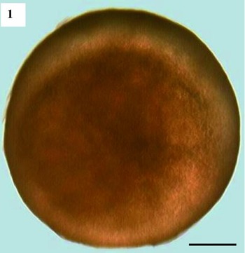
Embryonic developmental stages in Clarias gariepinus. Unfertilized egg. Scale bar: 1 mm. Magnification: ×40.

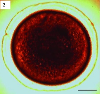
Embryonic developmental stages in Clarias gariepinus. Fertilized egg. Scale bar: 1 mm. Magnification: ×40.

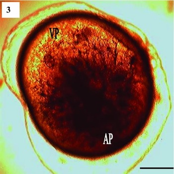
Embryonic developmental stages in Clarias gariepinus. Animal pole (AP) and vegetal pole (VP). Scale bar: 1 mm. Magnification: ×40.

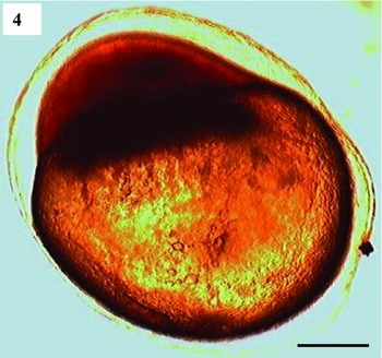
Embryonic developmental stages in Clarias gariepinus. One-cell stage. Scale bar: 1 mm. Magnification: ×40.

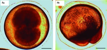
Embryonic developmental stages in Clarias gariepinus. Two-cell stage: (a) top view; (b) lateral view. Scale bar: 1 mm. Magnification: ×40.

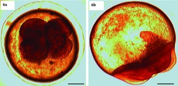
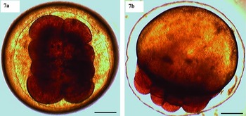
Embryonic developmental stages in Clarias gariepinus. Four-cell stage: (a) top view; (b) lateral view. Scale bar: 1 mm. Magnification: ×40.

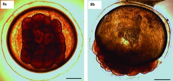
Embryonic developmental stages in Clarias gariepinus. Eight-cell stage: (a) top view; (b) lateral view. Scale bar: 1 mm. Magnification: ×40.

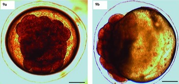
Embryonic developmental stages in Clarias gariepinus. Sixteen-cell stage: (a) top view; (b) lateral view. Scale bar: 1 mm. Magnification: ×40.

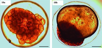
Embryonic developmental stages in Clarias gariepinus. Thirty-two-cell stage: (a) top view; (b) lateral view. Scale bar: 1 mm. Magnification: ×40.

Embryonic developmental stages in Clarias gariepinus. Morula: (a) top view; (b) lateral view. Scale bar: 1 mm. Magnification: ×40.

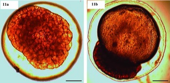
Embryonic developmental stages in Clarias gariepinus. Blastula: (a) top view; (b) lateral view. Scale bar: 1 mm. Magnification: ×40.

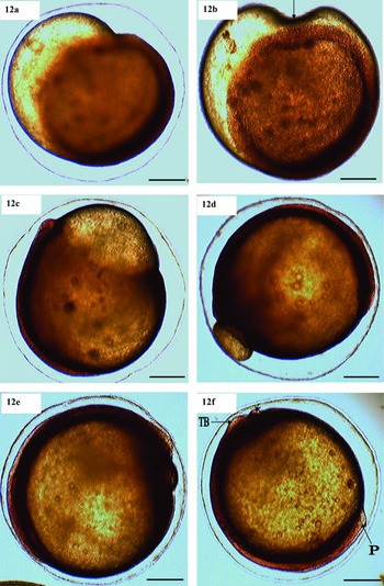
Embryonic developmental stages in Clarias gariepinus. Gastrula: (a) early gastrula; (b) 50% epiboly, arrow shows transitional wave movement; (c) 85% epiboly with blastopore wide open; (d) 95% epiboly with blastopore just about closing up; (e) epiboly complete; (f) gastrulation ends and early somite blocks formation. Arrow-head shows total closure of blastopore. P, polster; TB, tail bud. Scale bar: 1 mm. Magnification: ×40.

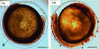
Embryonic developmental stages in Clarias gariepinus. Somite: (a) somite stage; (b) somite completed (moment before hatching). SO, somite block. Scale bar: 1 mm. Magnification: ×40.

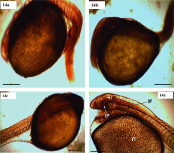
Embryonic developmental stages in Clarias gariepinus. Larva: (a) hatching larva; (b) hatched larva (agitating); (c) hatched larva (calm); (d) hatched larva (24 h). OP, optic primordium; OV, otic vesicle; YS, yolk sac. Scale bar: 1 mm. Magnification: ×40.

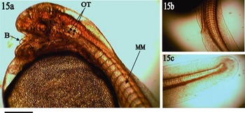
Post-embryonic developmental stages in Clarias gariepinus. (a) Barbells as diminutive knots at first day of hatching. (b) Rudimentary excretory system at first day of hatching. (c) Thick tail bud, fin yet to form at first day of hatching. B, barbell; MM, myotomal muscle; OT, otoliths. Scale bar: 1 mm. Magnification: ×40.

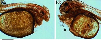
Post-embryonic developmental stages in Clarias gariepinus. (a) Eye coverings partly uncovered at the early hours of second day of hatching. (b) Mouth ajar, barbells developed to club shaped at early hours of second day of hatching. B, barbell. Scale bar: 1 mm. Magnification: ×40.

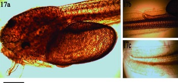
Post-embryonic developmental stages in Clarias gariepinus. (a) Melanophores spreads and reduced yolk sac are significant at late hours of second day of hatching. (b) Alimentary system is prominent at late hours of second day of hatching. (c) Early formation of caudal fin rays at late hours of second day of hatching. Scale bar: 1 mm. Magnification: ×40.

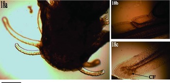
Post-embryonic developmental stages in Clarias gariepinus. (a) Eye musculature well developed at third day of hatching. (b) Developed alimentary canal at third day of hatching. (c) Developed caudal fins at third day of hatching. CF, caudal fin. Scale bar: 1 mm. Magnification: ×40.

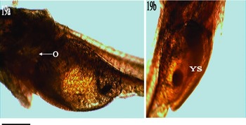
Post-embryonic developmental stages in Clarias gariepinus. (a) Reduced yolk sac and developed operculum at third day of hatching. (b) Well reduced yolk sac and developed operculum at third day of hatching. O, developed operculum; YS, reduced yolk sac. Scale bar: 1 mm. Magnification: ×40.

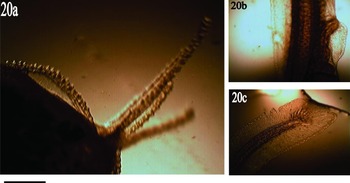
Post-embryonic developmental stages in Clarias gariepinus. Larval stages (fourth day of hatching) in Clarias gariepinus: (a) increased appendage barbells; (b) well developed alimentary canal; (c) well developed fins. Scale bar: 1 mm. Magnification: ×40.

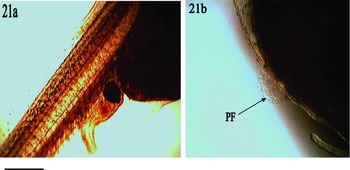
Post-embryonic developmental stages in Clarias gariepinus. Larval stages (fourth day of hatching) in Clarias gariepinus: (a) translucent larva with well developed alimentary canal; (b) well developed pectoral fin at second week old (depicted by arrow). PF, pectoral fin. Scale bar: 1 mm. Magnification: ×40.

The chronological study of embryological stages of development with respect to timing is a very significant aspect in C. gariepinus fish biology and subsequent manipulations, thereby explaining the critical importance of physical parameters such as temperature in successful hatching. Moreover, timing is helpful to determine the exact duration needed to block the extrusion of the cells for ploidy levels.
Unfertilized oocyte stage
The fully matured unfertilized egg was oval-shaped, brownish in colour, and opaque. It was adhesive in nature with absence of oil-like droplets; and about 1 ± 0.1 mm in diameter possessing a thin perivitelline membrane whose space was filled with protoplasmic layer (Fig. 1).
Fertilization stage
This photograph shows the entrance of the spermatozoon into the egg through the micropyle. The fusion of the pronuclei of both spermatozoon and the egg resulted in slight shrinkage/reduction of the yolk away from the membrane. Eggs adhesiveness increased to enable attachment to the substratum (Fig. 2).
Blastodisc/animal pole and vegetal pole stage
Fertilized egg was characterized with red spot (blastodisc) immediately following fertilization that was visible to naked eye. Then there was accumulation of yolk mass (vegetal pole) at one extreme pole that was entirely covered by granular cytoplasm (animal pole) at the other end pole (Fig. 3).
One-cell stage
This photograph shows the extrusion and formation of a cell stage that was characterized by accumulation of protoplasmic layer at the animal pole or submicropilar area (Fig. 4).
Two-cell stage
This photograph shows the first mitotic division, which was meridional and meroblastic forming two blastomeres, as it is the feature of telolecithal eggs (Fig. 5).
Four-cell stage
The formation of second mitotic meroblastic division was at right angle to the first division; hence four blastomeres (Fig. 6).
Eight-cell stage
Third mitotic division was similar to the first division thereby having two parallel rows resulting in four paired cells (Fig. 7).
Sixteen-cell stage
The fourth mitotic division was vertical to the first cell cleavage and resulted in 16 blastomeres. The cells were arranged in a 4 × 4 blocking layer (Fig. 8).
Thirty-two cell–sixty-four cell stage
These are early morula stages whereby continuous divisions and cleavages lead to heterogenous formation of many cells until they are really difficult to count. Major divisions here were latitudinal and gave rise to formation of second layer of cells (Fig. 9).
Morula stage
Several further cell divisions led to numerous blastomeres. This photograph shows the many celled stage referred to as mulberry (Fig. 10).
Blastula stage
This photograph shows proliferation of the blastocoele to form a dome-shaped structure that enveloped major part of the yolk (vegetal pole) (Fig. 11).
Gastrula stage
The blastoderm expanded, marking the transition of blastula to gastrula. Onset of epiboly started the gastrulation (Fig. 12 a), with the random moving transitional wave. The uniform and random quasi-peristaltic wave movement at this stage is a significant feature of living embryo. Embryos that do not engage in this movement are actually dead and will start darkening in colour. This movement occurs on the yolk sphere surface thereby allowing the embryo to move within the perivitelline spaces, and eventually leads to mixing of the perivitelline fluid mixture (Fig. 12 a–e). It leads to formation of epiblast and hypoblast. The closure of the blastopore (Fig. 12 f) marked the end of this movement and morphogenesis commenced, that is, the establishment of embryonic axis. At this stage, the polster (cephalic bud) and the tail bud were revealed.
Somite stage
Early somite formation can be noticed with the paired blocks of cells that developed along the back of the embryo forming the vertebral column. Somite formation then developed cephalocaudally (Fig. 13). Also, at this stage until the next (hatching) was characterized by a great offensive foul and chocking smell.
Hatching stage
This photograph shows the break-off of the embryo out of the chorion or egg capsule through the tail. Shortly before hatching, occasional embryonic peristaltic movement was noticed (average of nine twitching movements/min at the start then became more frequent). Then, first breathing was noticed with heartbeat range of 115–160/min, then the tail bud that encircles the yolk sac was noticed to be slightly disengaged from the middle part. The encephalic part/head is stuck well to the yolk sac, although the anterior part of the head was lengthening out and bulbed shaped. Successive frequent twitching eventually broke it off, i.e. at hatching, the tail bud beat and broke off the chorion or egg capsule/membranous covering (Fig. 14). The offensive and foul odour continues being the characteristic smell of the hatching stage. Further developmental stages such as pigmentation, and onset of circulatory system were then noted.
Hatched larvae/alevin stage (First day)
This stage was characterized by a series of continuous developmental activities such as pigmentation, circulatory system activation and continuous spreading of melanophore cephalocaudally. Upon hatching, the just-hatched larvae were translucent and of 5.0 ± 0.5 mm mean length. The embryos of bigger yolks had long and thick tailbuds compared with smaller egg-yolk embryos. As development progresses, the eye pigmentation was discernible, and as the eyes are still not yet formed the just-hatched larvae were photophobic. At almost an hour after hatching, there was an onset of blood circulation that only supplied the ventral part. The blood circulation first started through the posterior vitelline arteries and ran slowly back to the heart via posterior cardinal veins. Progressively, blood circulation was fully completed by coming from the aorta, and circulating through the ventral and dorsal parts and also going into the body cavities and trails into the just-forming caudal fins. Excretory system is rudimentary but starts developing gradually. There was an early development of melanophores, which were tiny, few and scattered at the anterior–dorsal part of the head, and gradually extended further towards the adjacent part of the head region. The mouth is closed while the notochord, heart and oval-shaped full yolk sac are obvious. The barbells can also be identified early as diminutive knots slightly above the heart, which is in front of the filled yolk sac (Fig. 15).
Second day post-embryonic stage
The larvae measured 6 ± 0.5 mm mean length by day 2, counting from the hour of hatching. The melanophore granules had spread considerably and were well pigmented towards dorsal areas. The larvae system for feeding had increased considerably and yolk content had decreased considerably. The mouth had also formed revealing upper and lower (jaw) parts; barbells had increased in development from earlier tiny knots to club-shaped size. Epicanthus of the eyes had been totally removed from very few (5%) of the larvae, while many were partially uncovered. The alimentary canal was distinct and had become more prominent. Blood circulation was now significantly vascularized and visible through the translucent nature of the larvae. Operculum and caudal rays are still very rudimentary but recognized visibly (Figs. 16 and 17).
Third day post-embryonic stage
The eye musculature was now well developed and fully uncovered by the ectodermal protective sclerotic coat. The operculum was fully developed and was visibly flapping the branchial arch. The yolk reserve had reduced significantly and led to vast swimming ability of the larvae. Very few larvae started exogenous feeding at the third day while the majority started external feeding on the fourth day. Larvae measurement recorded was 8.7 ± 0.5 mm (Figs. 18 and 19).
Four-day-old larvae/Larvae stage
The buccal and branchial systems are now fully vascularized and well developed. The barbells have increased in length and are segmented, viz. two pairs of mandibular and a pair of premaxillary and nasal. The melanophore has spread all over the body cephalocaudally but is concentrated heavily on the head parts. The yolk reserve had significantly reduced thereby making all larvae feed exogenously (Fig. 20). The digestive system is well developed and the urinogenital orifice resumed functionality and some larvae were even visible releaving themselves (Fig. 21 a). Larvae at this point measured 9.3 ± 0.5 mm mean length. The caudal fin rays are visible (about six rays) and the muscle folds thicken. At the second week, growth has progressed considerably and organs such as the pectoral fin are visibly developed (Fig. 21 b).
Discussion
Artificial breeding of C. gariepinus is practiced widely in many African and Asian countries. However, supply of fish seed cannot meet the growing aquaculture industry. This situation is usually due to low seed production and lack of adequate technical knowledge on artificial breeding. Hence, the detailed developmental biological studies of its embryonic and larval fish stages presented in this study will be useful in enhancing survival of C. gariepinus larvae.
The results obtained in this study corroborate earlier embryological works on C. gariepinus (Legendre & Teugels, Reference Legendre and Teugels1991; Haylor & Mollah, Reference Haylor and Mollah1995). Moreover, sequential and chronological photomicrographs of the embryonic and larval developmental stages shown in the present study revealed the detailed features of the developing C. gariepinus larvae.
Immediately after in vitro fertilization, C. gariepinus eggs became more adhesive as in many other order Siluriformes; Rhinelepis aspera (da Rocha Perini et al., Reference da Rocha Perini, Sato, Rizzo and Bazzoli2009), Heteropneustes fossilis (Puvaneswari et al., Reference Puvaneswari, Marimuthu, Karuppasamy and Haniffa2009; Korzelecka-Orkisz et al., Reference Korzelecka-Orkisz, Smaruj, Pawlos, Robakowski, Tanski, Szulc and Formicki2010), Heterobranchus longifilis (Legendre & Teugels, Reference Legendre and Teugels1991), Ictalurus punctatus (Goudie et al., Reference Goudie, Simco, Davis and Liua1995); and unlike Rhamdia quelen, Rhamdia hilarii, and Pseudoplatystoma coruscans eggs (Cardoso et al., Reference Cardoso, Alves, Ferreira and Godinho1995; Marques et al., Reference Marques, Nakaghi, Faustino, Ganeco and Senhorini2008) that are non-adhesive (Pereira et al., Reference Pereira, Barcellos, Kreutz, Quevedo, Ritter and Silva2006). The eggs were translucent and a greenish brown colour that actually depends on the individual parent stock. Some of the female breeders spawn deeply greenish- but faintly brownish-coloured eggs, while some have faintly greenish and deeply brownish oocytes, perhaps due to the strain. The eggs spawned were on average 1 ± 0.1 mm in diameter and possessed thin and translucent perivitelline membranes. This result is line with the matured oocyte size of 1–1.2 mm for C. gariepinus as given by de Graaf & Janssen (Reference de Graaf and Janssen1996); the value in this study is similar to that of Sule & Adikwu (Reference Sule and Adikwu2004), who reported a mean diameter of 1.3 ± 0.16 mm for C. gariepinus breeders that varied between mean 900–1099 g. Variation in egg size is a characteristic of fish and may be due to strain (Thakur, Reference Thakur1980; Puvaneswari et al., Reference Puvaneswari, Marimuthu, Karuppasamy and Haniffa2009), and the size of the breeders (Sule & Adikwu, Reference Sule and Adikwu2004). The size of the egg is a good feature that can be used to determine the quality of egg (Bromage & Roberts, Reference Bromage and Roberts1995). Egg size reported in this study is slightly smaller or almost the same size as that of the stinging catfish, Heteropneustes fossilis, of 1.3–1.5 mm (Puvaneswari et al., Reference Puvaneswari, Marimuthu, Karuppasamy and Haniffa2009), 1.4–1.6 mm (Thakur et al., Reference Thakur, Pal and Khan1974), 1.03 ± 0.03 mm (Korzelecka-Orkisz et al., Reference Korzelecka-Orkisz, Smaruj, Pawlos, Robakowski, Tanski, Szulc and Formicki2010), and contradicts findings by Korzelecka-Orkisz et al. (Reference Korzelecka-Orkisz, Smaruj, Pawlos, Robakowski, Tanski, Szulc and Formicki2010) who claimed that Heteropneustes fossils eggs, 1.03 ± 0.03 mm, are smaller compared with C. gariepinus.
The pattern of egg cleavage followed the established discoidal meroblastic mitotic division that commonly occurs in telolecithal eggs (Ninhaus-Silveira et al., Reference Ninhaus-Silveira, Foresti and Azevedo2006), containing large amounts of yolk in fish eggs but concentrated at one end. The meroblastic cleavage, restricted to the animal pole, is common in many teleost fish (Ninhaus-Silveira et al., Reference Ninhaus-Silveira, Foresti and Azevedo2006; da Rocha Perini et al., Reference da Rocha Perini, Sato, Rizzo and Bazzoli2009). The first mitotic cleavage observed at 38 min after fertilization (maf), occurred at a cell-staged protoplasmic bulge that emerged first, the cleavage furrow was meridional and undercuts the blastodisc, leaving the underlying yolky parts (review in Kimmel et al., Reference Kimmel, Ballard, Kimmel and Ullmann1995; Hall et al., Reference Hall, Smith and Johnston2004) and resulting in two equal blastomeres (Galman & Avtalion, Reference Galman and Avtalion1989; Kimmel et al., Reference Kimmel, Ballard, Kimmel and Ullmann1995). The second cleavage at 55 maf was also meroblastic, but at right angles to the first furrow and gave four equal blastomeres. While the third mitotic division, 67 maf, was similar to the first plane by having two parallel furrows that resulted in four paired blastomeres. Hence, the cleavages in operation were in relation to the furrow that firstly occurred (Kimmel et al., Reference Kimmel, Kane, Ho and Gerhart1991; Reference Kimmel, Ballard, Kimmel and Ullmann1995). Subsequent multiple divisions occurred and followed the embryonic developmental processes, passing through morula (many celled stage) to blastula and gastrulation, where involution of cells took place. At the completion of gastrulation, the polster and the tail bud were revealed that served as good identification for both the cranial and the caudal parts of the developing embryo respectively. Further body segmentation continued whereby blocks of somites were developed cephalocaudally, and finally hatching.
Hatching was facilitated by the muscular contraction, wriggling and twitching of the embryo within the egg membrane and eventual lashing of the caudal part against the chorion, thereby assisting the larvae to break out from the eggs’ capsule. The developmental process from fertilized egg to hatching, like other biological processes, is dependent upon temperature, that is the higher the water temperature the faster the eggs hatched and the better the survival of larvae (Haylor & Mollah, Reference Haylor and Mollah1995; de Graaf & Janssen, Reference de Graaf and Janssen1996). Constant supply of fresh water also facilitated hatching and survival due to cleansing of the developing embryo from the dirt of the hatching egg shell. Prior to hatching, breathing was noticed with heartbeat range of 115–160/min compared with 20–80/min in Heteropneustes fossilis (Korzelecka-Orkisz et al., Reference Korzelecka-Orkisz, Smaruj, Pawlos, Robakowski, Tanski, Szulc and Formicki2010). Also, during somite formation and shortly before hatching started, the characteristic foul and offensive odour increased and reflected active embryogenesis activity.
In the present study, hatching of C. gariepinus occurred at 17 h at 28.5 ± 0.5°C, and was earlier than that obtained by Hogendoorn & Vismans (Reference Hogendoorn and Vismans1980) who reported hatching 21 h at about the same temperature, 29°C. Legendre & Teugels (Reference Legendre and Teugels1991) reported that hatching normally occured at 14–18 h after fertilization at temperatures of 25 and 33°C. The hatching time recorded in this study supported previous studies that high temperatures lead to high hatching rates (Hogendoorn & Vismans, Reference Hogendoorn and Vismans1980; Galman & Avtalion, Reference Galman and Avtalion1989; Legendre et al., Reference Legendre, Linhart and Billard1996; Coppens, 2011).
Immediately after hatching, body pigmentation and onset of blood circulation were noticed. The mouth was closed and presence of large yolk sac observed. Optic primordia and the otic vesicle with its two otoliths are easily visible and develop to provide the needed visual and auditory functions of the developing larvae.
In the early hours after hatching, emergence of tiny bulbs or knots of growing barbells was recorded, while the mouth was still closed. In addition, body pigmentation starts becoming evident. As development progressed, the mouth opened by the second day and there was increase in length of the barbells as the larvae grew in size. The barbells are a necessary appendage and need to be well developed prior to active feeding (Korzelecka-Orkisz et al., Reference Korzelecka-Orkisz, Smaruj, Pawlos, Robakowski, Tanski, Szulc and Formicki2010). They will be utilized in searching for food as taste buds develop (Whitear Reference Whitear1990; Harvey & Batty, Reference Harvey and Batty1998; Sado & Kimura, Reference Sado and Kimura2002; Korzelecka-Orkisz et al., Reference Korzelecka-Orkisz, Smaruj, Pawlos, Robakowski, Tanski, Szulc and Formicki2010; Mukai et al., Reference Mukai, Tuzan, Lim and Yahaya2010). Sparse tiny melanophores were also noticed on the anterior part of the head and spreading caudally, then were more pronounced and condensed on the body parts as development continued. By the third day, significant body parts were well developed e.g. the operculum, the fins among others; the eyes were fully uncovered to be utilized in addition to the barbells in searching for food as the yolk had been reduced significantly. Feeding prior to this period influences survival and growth performances (Kamler, Reference Kamler1992; Haylor & Mollah, Reference Haylor and Mollah1995); at the fourth day, the alimentary canal was well developed and the larvae were seen even to pass excreta. At a week old the pectoral fin is readily visible.
Embryological studies are essential for sustainable breeding programs and are indispensable tools particularly in genome manipulation studies, for example in the induction of monosex fish production (androgenesis and gynogenesis) and polyploidy. The present study contributes further to knowledge on identification and behaviour of particular morphostructural stages of development. For the first time, this study presents sequential and chronological photomicrographs of the embryonic and larval developmental stages of C. gariepinus to allow better larviculture management. Moreover, this knowledge will be particularly crucial for correct timing of manipulation of gametes and zygotes.
Acknowledgements
This study was supported by a grant from World Bank/Federal Government Science and Technology Education Post Basic Project (Cr. 4304-UNI), Innovators of Tomorrow [STEPB (IOT)] Research and Technology Development, Abuja, Nigeria.