Atrial contraction contributes 20–30% to the stroke volume at rest in patients with heart failure and systemic ventricular systolic dysfunction. Reference Stanton, Hawkins, Hogg, Goodfield, Petrie and McMurray1 Impaired atrioventricular conduction reduces the cardiac output and the systolic blood pressure. Reference Auricchio, Fantoni and Steinberg2 A too-long atrioventricular interval is characterised by early atrial contraction, with fusion of the E and A waves, reduction of the systemic ventricular filling time, and possible induction of diastolic systemic atrioventricular valve regurgitation. A too-short atrioventricular interval results in early systemic ventricular contraction and atrioventricular valve closure, thereby reducing systemic atrial contribution to systemic ventricle filling. Reference Stanton, Hawkins, Hogg, Goodfield, Petrie and McMurray1,Reference Bertini, Delgado, Bax and Van de Veire3 That is why atrioventricular interval optimisation is important in patients with dual-chamber pacing, especially with heart failure.
Many different methods for atrioventricular interval optimisation have been reported. Reference Stanton, Hawkins, Hogg, Goodfield, Petrie and McMurray1,Reference Bertini, Delgado, Bax and Van de Veire3,Reference Ishikawa, Sumita and Kimura4 The atrioventricular interval wherein the end of the A wave on trans-systemic atrioventricular valve flow coincides with complete closure of the atrioventricular valve should be optimal. In the Ishikawa method, complete closure of the systemic atrioventricular valve was detected by the first cardiac sound using the phonocardiogram. Reference Ishikawa, Sumita and Kimura4
In patients with CHD, especially in those with Fontan circulation, the systemic atrial contraction is supposed to be more important than in patients without structural heart disease. Reference Ohuchi5,Reference Nothroff, Buchhorn and Ruschewski6 We should pay more attention to optimal atrioventricular interval in patients with CHD with dual-chamber pacemaker.
Here, we retrospectively evaluated two cases of patients that needed an extremely short setting of optimal sensed atrioventricular interval after Fontan procedure with dual-chamber pacemaker.
Materials and methods
Patients
We retrospectively evaluated two patients after Fontan procedure with dual-chamber pacemaker with a unique setting of optimal sensed atrioventricular interval.
Setting of atrioventricular interval
We examined the optimal sensed atrioventricular interval by echocardiography at rest. Optimal sensed atrioventricular interval was judged as the timing wherein the end of the A wave on trans-systemic atrioventricular valve flow coincides with complete closure of the atrioventricular valve, using the Ishikawa method by echocardiography. Reference Ishikawa, Sumita and Kimura4 We observed the diastolic filling time percentage [%diastolic filling time; diastolic filling time/RR interval] by trans-systemic atrioventricular valve flow by echocardiography, before and after setting of the optimal sensed atrioventricular interval. %diastolic filling time of <40% indicates atrioventricular dyssynchrony. Reference Cazeau, Bordachar and Jauvert7
The origin of patients’ own atrial rhythm and the site of the epicardial atrial lead
The origin of patients’ own atrial rhythm was estimated by P wave morphology in the 12-lead electrocardiogram, considering the anatomical atrial morphology. The site of the epicardial atrial lead was evaluated by the X-ray and the surgical records. The position of epicardial atrial lead was decided by the surgeon, possibility due to the field of view and easy access.
The intra-atrial conduction delay
The atrial sensing delay of the epicardial pacing lead was determined by the time differences of the actual P wave and ventricular pacing interval by 12-lead electrocardiogram and sensed atrioventricular interval. This also reflected the intra-atrial conduction delay from the origin of patient’s own atrial rhythm to the site of the epicardial atrial lead.
Results
Case 1
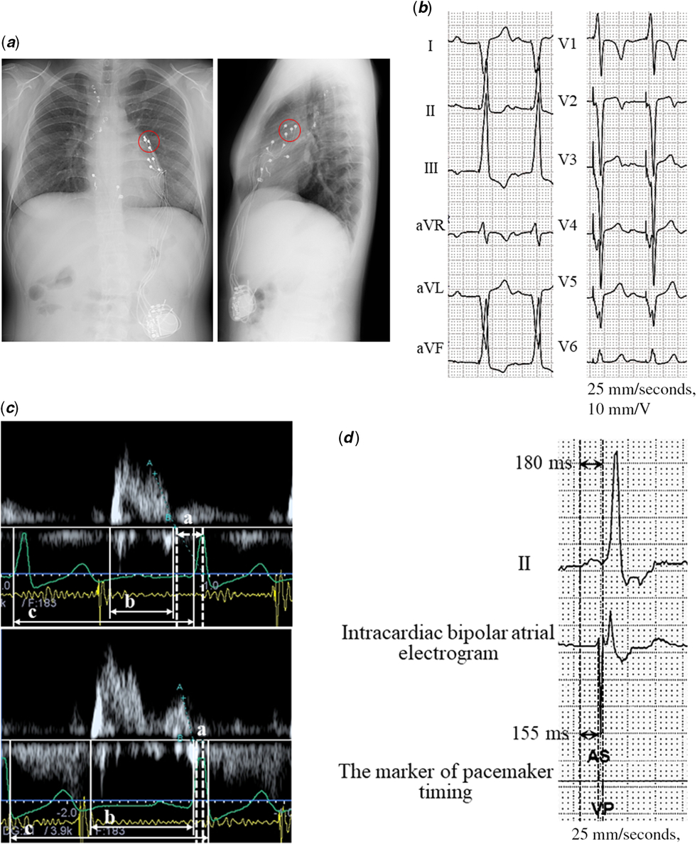
This was a case of an 18-year-old female patient with atrial situs solitus, double-inlet left ventricle, pulmonary atresia, and status post Fontan surgery using extracardiac graft. Complete atrioventricular block occurred at the age of 1 year. The first epicardial lead implantations were performed at the age of 4 years. At the age of 18 years, re-lead implantations (Fig 1a) were performed due to the ventricular pacing lead fracture, although atrial sensing and pacing threshold was stable. The protein-losing enteropathy was developed at the age of 12 years. She had no history of atrioventricular valvuloplasty and showed the slight atrioventricular regurgitation.
X-ray, electrocardiogram, and echocardiographic findings in case 1. ( a ) X-ray of anteroposterior view (right) and lateral view (left). Epicardial atrial lead (red circle) was implanted in the left-sided atrial appendage. ( b ) Twelve-lead electrocardiogram at setting of sensed atrioventricular interval (sAVI) at 150 ms. The origin of the atrial rhythm (supposed to be sinus rhythm) was estimated to be the right-sided anatomical right atrium. ( c ) Trans-mitral inflow pattern at the setting of sAVI at 150 ms (upper) and 25 ms (lower). The time differences between the end of the A wave and the first cardiac sound (a) changed from 136 to 45 ms after changing sAVI from 150 to 25 ms. Consequently, the %DFT (diastolic filling time [b]/cardiac cycle [c]) improved from 34 to 52%. ( d ) Lead II electrocardiogram (upper), intracardiac bipolar atrial electrogram measured by the pacemaker device (middle) and the marker of pacemaker timing (lower) at the setting of sAVI at 25 ms. The actual P wave and ventricular pacing interval of lead II was 180 ms. The atrial sensing was delayed 155 ms from the onset of the P wave on lead II.

The pacemaker mode was DDD with lower rate 50 bpm and upper track rate 130 bpm. From the heart rate histogram of the pace maker device, the mean heart rate was around 70–80 bpm and %A and %V paces were <1 and >99%, respectively. We initially set the sensed atrioventricular interval at 150 ms (Fig 1b). However, with this setting, %diastolic filling time showed 34% by trans-mitral valve flow by echocardiogram, indicating atrioventricular dyssynchrony (Fig 1c). Reference Cazeau, Bordachar and Jauvert7 Therefore, we used the Ishikawa method and changed sensed atrioventricular interval to 25 ms, which was the shortest sensed atrioventricular interval with the pacemaker device. This resulted in the improvement of the atrioventricular dyssynchrony, revealing a %diastolic filling time of 52%. After changing sensed atrioventricular interval, her clinical situation was not dramatically changed with the NYHA Functional Classification class II; however, protein-losing enteropathy gradually became easier to control.
The patient’s own atrial rhythm was supposed to originate from the sinus node located in the right-sided anatomical right atrium (Fig 1b). The atrial epicardial lead was implanted in the left-sided atrial appendage (Fig 1a). At the setting of sensed atrioventricular interval at 25 ms, the actual P wave and ventricular pacing interval was 180 ms, as determined by electrocardiogram, and a local atrial electrogram at the site of epicardial lead occurred after 155 ms from the onset of P wave, according to the intracardiac atrial electrogram (Fig 1d); hence, the atrial sensing delay of the epicardial pacing was 155 ms.
Case 2
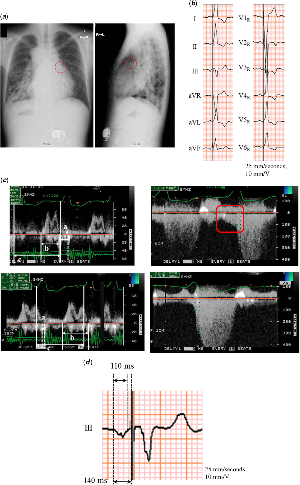
This was a case of a 26-year-old male patient with left atrial isomerism, dextrocardia, mitral atresia, double-outlet right ventricle, and status post Fontan operation using an intra-atrial graft. He had no history of tricuspid valvuloplasty. The epicardial leads were implanted because of a high-grade atrioventricular block at the age of 25 years (Fig 2a). The pacemaker mode was VDD with lower rate 70 bpm and upper track rate 140 bpm. The %V pace was 100%. In addition, the patient showed moderate tricuspid regurgitation. We initially set the sensed atrioventricular interval at 120 ms. However, with this setting, %diastolic filling time was 38% by trans-tricuspid valve flow by echocardiogram (Fig 2c). Therefore, using the Ishikawa method, we changed sensed atrioventricular interval to 30 ms, which was the shortest sensed atrioventricular interval with the pacemaker device. This resulted in the improvement of the atrioventricular dyssynchrony, revealing a %diastolic filling time of 58%. Furthermore, the diastolic tricuspid regurgitation observed during the setting of the sensed atrioventricular interval at 120 ms disappeared during setting of the sensed atrioventricular interval to 30 ms, although his clinical situation was not dramatically changed with NYHA class III.
X-ray, electrocardiogram, and echocardiographic findings in case 2. ( a ) X-ray of anteroposterior view (right) and lateral view (left). The epicardial atrial lead (red circle) is implanted in the left-sided atrial appendage. ( b ) Twelve-lead electrocardiogram at the setting of sAVI at 30 ms. The origin of the atrial rhythm is estimated to be the lower part of the right-sided anatomical left atrium. ( c ) Trans-tricuspid inflow pattern (left) and flow pattern of tricuspid regurgitation (right) at the setting of the sAVI interval at 120 (upper) and 30 ms (lower). The time differences between the end of the A wave and the first cardiac sound (a) changed from 105 to 20 ms after changing sAVI from 120 to 30 ms. Consequently, the %DFT (diastolic filling time [b]/cardiac cycle [c]) improved from 38 to 58%, and the diastolic tricuspid regurgitation disappeared. ( d ) Lead III electrocardiogram at the setting of sAVI at 30 ms. The actual P wave and ventricular pacing interval of lead III was 140 ms. Pacemaker atrial sensing timing was 30 ms before the ventricular pacing spike. The time differences between the actual P wave and ventricular pacing interval and sAVI, 110 ms, showed the atrial sensing delay of the epicardial pacing lead.

The patient’s own atrial rhythm was estimated arising from the inferior site of the right-sided anatomical left atrium (Fig 2b). The atrial epicardial lead was implanted in the left-sided atrial appendage (Fig 2a). At the setting of sensed atrioventricular interval at 30 ms, the actual P wave and ventricular pacing interval measured by electrocardiogram was 140 ms (Fig 2d). The atrial sensing delay of the epicardial pacing lead was 110 ms.
The epicardial pacing lead position and the atrial sensing delay
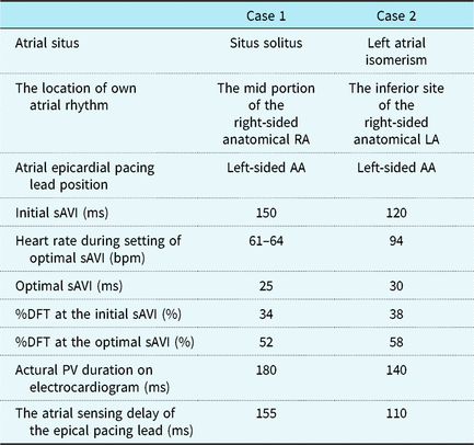
In both patients, the atrial epicardial leads were implanted on the opposite site of the origin of the patient’s own atrial rhythms (Table 1). The atrial sensing delay of the epicardial pacing lead was 155 and 110 ms. This was also due to intra-atrial conduction delay from the origin of their own atrial rhythms to the site of the epicardial atrial lead. The optimal atrioventricular interval needed to be extremely short because of the epicardial lead position and intra-atrial conduction delay.
The clinical results of setting of optimal sAVI.

AA = atrial appendage; %DFT = %diastolic filling time; RA = right atrium; sAVI = sensed atrioventricular interval.
Discussion
Here, we described two patients with Fontan circulation that needed to be set with an extremely short sensed atrioventricular interval because of the epicardial lead position and intra-atrial conduction delay. These two cases indicated the important three unique aspects for patients with CHD when the optimal sensed atrioventricular interval is set: the origin of the atrial rhythm, the site of the epicardial atrial lead, and the atrial conduction delay.
First, the basic own rhythm does not always originate on the right atrium or sinus node. Knowing the position of the basic own rhythm in the atrial situs is important. The sinus node in situs solitus is located at the superior rim of the crista terminalis in the right-sided anatomical right atrium. The crista terminalis and sinus nodes are in the left-sided anatomical right atrium in situs inversus. Both-sided atria show right atrial morphology in the right atrial isomerism heart, and the crista terminalis and sinus nodes often exist in both-sided atria. Both-sided atria show left atrial morphology in the left atrial isomerism heart, and the crista terminalis often does not exist. Subsequently, the sinus nodes are hypoplastic and, sometimes, do not exist or exist in multiples. Reference Dickinson, Wilkinson, Anderson, Smith, Ho and Anderson8,Reference Smith, Ho and Anderson9 The P wave morphology of the 12-lead electrocardiogram roughly shows the location of the origin of the atrial rhythm.
Second, the atrial lead is not always implanted in the same side of the origin of the patients’ atrial rhythm. In patients with CHD, the epicardial rather than endocardial leads are often implanted because of difficulty of venous access to the heart and the risk of thromboembolism due to the presence of an intracardiac shunt. Reference Khairy, Van Hare and Balaji10 When the epicardial atrial lead is used, the site of implantation is not needed to be in the atria where the systemic vein drainage is located. In these two cases, the reason why the surgeon implanted the lead at the left-sided atria was unclear by the surgical records. We should check the site of the epicardial atrial lead by using X-ray or reviewing the surgical record.
Third, intra-atrial conduction delay is sometimes observed because of the surgical scar due to the previous atriotomy, and atrial enlargement and fibrosis due to atrial overload. In the histopathologic analysis of the atria from post-mortem specimens of Fontan hearts, the increments in wall thickness and interstitial fibrosis were observed in both the right and left atria. Reference Wolf, Seslar and den Boer11 The 12-lead electrocardiogram and intracardiac atrial electrogram allow us to estimate the conduction time from the origin of the atrial rhythm to the site of the atrial lead.
These three aspects can be observed in the patients with other complicated CHD, not only with Fontan circulation, who are implanted atrial pacing lead at the opposite side of own atrial rhythm or with atrial conduction delay. We must pay attention to these aspects for every patient with complicated CHD with epicardial atrial lead. In this study, we did not evaluate hemodynamic by catheterisation or the serum level of brain natriuretic peptide before and after changing sensed atrioventricular interval and the clinical situation was not dramatically changed in both patients. Further, although optimal AVI should be shorter at the faster heart rate, Reference Mehta, Gilmour, Ward and Camm12 we did not the evaluate the optimal sAVI and % DFT during heart rate increased. It was because optimal sAVI at rest in these patients was the shortest sAVI that we could set with the pacemaker device. As the result, %DFT in these patients was supposed to be shorter at the faster heart rate than those at rest. However, we should be able to expect the chronic hemodynamic effects of the atrioventricular synchronisation.
Conclusion
We need to consider the origin of the atrial rhythm, the site of the epicardial atrial lead, and the atrial conduction delay by using electrocardiogram and X-ray when we set the optimal sensed atrioventricular interval in complicated CHD.
Acknowledgements
The authors would like to thank Enago (www.enago.jp) for the English language review.
Financial Support
This research received no specific grant from any funding agency, commercial or not-for-profit sectors.
Conflicts of Interest
None.
Ethical Standards
All procedures performed in studies involving human patients were in accordance with the ethical standards of the institutional and/or national research committee and with the 1964 Helsinki declaration and its later amendments or comparable ethical standards. Informed consent for the procedures was obtained from all individual patients included in the study.