Introduction
Spermatogenesis is a complex and male-specific process supported by spermatogonial stem cells (SSCs), the only cells that contribute genes to subsequent generations (Huckins, Reference Huckins1971; Clermont, Reference Clermont1972). Testis cells are made up of somatic cells and reproductive cells. In the mouse, it is estimated that there are 2 × 10⁴ stem cells in the total 108 cells, approximately, in the testis (Brinster & Nagano, Reference Brinster and Nagano1998). Furthermore, a large percentage of germ cells are mitotic spermatogonia, spermatocytes or highly differentiated terminal stages cells (Uchida et al., Reference Uchida, Fleming, Alpern and Weissman1993; Morrison et al., Reference Morrison, Uchida and Weissman1995). There is enormous value in understanding the biological characteristics of SSCs through the study of maintenance and proliferation of SSCs in vitro. At this time, many studies are focusing on understanding cultivation, self-renewal and differentiation of SSCs (Nagano et al., Reference Nagano, Avarbock, Leonida, Brinster and Brinster1998; Lee et al., Reference Lee, Parks, Lim, Yoon, Ko and Kim2003; Kubota et al., Reference Kubota, Avarbock and Brinster2004; Kanatsu-Shinohara et al., Reference Kanatsu-Shinohara, Inoue, Ogonuki, Miki, Yoshida, Toyokuni, Lee, Ogura and Shinohara2007; Oatley et al., Reference Oatley, Avarbock and Brinster2007; Araki et al., Reference Araki, Sato, Katagiri, Kubota, Araki and Ogawa2010; Nolte et al., Reference Nolte, Michelmann, Wolf, Wulf, Nayernia, Meinhardt, Zechner and Engel2010).
Stem cells are considered to grow in a special environmental niche supplemented with various factors that contribute to regulating proliferation and differentiation (Spradling et al., Reference Spradling, Drummond-Barbosa and Kai2001). Although the essential roles of growth factors have been explored, little information is known about the exact mechanism of proliferation or differentiation of SSCs. Analysis of SSCs directly in vivo has proved to be very difficult, as the SSC number in a testis is too small for easy study (Tegelenbosch & Derooij, Reference Tegelenbosch and Derooij1993). To overcome this problem, some in vitro culture systems for SSCs have been developed (Kanatsu-Shinohara et al., Reference Kanatsu-Shinohara, Ogonuki, Inoue, Miki, Ogura, Toyokuni and Shinohara2003). It has been demonstrated that glial cell line-derived neurotrophic factor (GDNF) is beneficial to maintaining SSCs in vitro (Tegelenbosch & Derooij, Reference Tegelenbosch and Derooij1993; Nagano et al., Reference Nagano, Ryu, Brinster, Avarbock and Brinster2003). A complex and undefined medium was shown to be beneficial for the proliferation SSCs derived from DBA/2 mice, but not for other mouse strains (Kanatsu-Shinohara et al., Reference Kanatsu-Shinohara, Ogonuki, Inoue, Miki, Ogura, Toyokuni and Shinohara2003). Although the growth requirements of mouse SSCs still remain unclear, several laboratories have attempted to culture SSCs for long or short time periods in vitro (Jeong et al., Reference Jeong, McLean and Griswold2003; Kanatsu-Shinohara et al., Reference Kanatsu-Shinohara, Ogonuki, Inoue, Miki, Ogura, Toyokuni and Shinohara2003; Nagano et al., Reference Nagano, Ryu, Brinster, Avarbock and Brinster2003).
Germ cells and somatic cells, such as Sertoli cells and Leydig cells, in the testis can support spermatogenesis via interactions between these cells and actions of hormones secreted by the pituitary gland and the testis (Jegou, Reference Jegou1993; Parvinen et al., Reference Parvinen, Vihko and Toppari1986; Skinner, Reference Skinner1991). It has been claimed that some growth factors have important functions in mammalian testis. Insulin-like growth factors (IGFs) can stimulate mitotic DNA synthesis (Soder et al., Reference Soder, Bang, Wahab and Parvinen1992) and the stem-cell factor (SCF)/c-kit system is essential for survival (Packer et al., Reference Packer, Besmer and Bachvarova1995; Hakovirta et al., Reference Hakovirta, Yan, Kaleva, Zhang, Vanttinen, Morris, Soder, Parvinen and Toppari1999), proliferation and for differentiation of germ cells (Yoshinaga et al., Reference Yoshinaga, Nishikawa, Ogawa, Hayashi, Kunisada and Fujimoto1991; Manova et al., Reference Manova, Huang, Angeles, De Leon, Sanchez, Pronovost, Besmer and Bachvarova1993; Vincent et al., Reference Vincent, Segretain, Nishikawa, Nishikawa, Sage, Cuzin and Rassoulzadegan1998; Sette et al., Reference Sette, Dolci, Geremia and Rossi2000). However, little information is known about the effects of testicular interstitial fluid on mouse SSCs proliferation in vitro. In this study, an original, undefined and complex culture system was designed to culture mouse SSCs in order to obtain more information about the use of testicular interstitial fluid as a supplement for proliferation of mouse SSCs in vitro.
Materials and methods
All chemical agents used in the study were purchased from Sigma Chemical Co. (St. Louis, MO, USA).
Experimental animals
Eighty Institute for Cancer Research (ICR) male mice (6-day-old) were obtained from the Experimental Animal Center of Medical College of Xi'an Jiaotong University, China for use in the study.
Preparation of testicular interstitial fluid
Forty testes were excised from mice and cut into small fragments. Subsequently, the testicular interstitial fluid was extracted by tissue homogenization.
Preparation of culture medium
Components of M1 medium were Dulbecco's modified Eagle's medium (DMEM), 4 mol/l l-glutamine, 1% non-essential amino acids, 4 × 10⁻² mg/ml gentamicin and 10% fetal bovine serum (FBS). M2 medium was composed of DMEM, 4 mol/l l-glutamine, 1% non-essential amino acids, 4 × 10⁻² mg/ml gentamicin, 10% FBS and testicular interstitial fluid. The final concentrations of testicular interstitial fluid in M2 medium were 0, 5, 10, 20, 30 or 40% (v/v), respectively.
Isolation and purification of mouse SSCs
Two-step enzymatic digestion was used to prepare single cell suspensions from testis tissues (Herrid et al., Reference Herrid, Davey and Hill2007). The dispersed cells were filtrated with a four-hundred-eye mesh and rinsed twice with DMEM, then the pellet was suspended in M1 medium. The cells were cultured by a two-step differential plating process to enrich for SSCs. Briefly, 106 cells/ml were cultured in 60-mm dishes for 12 h at 37°C in an atmosphere of 5% CO2 in air, then the non-adherent cells were transferred to a new plate and cultured for 4 h under the same conditions. Suspended cells were collected and divided into six aliquots. The first aliquot was cultured in vitro as a control; the other five aliquots were cultured in parallel in 48-well plates at 5 × 10⁴ cells per well in M2 medium. The cells were sub-cultured every day by replenishing half the medium. The cell suspension was placed into cell culture flasks and maintained at 37°C in a humidified 5% CO2 and 95% air atmosphere. As a result of the differential plating, a final proportion of about 80–85% SSCs was gained by eliminating adherent Sertoli cells every 6–8 h. The medium was changed every 2–3 days.
Alkaline phosphatase (AKP) assay
Mouse SSCs were fixed in 4% paraformaldehyde and then stained with NBT/BCIP AKP substrate. The staining reaction was stopped after 20–40 min incubation in light by rinsing the cells with phosphate-buffered saline (PBS). Stained cells were observed and photographed under an inverted phase contrast microscope (Nikon Imaging Sales Co Ltd., Tokyo, Japan).
RT-PCR
Total cellular RNA was extracted from mouse SSCs and Sertoli cells using the RNA Simple Total RNA Kit (TianGen Co. Ltd, China). Reverse-transcription was performed using the SuperScript™ First-Strand Synthesis System and cDNAs were used to analyze the expression of SSC-specific genes by PCR. β-Actin was used as the housekeeping gene. The primer sequences and GenBank accession numbers are as follows: Ngn3 (GenBank No. NM_009719.6) Forward Primer: 5′-TTGGCACTCAGCAAACAGC-3′, Reverse Primer: 5′-TCCCTTTCCACTAGCACCC-3′, 467 bp; Oct4 (GenBank No. NM_013633.2) Forward Primer: 5′-CCCCAATGCCGTGAAGTT-3′, Reverse Primer: 5′-GAAAGGTGTCCCTGTAGCC-3′, 556 bp; β-actin (GenBank No. NM_007393.3) Forward Primer: 5′-GCCTTCCTTCTTGGGTAT-3′, Reverse Primer: 5′-CCTT-CACCGTTCCAGTTT-3′, 549 bp; TH2B (GenBank No. NM_175663.1) Forward Primer: 5′-CGGTAAAGGGTGCTACTAT-3′, Reverse Primer: 5′-CACTTGTTTCAGCACCTTA-3′, 137 bp; Integrin alpha 6 (GenBank No. NM_008397.3) Forward Primer: 5′-ATGATGAAAGTCTCGTGC-3′, Reverse Primer: 5′-CATAGCCAAACGAGGAAG-3′, 222 bp; Integrin beta 1 (GenBank No. NM_010578.2) Forward Primer: 5′-TTGATGAATGAAATGAGGAG-3′, Reverse Primer: 5′-TCCAGATATGCGTTGCTG-3′, 225 bp; and Sycp3 (GenBank No. NM_011517.2) Forward Primer: 5′-TCAGAGCCAGAGAATGAAAG-3′, Reverse Primer: 5′-CTGCTGAGTTTCCATCATAAC-3′, 163 bp. The reaction products were separated and visualized by 1.0% agarose gel electrophoresis.
Indirect immunofluorescence cell analysis
The mouse SSCs obtained were evaluated by staining with CD9, which is a marker for undifferentiated type A spermatogonia (Abu Elhija et al., Reference Abu Elhija, Lunenfeld, Schlatt and Huleihel2012). Briefly, cells were fixed in 4% paraformaldehyde for 15 min and then were rinsed three times in PBS plus 0.1% Tween-20. Subsequently, the cells were re-suspended in PBS plus bovine serum albumin (BSA) for 1 h at 37°C. The cells were then incubated with anti-CD9 antibody (final concentration 1:1000) for 12 h at 37°C. After three washes in PBS, cells were incubated with the Cy3 AffiniPure Goat Anti-Rabbit IgG (H+L) (final concentration 1:1000) for 1 h at 37°C. Finally, the solution was incubated with Hoechst stain (1:800) for 8 min and then washed three times with PBS. Cells were re-suspended in 0.5 ml PBS–BSA. Fluorescence was monitored using the 8- and 644-nm double band-pass filter and fluorescence microscopy (Nikon Imaging Sales Co Ltd., Tokyo, Japan).
Proliferation and diameter of SSCs colony assay
The mouse SSCs obtained were treated with 5 mg/ml 3-(4,5-dimethylthiazol-2-yl)-2,5-diphenyl-tetrazolium bromide (MTT) reagent for 4 h at 37°C to measure cell proliferation and placed in a shaker for 20 min. For quantitation, dimethyl sulfoxide (DMSO) was added to dissolve water-insoluble blue-violet MTT formazan formed in the cells. The diameters of cell colonies were measured using the microscope software.
Statistical analysis
All statistical analyses were performed using Statistical Product and Service Solutions software (SPSS 11.5 for Windows; SPSS, Chicago, IL, USA). For culture assays of the mouse SSCs with the testicular interstitial fluid in vitro, each data point was represented by the average of three separate experiments and presented as mean ± standard deviation (SD). Statistical significance among the proliferation rate and diameter of SSCs colony were determined using Duncan's multiple range test and by analysis of variance (ANOVA). A P-value <0.05 indicated statistical significance.
Results
Characterization of SSCs during short-term culture
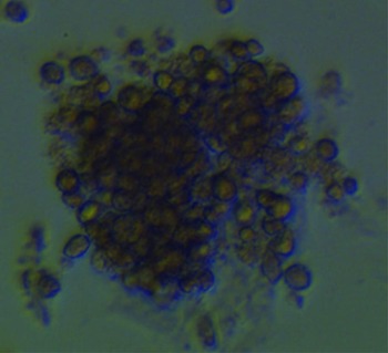
Isolated cells were enriched by the anchorage velocity-dependent separation method. After 6–8 days of culture, cell colonies formed in the dishes (Fig. 1). SSC cells appeared round, and began to divide and proliferate at 2 days and cell colonies emerged after culture for 6 days (Fig. 1).
Cell colonies (×250 magnification).

Alkaline phosphatase (AKP) assay
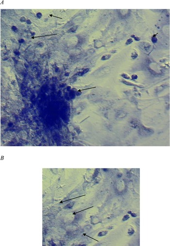
The control group in the assay was represented by purified Sertoli cells. Stem cells stained bluish violet by AKP staining. In two images (Fig. 2 A, B), SSCs and Sertoli cells were clear differentiated; SSCs were round and stained bluish violet, while Sertoli cells had an irregular form and stained minimally. The AKP assay and cell colony imaging (Fig. 1) demonstrated that the round cells could be considered to be SSC colonies.
(A) Cells with alkaline phosphatase staining (×200 magnification). Arrows indicate that the cells stained bluish violet were considered to be spermatogonial stem cells (SSCs). (B) Cells with alkaline phosphatase staining (×200). The arrows indicate the Sertoli cells. The SSCs stained bluish violet (and were round), but the Sertoli cells were minimally stained (and were irregular in shape).

Characteristic genes expression
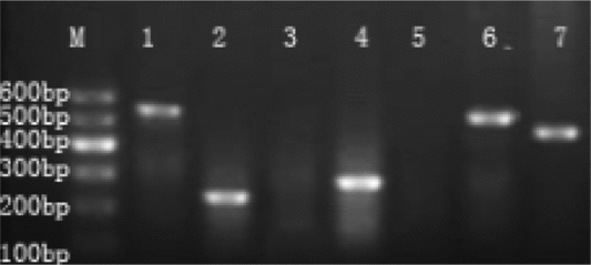
Some genes characteristic of germ cells were selected to identify SSCs and haploid male germ cells. The Ngn3 gene is typical of SSCs (Yoshida et al., Reference Yoshida, Takakura, Ohbo, Abe, Wakabayashi, Yamamoto, Suda and Nabeshima2004) and Oct4 expression marks the pluripotency and self-renewal of stem cells (Loh et al., Reference Loh, Wu, Chew, Vega, Zhang, Chen, Bourque, George, Leong, Liu, Wong, Sung, Lee, Zhao, Chiu, Lipovich, Kuznetsov, Robson, Stanton, Wei, Ruan, Lim and Ng2006). The β-actin gene was used as the housekeeping gene for SSCs and Sertoli cells (Thellin et al., Reference Thellin, Zorzi, Lakaye, De Borman, Coumans, Hennen, Grisar, Igout and Heinen1999). Integrin alpha 6 and Integrin beta 1 gene products are expressed as surface markers on SSCs (Shinohara et al., Reference Shinohara, Avarbock and Brinster1999). Expression of Sycp3 and TH2B indicates the presence of haploid male germ cells (Lim et al., Reference Lim, Sung, Kim, Song, Hong, Yoon, Kim, Kim and Lee2010). The mouse SSCs obtained expressed OCT4, Integrin alpha 6, Integrin beta 1, β-actin, Ngn 3 (Fig. 3). Sertoli cells only expressed β-actin, data not shown.
Reverse transcriptase polymerase chain reaction (RT-PCR) analysis. The characteristic genes of spermatogonial stem cells (SSCs). The M lane indicates the DNA marker; lane 1 indicates Oct4; lane 2 is Integrin alpha 6; lanes 3, 4, 5, 6 and 7 indicate the expression of TH2B, Integrin beta 1, Scyp3, β-actin and Ngn 3, respectively.

Indirect immunofluorescence cell analysis
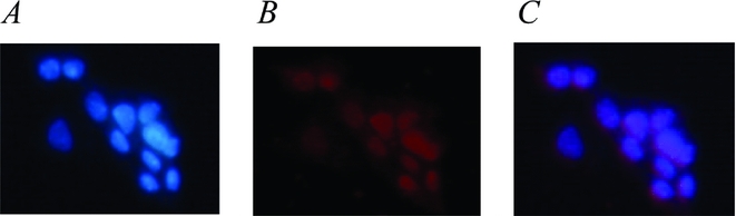
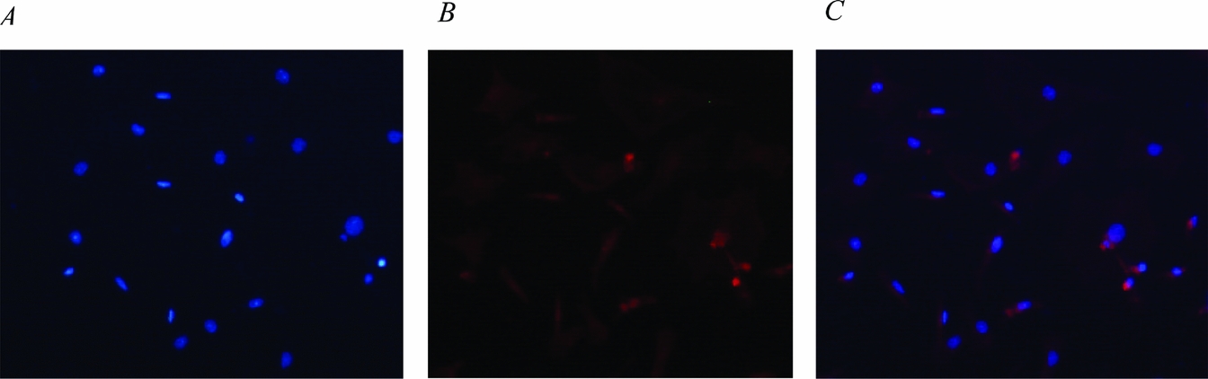
It is usual to consider that As (a single) and Apr (a paired) spermatogonia have stem-cell properties. As no specific markers have been reported, CD9 was selected as the surface maker for SSCs. The results of indirect immunofluorescence analysis of Sertoli cells and SSCs are shown in Fig. 4. It was clear that the SSCs colonies expressed CD9 on their cell surfaces after culture for 3–5 days (Fig. 4). Although Sertoli cells also expressed CD9, staining showed a small spherical nucleus with a thick rim of cytoplasm (Fig. 5). In contrast, SSCs had a large spherical nucleus with a thin rim of cytoplasm (Fig. 4 A, C. Therefore we demonstrated here again that the cell colonies were SSCs.
Spermatogonial stem cells (SSCs) were identified by CD9. (A) SSCs were stained with Hoechst (×200). (B) SSCs were defined by CD9 indirect immunofluorescence staining (×200 magnification). (C) SSCs were defined by CD9 and by Hoechst stain combined with indirect immunofluorescence staining (×200), (C) the merging of images (A) and (B).

Sertoli cells were identified by CD9. (A) Sertoli cells were stained with Hoechst (×400). (B) Sertoli cells were detected by CD9 by indirect immunofluorescence staining (×400). (C) Sertoli cells were detected by CD9 by Hoechst combined with indirect immunofluorescence staining (×400), (C) is the merging of images (A) and (B). The control group was Sertoli cells.

Effects of testicular interstitial fluid on mouse SSCs proliferation in vitro
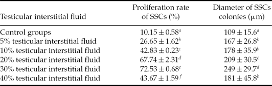
After overnight culture, most SSCs were scattered singly and some had begun to divide. It is clearly shown in Table 1 that the optimal proliferation rate and the diameter of the SSC clusters were 72.53% and 249 μm, respectively, in the 30% testicular interstitial fluid treated group after 8 days, values that were significantly higher (P < 0.05) than other groups. This finding demonstrated that medium that contained 30% testicular interstitial fluid could significantly promote mouse SSC proliferation in vitro.
The effects of testes liquid on mouse spermatogonial stem cells (SSCs) proliferation in vitro

Note: Values in the same row with different letters a–f indicate significant difference (P < 0.05).
Discussion
Testicular interstitial fluid is a good resource for SSCs culture in vitro because it is rich in growth factors and able to support a specific niche for SSCs (Yoshinaga et al., Reference Yoshinaga, Nishikawa, Ogawa, Hayashi, Kunisada and Fujimoto1991; Soder et al., Reference Soder, Bang, Wahab and Parvinen1992; Vincent et al., Reference Vincent, Segretain, Nishikawa, Nishikawa, Sage, Cuzin and Rassoulzadegan1998; Hakovirta et al., Reference Hakovirta, Yan, Kaleva, Zhang, Vanttinen, Morris, Soder, Parvinen and Toppari1999; Sette et al., Reference Sette, Dolci, Geremia and Rossi2000). In particular, it is assumed that the early differentiating spermatogonia and SSCs emerge at the time when germ cells locate in the lumen of immature seminiferous tubules in the neonatal testis (de Rooij, Reference de Rooij2006). Hence, it is possible to efficiently recover SSCs from testis by a differential plating method. The optimal proliferation rate and diameter of the SSCs colony was 72.53% and 249 μm, respectively, when the culture medium was supplemented to 30% with the testicular interstitial fluid.
AKP assay, RT-PCR and indirect immunofluorescence cell analysis were performed to verify the mouse SSCs in the study. AKP is a pluripotency cell marker (Neri et al., Reference Neri, Monti, Rebuzzini, Merico, Garagna, Redi and Zuccotti2007; Park et al., Reference Park, Zhao, West, Yabuuchi, Huo, Ince, Lerou, Lensch and Daley2008). The AKP-positive cells have the properties of stem cells and are stained bluish violet. Our results were similar to those in previous studies (Guan et al., Reference Guan, Nayernia, Maier, Wagner, Dressel, Lee, Nolte, Wolf, Li and Engel2006; Goel et al., Reference Goel, Sugimoto, Minami, Yamada, Kume and Imai2007). Oct4, Integrin alpha 6, Integrin beta 1, and Ngn3 genes were selected to identify the SSCs and the Sertoli cells (Shinohara et al., Reference Shinohara, Avarbock and Brinster1999). In male mice, the Oct-4 gene is expressed before spermatogenesis and is confined to type A spermatogonia (Feng et al., Reference Feng, Chen, Dettin, Pera, Herr, Goldberg and Dym2002). The Ngn3 gene is predominantly expressed in the As, Apr and Aal stages of c-Kit-negative spermatogonia in adults and the c-Kit-negative fraction of the pre-pubertal prespermatogonia (Yoshida et al., Reference Yoshida, Takakura, Ohbo, Abe, Wakabayashi, Yamamoto, Suda and Nabeshima2004). Integrin alpha 6 and Integrin beta 1 are considered to be surface markers for SSCs (Shinohara et al., Reference Shinohara, Avarbock and Brinster1999). Thus, the expression of the Ngn3, Oct4, Integrin alpha 6 and Integrin beta 1 genes can be considered as a marker of SSCs and Sertoli cells. In this study, the mouse SSCs obtained expressed OCT4, Integrin alpha 6, Integrin beta 1, β-actin and Ngn 3 (Fig. 3). The expression of Sycp3 and TH2B is considered the specific marker of haploid male germ cells (Lim et al., Reference Lim, Sung, Kim, Song, Hong, Yoon, Kim, Kim and Lee2010; Li et al., Reference Li, Bolcun-Filas and Schimenti2011), thus the expression of the TH2B and Scyp3 genes can be considered to indicate differentiated SSCs and haploid male germ cells. In Fig. 3, we can see that the SSCs did not express the TH2B and Scyp3, which indicated that the SSCs had not started to differentiate. As no specific markers were reported, CD9 was selected as the surface maker of SSCs. It was clear that the cell colonies expressed CD9 on the cell surface after culture for 3–5 days (Fig. 4). Although the Sertoli cells were also expressed CD9, they showed a small spherical nucleus with a thick rim of cytoplasm (Fig. 5). In contrast, mouse SSCs had a large spherical nucleus with a thin rim of cytoplasm (Fig. 4 A, C), whose morphology was the same as the mouse SSCs. Therefore, it was obvious that the cell colonies were indeed undifferentiated mouse SSCs.
The specific biochemical and cytoarchitectural microenvironment in the adluminal compartment where germ cells proliferate and differentiate are constituted the region for tissue remodeling and the production of Sertoli cell factors during testis development (Griswold, Reference Griswold1995). Until a niche is available, only one in two daughter cells can remain and the other cell will be committed to differentiate (Schofield, Reference Schofield1978). The culture system for SSCs consisted of serum medium and mitosis inactivated Sertoli cell feeders; purified mouse SSCs were seeded on the feeders at densities of 6–10 × 104 cells/ml in this study. It was demonstrated that proliferation of the mouse SSCs could be largely promoted by supplementing the M2 medium with 30% testicular interstitial fluid. The optimal diameter of the mouse SSCs colony was 249 μm at 8 days. Some factors have an important role in germ cell proliferation in the testis. Neuregulin 1 (NRG1) belonging to the epidermal growth factor (EGF) family can induce growth and differentiation of some types of cells during cultivation (Falls, Reference Falls2003); SCF is produced by the Sertoli cells (Rossi et al., Reference Rossi, Dolci, Albanesi, Grimaldi, Ricca and Geremia1993) and can promote germ cell proliferation and survival (Tajima et al., Reference Tajima, Onoue, Kitamura and Nishimune1991); interleukin (IL)-α has been considered to stimulate the proliferation of germ cells, Sertoli cells and peritubular cells (Pollanen et al., Reference Pollanen, Soder and Parvinen1989; Parvinen et al., Reference Parvinen, Soder, Mali, Froysa and Ritzen1991; Svechnikov et al., Reference Svechnikov, Sultana and Soder2001; Petersen et al., Reference Petersen, Boitani, Froysa and Soder2002); testosterone is necessary for the completion of spermatogenesis and maintenance of male reproductive function. Testis is rich in growth factors such as acidic fibroblast growth factor (aFGF), nerve growth factor (NGF), seminiferous growth factor (SGF), IGF-1, and transforming growth factors (TGF) alpha and beta (Braunhut et al., Reference Braunhut, Rufo, Ernisee, Zheng and Bellve1990; Skinner, Reference Skinner1991). According to our results, it is hypothesized that culture medium supplemented with 30% testicular interstitial fluid contains the appropriate concentration of hormones and growth factors to promote mouse SSC growth. In the testes of 6-old-day mice, this niche is available for SSCs maintainance rather than differentiation. These original, undefined and complex testicular interstitial fluid conditions make it possible to culture SSCs in vitro, but more details are needed to expound this result.
The successful culture of SSCs in vitro is beneficial to SSC research. The present study indicated that culture medium containing 30% of the testicular interstitial fluid could significantly enhance mouse SSCs proliferation in vitro. The use of this culture system is valuable for subsequent experiments, such as in pluripotency of SSCs, production of genetically modified animals, gene therapy, and so on. However, more research is need to evaluate and understand the precise physiological role of testicular interstitial fluid in cultured SSCs in vitro.
Acknowledgements
This research was supported by the Youth Extra Fund of Northwest A&F University (Z111020905), the Special Research Subsidy Project of Northwest A&F University (07ZR002) and the Basic Scientific Research Expense of Sci-Tech Innovation Major Project of Northwest A&F University (QN2011061).