Calcifying fibrous tumour is a rare benign fibrous lesion. It is paucicellular, with fibroblasts, dense collagenisation, psammomatous and dystrophic calcification, and patchy lymphoplasmacytic infiltrates.Reference Fletcher, Unni and Mertens 1 Calcifying fibrous tumour was first described in 1988 by Rosenthal and Abdul-KarimReference Rosenthal and Abdul-Karim 2 under the name of “childhood fibrous tumour with psammoma bodies” in subcutaneous and deep soft tissues, and recently has been reported all over the body. By 2006, no more than 80 cases had been reported in the literature.Reference Kirby, Sato, Tannous and Dehner 3 Calcifying fibrous tumour originating from the heart is extremely rare. To the best of our knowledge, only two cases have been reported, both affecting the left heart in young women.Reference Kirby, Sato, Tannous and Dehner 3 , Reference Shigematsu, Sano, Kasahara, Yanai and Date 4 This article describes the first case of a giant calcifying fibrous tumour arising from the right ventricle in a child, where the tumour was totally resected and no recurrence was observed during a 4-year follow-up period.
Clinical summary
The patient was an 8-year-old boy, who presented with palpitation and shortness of breath after activity for 18 months. A year before admission to our hospital, he received an open heart surgery in another hospital, but the tumour was too large to be resected. Biopsy in that hospital showed hyalinised fibrous tissue with calcifications and psammomatous bodies.
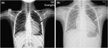
A precordial bulge was noticed and grade 2/6 systolic heart murmurs were audible at the 2nd and 3rd intercostal space near the left sternal border. Chest X-ray showed a limited bulge of the left cardiac border (Fig 1a). Electrocardiogram showed I, aVL, and V2-5 ST segment depression with different degrees, incomplete right bundle branch block, and left anterior hemiblock. 2D-echocardiography revealed a large solid space-occupying lesion with diffuse calcifications in the free wall of the right ventricle, and mild right ventricular outflow tract obstruction. Echoes inside were heterogeneous and hyperintense compared with the adjacent myocardium. The border of the tumour was indistinct.
Chest X-ray: ( a ) pre-operative chest X-ray showing limited bulge of the left cardiac border (black arrow) and ( b ) post-operative chest X-ray showing the disappearance of the pre-operative bulge of the left cardiac border (white arrow).

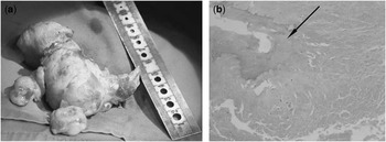
An operation was performed in December, 2007, including full sternotomy, cardiopulmonary bypass and incision of both the right atrium and right ventricular outflow tract. The base of the tumour measured 3.0 × 2.1 cm2 and was located in the anterolateral wall of the right ventricle, close to the epicardium and the left anterior descending coronary artery. The tumour, extending from the right ventricular outflow tract to near the heart apex, was covered with a thin layer of the myocardium at the right ventricular chamber, and the border was clear. The tumour was first cut into two pieces, and then pulled out with some surrounding myocardium via the tricuspid valve or the right ventricular outflow tract incision after sharp dissection. The tumour base was totally resected together with the adjacent wall of the right ventricle. The defect was repaired with an autologous pericardial patch. The tumour, measuring 7.8 × 4.9 × 4.1 cm3, was firm, elliptical, lobulated, oyster white, elastic, and partially calcified (Fig 2a).
Pathology: ( a ) the tumour was totally resected, and it was firm, elliptical, lobulated, oyster white, elastic, and partially calcified and ( b ) pathological sections of the tumour showing diffuse collagenous tissues, fibrocytes, and scattered calcifications (black arrow) (HE, ×100).

Pathological examination revealed that the tumour was composed of diffuse collagenous tissues containing fibrocytes, scattered calcifications, and infiltrates with some lymphocytes, without evidence of necrosis and malignancy (Fig 2b). Immunohistochemistry of the tumour cells was positive for vimentin and actin, and negative for desmin and S-100 protein. In addition, the result of specific staining for elastic fibres was positive. These findings were consistent with the diagnosis of calcifying fibrous tumour.
No post-operative complication occurred. The patient was free of symptoms during the 4-year follow-up period. Chest X-ray re-examination showed that the pre-operative bulge of the left cardiac border had disappeared (Fig 1b). No pathological finding was elicited from the echocardiograms.
Discussion
Calcifying fibrous tumour is a pseudotumour,Reference Fletcher, Unni and Mertens 1 histologically characterised by hypocellular, hyalinised, and collagenous tissue with calcifications and lymphoplasmacytic infiltrates. Dystrophic calcifications and psammomatous bodies are specific characteristics of this entity, which makes it distinct from inflammatory myofibroblastic tumour, another benign lesion that is usually more cellular and less hyalinised.Reference Shigematsu, Sano, Kasahara, Yanai and Date 4 Although examples have followed trauma and have occurred in association with Castleman's disease and inflammatory myofibroblastic tumour, the pathogenesis of calcifying fibrous tumour remains elusive.Reference Fletcher, Unni and Mertens 1
Calcifying fibrous tumour mainly affects children and young adults without gender predilection.Reference Fletcher, Unni and Mertens 1 The calcifying fibrous tumour size varies from 1.2 cm to 25 cm.Reference Kirby, Sato, Tannous and Dehner 3 The other reported cases of cardiac calcifying fibrous tumour occurred in young female patients without significant symptoms and were diagnosed by accident.Reference Kirby, Sato, Tannous and Dehner 3 , Reference Shigematsu, Sano, Kasahara, Yanai and Date 4 Both of them were large, one arising from the left ventricle and the other from the left auricle.
Calcifying fibrous tumours are usually positive for vimentin, actin, and CD34, and negative for desmin, S-100 protein, and anaplastic lymphoma kinase, which is thought of as an important immunophenotype that differentiates calcifying fibrous tumour from inflammatory myofibroblastic tumour, which is generally positive for the anaplastic lymphoma kinase,Reference Kirby, Sato, Tannous and Dehner 3 , Reference Shigematsu, Sano, Kasahara, Yanai and Date 4 although not all cases demonstrate these features. Actin was negative in the patient reported by Shigematsu et al.Reference Shigematsu, Sano, Kasahara, Yanai and Date 4
In view of continued growth and possible local recurrence as reported in two cases,Reference Rudez, Legac, Baric, Manojlović, Unic and Sutlic 5 complete resection appears to be the treatment of choice as long as the tumour is surgically resectable. Of the three tumours arising from the heart, two were totally resected, while the remaining one was not resectable because it was in the myocardium of the left ventricle encasing the left anterior descending coronary artery. Cardiac transplantation may be the only option for such a case at present.
The biological and clinical behaviours of calcifying fibrous tumour remain indeterminate because of the limited number of cases, and regular follow-up and review are obligatory.