Introduction
Avian mycoplasmosis caused by Mycoplasma species was first described in turkeys in 1926 and in chickens in 1936 (Nascimento et al., Reference NASCIMENTO, PEREIRA, NASCIMENTO and BARRETO2005; Kleven, Reference KLEVEN2008). There are more than 120 named Mycoplasma spp. and more than 20 are known to infect avian hosts (Nascimento et al., Reference NASCIMENTO, PEREIRA, NASCIMENTO and BARRETO2005; Purswell et al., Reference PURSWELL, EVANS and BRANTON2011). Of these, M. gallisepticum (MG) and M. synoviae (MS) are the major pathogens, and M. meleagridis (MM), and M. iowae (MI) are important in turkeys (Sprygin et al., Reference SPRYGIN, ELATKIN, KOLOTILOV, VOLKOV, SOROKINA, BORISOVA, ANDREYCHUK, MUDRAK, IRZA, BORISOV and DRYGIN2011). MG infection is a chronic respiratory disease of chickens and causes infectious sinusitis in turkeys. Symptoms include respiratory rales, coughing, nasal discharges and , in turkeys, frequently include sinusitis. MS infection is commonly known as infectious synovitis, an acute-to-chronic disease for chickens and turkeys involving primarily the synovial membranes of joints and tendons sheaths. However, during recent years, MS has been less frequently associated with synovitis, but more with airsacculitis in chickens and sometimes in turkeys (Khalifa et al., Reference KHALIFA, ABDELRAHIM, BADWI and MOHAMED2013).
Transmission may be transovarian or lateral via respiratory aerosols and direct contact. Infection occurs via the respiratory tract and can affect 100% of the birds. Following infection birds become persistently infected with MS and remain carriers for life. As a result of the expansion of poultry farms and the concentration of large, multiage production complexes within a restricted geographic area, it is becoming more difficult to maintain flocks that are free of MS. Both diseases are economically important, being egg transmitted and hatchery disseminated. They lead to tremendous economic losses in poultry production as a result of decreased hatchability and egg production, reduced quality of day-old chicks, reduced growth rate, increased costs of eradication procedures (site cleaning and depopulation), and increased costs of medication and vaccination (Ferguson-Noel and Williams, Reference FERGUSON-NOEL and WILLIAMS2015).
For many years, diagnosis of avian mycoplasmosis was based on serological assays to detect antibody production and/or on isolation and identification of the organism. Serological tests include serum plate agglutination test (SPA), haemaglutination inhibition test (HI) and enzyme linked immunosorbant assay (ELISA) for MG, MS, or MM (Khalifa et al., Reference KHALIFA, ABDELRAHIM, BADWI and MOHAMED2013). Difficulties encountered with the use of serologic tests for Mycoplasma have been described extensively, which is related to imperfections in the interpretation of results (Kleven, Reference KLEVEN2008). Problems arise primarily from the multiplicity of serotype strains isolated and their coexistence in the same isolate. Cultivation techniques are laborious, slow, expensive and require sterile conditions. Problems experienced with culturing include overgrowth by faster-growing Mycoplasma species and other organisms or no growth in subcultures. It can take up to four weeks, and even then, the result can be negative or be hampered by mixed infections (Ferguson-Noel and Williams, Reference FERGUSON-NOEL and WILLIAMS2015). For these reasons, use of rapid and sensitive detection methods, e.g. polymerase chain reaction (PCR) methods, can be advantageous because they provide better sensitivity and specificity facilitating the detection of pathogens in clinical samples collected from asymptomatic animals, or those who are under treatment with antibiotics (Evens et al., 2005; Peebles et al., Reference PEEBLES, JACOB, BRANTON and GERARD2014).
Control of avian mycoplasma infections is, in theory, quite simple and straightforward, especially because the pathogenic avian mycoplasmas are egg transmitted and lack a cell wall, rendering them susceptible to environmental influences. Good control begins with mycoplasma-free replacement stock that is placed on a single-age farm with all-in, all-out type management, good biosecurity and a consistent monitoring programme. Multi-age commercial egg producers are mostly positive for MG and MS, and in some parts of the world, both infections are widespread in commercial broiler production. MG vaccines are used with increasing frequency in areas where control is not feasible, such as large poultry populations in small geographic areas and multiple-age farms that never depopulate (Kleven, Reference KLEVEN2008). Vaccines have been used extensively; it is reported to be very immunogenic and mildly virulent in chickens and is effective in displacing virulent (field) strains from poultry operations. Control of poultry mycoplasmas consists of three general aspects: prevention, medication, or vaccination. In this review, a general summary of the pathogenesis of the mycoplasmas, followed by discussions on diagnosis and control by eradication, medication, and vaccination.
Pathogenesis of Mycoplasma gallisepticum
Mycoplasmas tend to be host-specific, but some species may have the ability to colonise several species (Rottem, Reference ROTTEM2003). Mycoplasmas are generally flask shaped with a narrowed tip structure that enables gliding motility and mediates tight attachment to host cell surface. MG attachment is mediated by their primary cytadhesion molecule GapA, whereas another molecule CrmA facilitates attachment. Cytadhesion to host cells is a necessary step towards disease pathogenesis and supported by P1 and P30 surface proteins (Rottem, Reference ROTTEM2003; Fürnkranz et al., Reference FÜRNKRANZ, SIEBERT-GULLE, ROSENGARTEN and SZOSTAK2013; Majumder, Reference MAJUMDER2014). Protein P1 is primary adhesion molecule densely aggregated near the tip structure along with an associated molecule P30, which enables P1 to function. Gliding motility is yet another unique phenomenon observed in mycoplasma and involves the polarised tip structure, which enables them to bind and move on solid surfaces (Manafi et al., Reference MANAFI, PIRANY, NOOR ALI, HEDAYATI, KHALAJI and YARI2015). Gliding motility allows mycoplasmas to escape mucocilary clearance, enabling them access the mucosal epithelial cells (Indiková et al., Reference INDIKOVÁ, VRONKA and SZOSTAK2014; Wijesurendra et al., Reference WIJESURENDRA, KANCI, TIVENDALE, BACCI, NOORMOHAMMADI, BROWNING and MARKHAM2015). Therefore these properties across various species of mycoplasma play a significant role in virulence and disease pathogenesis upon infection.
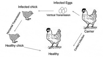
Upon attachment they can cause the release of mucus from goblet cells (Rottem, Reference ROTTEM2003; Majumder, Reference MAJUMDER2014; Xu et al., Reference XU, TENG, JIANG, ZHANG, EL-ASHRAM, WANG, SUN, HE, SHEN, WU and LI2015). Pathogenicity of MG may be further complicated by its ability to penetrate and survive within non-phagocytic host cells resulting in evasion of host immune response and dissemination throughout the host (Gharaibeh and Al-Rashdan, Reference GHARAIBEH and AL-RASHDAN2011; Indiková et al., Reference INDIKOVÁ, MUCH, STIPKOVITS, SIEBERT-GULLE, SZOSTAK, ROSENGARTEN and CITTI2013). Mycoplasma infections are known to be associated with damage to host cells and tissues due to reactive oxygen species generated by both the immune system of the host and the bacterium as its primary virulence factor (Rottem, Reference ROTTEM2003; Szczepanek et al., Reference SZCZEPANEK, BOCCACCIO, PFLAUM, LIAO and GEARY2014; Xu et al., Reference XU, TENG, JIANG, ZHANG, EL-ASHRAM, WANG, SUN, HE, SHEN, WU and LI2015). MGA-0676 (putative lipoprotein) and cytokines play a major contributory role in manifestation of disease during mycoplasma infection by recruitment and activation of leucocytes and may play a significant role in the immunopathology observed during mycoplasma infection (Majumder, Reference MAJUMDER2014; Xu et al., Reference XU, TENG, JIANG, ZHANG, EL-ASHRAM, WANG, SUN, HE, SHEN, WU and LI2015). A brief description of transmission and pathogenicity is shown in Figures 1 and 2.
Transmission of Mycoplasma gallisepticum.

Mechanism of pathogenesis: 1. Mycoplasma enter airways (ciliastasis, colonisation), 2. mycoplasma destroy cilia (reduce mucus, inhibit protein and DNA synthesis), 3. Systemic invasion by mycoplasma and E.coli (immunosuppression and diseases, production loss and health).

Diagnosis
Laboratory methods are essential to diagnose MG, since clinical signs and pathological lesions cannot reliably diagnose the source of infection. Rapid and early diagnostic detection of mycoplasma infections is important to prevent the spread of infection and to limit economic losses to the poultry industry. There are three approaches to diagnose MG: isolation and identification of organism, detection of its DNA and identification of specific antibodies (Dufour-Zavala et al., Reference DUFOUR-ZAVALA, SWAYNE, GLISSON, PEARSON, REED, JACKWOOD and WOOLCOCK2008; OIE, 2008; Qasem et al., Reference QASEM, AL-MOUQATI, AL-ALI and BEN-HAJI2015). SPA, HI and ELISA are the most common serological techniques. Serologic testing and isolation are used for the diagnosis of mycoplasma infections; however, these methods are laborious and time-consuming, and have low sensitivity. Moreover, such analysis is often plagued by non-specific reactions and problems with reagent cross-reaction (Heleili et al., Reference HELEILI, AYACHI, MAMACHE and CHELIHI2012). Serological procedures are useful for flock monitoring in MG control program and aid in diagnosis when infection is suspected (Ley, Reference LEY2008; Kahya et al., Reference KAHYA, TEMELLI, EYIGOR and CARLI2010). ELISA has been used to detect MG antibodies in respiratory tract and egg yolk samples. Serum agglutination reactions (SAR) can be performed with egg yolk samples and results are considered positive, suspicious and negative for titres equal or higher than 1:10, 1:5 and lower than 1:5, respectively (Heleili et al., Reference HELEILI, AYACHI, MAMACHE and CHELIHI2012; Nadeem et al., Reference NADEEM, YOUSAF, IQBAL, AWAIS and PERVEZ2014). Isolation and identification of the organism is the ‘gold standard’ for diagnosis of mycoplasma infections.
Procedures for isolation and identification, including formulations of commonly used media, are available. However, mycoplasmas are slow growing and are commonly overgrown by commensals such as M. gallinarum and M. gallinaceum. PCR testing is rapid, sensitive, and specific, and is often used instead of culturing to detect the presence of specific mycoplasmal DNA. PCR procedures including real-time procedures for MG, are available for MG or MS and procedures for MM and MI have been published (Bagheri et al., Reference BAGHERI, DOOSTI and ARSHI2011; Fraga et al., Reference FRAGA, DE VARGAS, IKUTA, FONSECA, CELMER, MARQUES and LUNGE2013).
MG strains showed seroconversion as measured by SAR, but only when serum samples were not diluted (Qasem et al., Reference QASEM, AL-MOUQATI, AL-ALI and BEN-HAJI2015). HI interpretation should be standardised, and positive results for MG and MS should consider a cut-off point of 1:40, as previously suggested for MG (Kleven, Reference KLEVEN2008). Other serologic tests can be used, and ELISA is the most promising technique to substitute for SAR (Heleili et al., Reference HELEILI, AYACHI, MAMACHE and CHELIHI2012) or even HI (Khalifa et al., Reference KHALIFA, ABDELRAHIM, BADWI and MOHAMED2013; Nadeem et al., Reference NADEEM, YOUSAF, IQBAL, AWAIS and PERVEZ2014). However, according to a previous field study (Nascimento et al., Reference NASCIMENTO, PEREIRA, NASCIMENTO and BARRETO2005), ELISA and SAR gave negative results whereas HI and PCR were positive. Serologic diagnosis can be influenced by a number of factors.
It is worth noting that pullets exposed to two vaccinations responded serologically better when analysed using SAR and HI than those exposed to a single vaccination, regardless of the vaccine used (Roussan et al., Reference ROUSSAN, KHAWALDEH and SHAHEEN2015), although this was not related to protection. Furthermore, commercial layers vaccinated twice with MG-F and infected with wild-type MG yielded the highest titres (HI and SAR) and percentage of positive birds. Birds vaccinated and infected with MG had intermediate titres, and non-vaccinated birds had the lowest titres. These results indicated that the serologic response was higher when birds were exposed more times to vaccine and/or wild-type MG strains (Qasem et al., Reference QASEM, AL-MOUQATI, AL-ALI and BEN-HAJI2015). Conflicting results from the same and/or different serologic tests may be ascribed to changes in mycoplasma surface antigens due to mutations (Heleili et al., Reference HELEILI, AYACHI, MAMACHE and CHELIHI2012). The problems arise primarily from the multiplicity of serotype strains isolated and their coexistence in the same isolate. Cross-reactions between MG and MS were not seen under controlled experimental conditions (Roussan et al., 215), indicating that this phenomenon depends on environmental conditions. These conflicting reports for serologic test results favour the idea of considering the SAR reaction at undiluted sera as suspicious result, and the adoption of 1:40 HI title as positive diagnosis. For these reasons, use of rapid and sensitive detection methods, e.g. PCR, can be advantageous because it provides a better sensitivity and specificity, facilitating the detection of pathogens in clinical samples collected from asymptomatic animals, or those who are under treatment with antibiotics (Raviv et al., Reference RAVIV, CALLISON, FERGUSON-NOEL and KLEVEN2008).
Mycoplasma can be detected in tissue fragments of affected organs like the trachea, air sacs and lungs. Besides synovial, ocular and infraorbital sinus exudates, and swabs from trachea and air sacs, and pipped embryos (Raviv and Kleven, Reference RAVIV and KLEVEN2009; Ferguson-Noel et al., Reference FERGUSON-NOEL, VICTORIA and FARRAR2012b). Swabs from trachea and choanal cleft constitute excellent specimens, mainly for isolation or PCR, which are used as confirmation tools for monitoring MG and MS infections in live birds (Nascimento et al., Reference NASCIMENTO, PEREIRA, NASCIMENTO and BARRETO2005; Kleven, Reference KLEVEN2008). An alternative to non-specific PCR for strain identification is direct sequencing of the amplified products (amplicons) from a specific PCR with primers directed to the 16S rRNA gene (Raviv and Kleven, Reference RAVIV and KLEVEN2009), but this technique requires certain laboratory capabilities. Sequencing is more accessible nowadays, and mycoplasma sequencing, including MG, has been facilitated because they have such a small genome (Bagheri et al., Reference BAGHERI, DOOSTI and ARSHI2011). Gross and microscopic examinations help the diagnosis of avian mycoplasmosis in naturally infected birds and are similar to the lesions described in experimentally infected birds (Qasem et al., Reference QASEM, AL-MOUQATI, AL-ALI and BEN-HAJI2015). During MG and E.coli infections gross lesions were observed, denoted by oedematous airsacculitis with fibrin deposition, leading to pericarditis and perihepatitis. In addition, it has been reported that haemorrhagic tracheitis with strong mononuclear cell infiltration can obliterate bronchiolitis with mononuclear cell infiltration, diffuse airsacculitis and air sac hyperplagia with monophil and heterophil cell infiltration and multiple granulomas in the lungs rich in multinucleated giant cells, as well as necrosis (Kleven, Reference KLEVEN2008).
Preventive measures
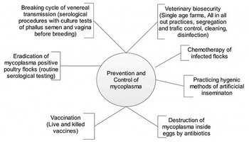
Because all mycoplasmas which are pathogenic for poultry are vertically transmitted, obtaining replacement stock from uninfected sources is essential. The first step towards control is the acquisition of fertile eggs and birds free from MG, MS and/or MM, which have been produced by treatment of fertile eggs, such as heat at 46°C for 12-14 hours or, more efficiently, by antibiotic treatment, either by in ovo injection or by dipping eggs in antimicrobial solutions (Nascimento et al., Reference NASCIMENTO, PEREIRA, NASCIMENTO and BARRETO2005; Kleven, Reference KLEVEN2008). An effective biosecurity program should be adequate for maintaining flocks free of mycoplasma infection. However, there are now large concentrations of poultry in small geographical areas, thereby increasing the probability of exposure, especially when there are lapses in biosecurity (Purswell et al., Reference PURSWELL, EVANS and BRANTON2011). A consistently applied monitoring system is essential for prevention of infections, and in the event of an outbreak in a flock, early detection is of utmost importance in order to prevent contamination of other flocks ( Figure 3 ).
Prevention and control of Mycoplasma species in poultry.

Medication
The treatment of mycoplasma-infected breeders with antimicrobials decreases the rate of clinical manifestations and the risk of transovarian transmission to a level less than 0.1% (Gerchman et al., Reference GERCHMAN, LYSNYANSKY, PERK and LEVISOHN2008; Kleven, Reference KLEVEN2008). Since mycoplasmas lack a cell wall, they are resistant to β-lactamic antibiotics such as penicillins or cephalosporins. However, they tend to be sensitive to macrolides, tetracyclines, fluoroquinolones and others. There are numerous reports of efficacy testing of various antibiotics in infection models and in naturally infected birds (Gerchman et al., Reference GERCHMAN, LEVISOHN, MIKULA and LYSNYANSKY2009; Mavromati et al., Reference MAVROMATI, MAVROMATI and GJETA2011; Xiao et al., Reference XIAO, SUN, YANG, FANG, WU, XIONG, CHENG, CHEN, SHI and LIU2015), however the problem of antibiotic resistance has been reported (Gerchman et al., Reference GERCHMAN, LEVISOHN, MIKULA, MANSO-SILVÁN and LYSNYANSKY2011; Gharaibeh and Al-Rashdan, Reference GHARAIBEH and AL-RASHDAN2011).
Antibiotic medication either by dipping or injecting individual eggs has been used to reduce egg transmission and to improve hatchability and chick health in progeny from MG and MM infected layers (Nascimento et al., Reference NASCIMENTO, PEREIRA, NASCIMENTO and BARRETO2005; Forrester et al., Reference FORRESTER, BRADBURY, DARE, DOMANGUE, WINDSOR, TASKER and MOCKETT2011; Nadeem et al., Reference NADEEM, YOUSAF, IQBAL, AWAIS and PERVEZ2014). Valnemulin (a semi synthetic pleuromutilin antibiotic derivative), has recently been found to be safe and effective against mycoplasma (Xiao et al., Reference XIAO, SUN, YANG, FANG, WU, XIONG, CHENG, CHEN, SHI and LIU2015). However, tylosin or tetracyclines are the most commonly used products for preventing egg transmission or prophylactic treatment for respiratory disease in broilers or commercial turkeys. Highly effective products, such as enrofloxacin or tilmicosin, are not approved for use in poultry in most of the countries. A typical treatment program in infected breeding stock may consist of continuous medication in the feed or treatment for five to seven days each month (Hong et al., Reference HONG, KWON, LEE, SONG and LEE2015) Treatment can reduce populations of MG in the respiratory tract (Mavromati et al., Reference MAVROMATI, MAVROMATI and GJETA2011), potentially reducing the risk of spread to neighbouring flocks. Nevertheless, even though antibiotic medication can be an effective tool for the reduction of egg transmission, clinical signs, and lesions, medication cannot be depended upon to completely eliminate infection from a flock, and continuous use may result in the development of resistance. Antibiotic medication can be effective and useful in preventing economic losses associated with avian mycoplasma infections, but it should not be considered to be a long-term solution (Uemura et al., Reference UEMURA, SUEYOSHI and NAGATOMO2010; Forrester et al., Reference FORRESTER, BRADBURY, DARE, DOMANGUE, WINDSOR, TASKER and MOCKETT2011).
Vaccination
Vaccination can be a viable option especially in multi-age commercial poultry farms where maintaining flocks free of MG or MS infection is not feasible. Vaccines that can be used against virulent field-strain infections include inactivated oil-emulsion bacterins (MGBac) and live attenuated vaccines (Kleven, Reference KLEVEN2008). The latter that are now commercially available are F (FMG), ts-11 (ts11MG), and 6/85 (Peebles et al., Reference PEEBLES, JACOB, BRANTON, EVANS, LEIGH and GERARD2015a). Currently, inactivated, oil-emulsion bacterins, live vaccines, or a recombinant live poxvirus vaccine (rFP-MG) containing and expressing key protective MG antigens are available for poultry (Leigh et al., Reference LEIGH, BRANTON, EVANS and COLLIER2012; Reference LEIGH, BRANTON, EVANS and COLLIER2013). There are reports of the use of MG bacterins (MG-Bac) in chickens to protect against respiratory signs, airsacculitis, egg production losses, ovarian regression and reducing vertical transmission (Ferguson-Noel et al., Reference FERGUSON-NOEL, COOKSON, LAIBINIS and KLEVEN2012a; Ferguson-Noel and Williams, Reference FERGUSON-NOEL and WILLIAMS2015). Bacterin-vaccinated chickens showed minimal resistance against infection in a contact-challenge model (Feberwee et al., Reference FEBERWEE, LANDMAN, BANNISEHT-WYSMULLER, KLINKENBERG, VERNOOIJ, GIELKENS and STEGEMAN2006), although there have been reports of local inflammatory reactions at the injection site (Jacob et al., Reference JACOB, BRANTON, EVANS, LEIGH and PEEBLES2014; Reference JACOB, BRANTON, EVANS, LEIGH and PEEBLES2015; Olanrewaju et al., Reference OLANREWAJU, COLLIER and BRANTON2011).
The major advantage of oil-emulsion bacterins is that protection against economic losses can be obtained without the introduction of a live-vaccine strain. Disadvantages include high cost, requirement for handling individual birds and the relative lack of protection against colonisation of field challenge strains of MG. A recombinant fowl pox (rFP-MG) vaccine that contains and expresses protective MG proteins has been introduced but is less efficacious than MG-Bac and F-strain vaccines (Ferguson-Noel et al., Reference FERGUSON-NOEL, VICTORIA and FARRAR2012b; Leigh et al., Reference LEIGH, BRANTON, EVANS and COLLIER2013). However, it has the advantage of not introducing a live MG vaccine strain into a flock. Since no circulating antibodies are detected after vaccination, a serologic response would be an excellent indicator of colonisation by a wild-type field strain.
There are five live MG vaccine strains commonly used in poultry sector: F-strain (F Vax-MGH); 6/85 (Mycovac-LH); K-strain, MS-H strain (Vaxsafe MS) and ts-11 (MG ts-11H). F-strain vaccine has been used widely and is effective in displacing virulent (field) strains from poultry operations. F-strain is very immunogenic but only mildly virulent in chickens, although it is virulent to turkeys and has been shown to be responsible for outbreaks of clinical MG infection in turkeys in the field; whereas ts-11 or 6/85 may induce a milder respiratory post-vaccination reaction and result in lower immunity than F-strain. The ts-11 vaccine has been reported to have minimal or no virulence for chickens and turkeys, and to induce protection against MG in experimental and field situations. Ferguson-Noel and Williams (Reference FERGUSON-NOEL and WILLIAMS2015) conducted a vaccination trial in broiler and layers type chickens, they found that the K-type strain was highly efficacious when used in comparison with t-strain or F-strain. K-strain has a low rate of horizontal transmission; it remains primarily in the respiratory system of inoculated birds where it persists in the upper respiratory tract. No vertical transmission of K-strain has been observed in chickens and it has potential as a safe and effective live MG vaccine (Ferguson-Noel and Williams, Reference FERGUSON-NOEL and WILLIAMS2015).
F-strain has been shown to be egg transmitted when chickens were challenged during lay, but if challenged prior to the onset of production, hens did not shed via the egg (Evans et al., Reference EVANS, LEIGH, BRANTON, COLLIER, PHARR and BEARSON2005; Nascimento et al., Reference NASCIMENTO, PEREIRA, NASCIMENTO and BARRETO2005). When administered before lay, F-strain MG reduced egg production, but the ts11MG did not exert this effect. It has been suggested that pre-laying vaccinations of either ts11MG or the 6/85 strain of MG applied in conjunction with an overlay vaccination of FMG during lay may provide effective continual protection for commercial flocks against field strain MG infections, while avoiding the adverse effects of an individual pre-lay vaccination of FMG on performance (Peebles et al., Reference PEEBLES, JACOB, BRANTON, EVANS, LEIGH and GERARD2015a). Vance et al. (Reference VANCE, BRANTON, COLLIER, GERARD and PEEBLES2009) reported that the vaccination of layers with ts11MG at 10 weeks of age increased vagina length as a percentage of total oviduct length, but had no influence on any of the other gross characteristics of their digestive and reproductive tracts. Similarly, no effects of a 6/85 strain of MG vaccination at 10 weeks of age on the gross characteristics of the reproductive and digestive organs of layers were observed (Peebles et al., Reference PEEBLES, JACOB, BRANTON, EVANS, LEIGH and GERARD2015a).
The 6/85 vaccine strain induces protection against air sac lesions, and is genetically stable (Evans et al., Reference EVANS, LEIGH, PURSWELL, JACOB, PEEBLES, COLLIER and BRANTON2012). It was detectable in the upper respiratory tract for four to eight weeks after vaccination and did not induce a detectable serologic response. The strain was very poorly transmissible in chickens and turkeys in a floor-pen trial (Ley, Reference LEY2008). Bird-to-bird transmission of a challenge strain was lower in vaccinated birds than in controls, but the reduction in transmissibility was judged to be inadequate for stopping the spread of wild-type strains in vaccinated birds in the field (Feberwee et al., Reference FEBERWEE, LANDMAN, BANNISEHT-WYSMULLER, KLINKENBERG, VERNOOIJ, GIELKENS and STEGEMAN2006). Vaccination prior to the onset of lay had no effect on egg production and egg quality in unchallenged chickens (Evans et al., Reference EVANS, LEIGH, BRANTON, COLLIER, PHARR and BEARSON2005; Peebles et al., Reference PEEBLES, JACOB, BRANTON, EVANS, LEIGH and GERARD2015b) and did not exhibit increased virulence after back passage in chickens or turkeys (Evans et al., Reference EVANS, LEIGH, PURSWELL, JACOB, PEEBLES, COLLIER and BRANTON2012). The 6/85 vaccine is available as a lyophilised product and vaccination by spray is recommended. Recently, K-strain, MS-H showed great potential as a safe and effective live MG vaccine (Ferguson-Noel et al., Reference FERGUSON-NOEL, VICTORIA and FARRAR2012b; Ferguson-Noel and Williams, Reference FERGUSON-NOEL and WILLIAMS2015). Live, attenuated, temperature sensitive MS-H vaccine is used in many countries to control MS infection in chicken flocks and it has been proven safe in turkeys when given through aerosol (Noormohammadi et al., Reference NOORMOHAMMADI, HEMMATZADEH and WHITHEAR2007).
The ts-11 vaccine strain originated from field strains which were chemically mutagenised and selected to generate a clone which would grow better at 33°C than at 37°C and was shown to be safe and efficacious in chickens. The ts-11 strain has been shown to be safe, induce a slow antibody response, poorly transmissible from bird to bird and demonstrates efficacy in both laboratory and field situations (Peebles et al., Reference PEEBLES, JACOB, BRANTON, EVANS, LEIGH and GERARD2015a). The poor systemic antibody response after vaccination is not associated with lack of protection against challenge (Noormohammadi et al., Reference NOORMOHAMMADI, HEMMATZADEH and WHITHEAR2007). The ts-11 strain persists in the upper respiratory tract for the life of the flock and induces long-lived immunity. There are no deleterious effects on egg size, laying, or egg quality in vaccinated, unchallenged chickens (Bayatzadeh et al., Reference BAYATZADEH, POURBAKHSH, ASHTARI, ABTIN and ABDOSHAH2014). In a field study, vaccinating broiler breeders resulted in improved egg production and decreased respiratory disease in broiler progeny (Béjaoui et al., Reference BÉJAOUI, LANDOULSI, AISSA, MLIK, AMOUNA, EJLASSI and MARDASSI2011). The ts-11 vaccine is provided as a frozen product, and is administered by eye drop (Ferguson-Noel and Williams, Reference FERGUSON-NOEL and WILLIAMS2015).
Generally, ts-11 and 6/85 induces a milder post-vaccination reaction than F-strain and does not persist for as long as the F-strain in the upper respiratory tract (Kleven, Reference KLEVEN2008; Peebles et al., Reference PEEBLES, JACOB, BRANTON, EVANS, LEIGH and GERARD2015a). In comparing serologic responses to vaccination, ts-11, F-strain, and 6/85 all induced a slow or negative antibody response, as measured by serum plate agglutination and HI, but antibodies were detected earlier by ELISA (Nascimento et al., Reference NASCIMENTO, PEREIRA, NASCIMENTO and BARRETO2005; Raviv et al., Reference RAVIV, CALLISON, FERGUSON-NOEL and KLEVEN2008).
When live vaccines are used, it becomes important to be able to detect challenge with wild-type strains and to distinguish between wild-type strains and the vaccine source. It is also important to examine whether the vaccine strain has successfully colonised the respiratory mucosa and thus produced an efficient immune response against wild-type strains. The sequence analysis of the vlhA gene was widely used to differentiate the MS-H vaccine strain from clinical isolates (Bayatzadeh et al., Reference BAYATZADEH, POURBAKHSH, ASHTARI, ABTIN and ABDOSHAH2014). Unfortunately, it turned out that the vlhA gene sequence profile of the MS-H vaccine strain is not unique and several Australian and European field strains share the same vlhA gene sequence. Shahid and co-workers (2014) discovered two single nucleotide polymorphisms (SNP) on the obg gene sequence, which are able to differentiate the vaccine and wild type strain by developing PCR with high-resolution melting (HRM)-curve analysis for the differentiation of strains based on SNPs. More recently, simplified PCR assays with melt-curve and agarose gel based mismatch amplification mutation assays (MAMA) have been designed to distinguish the wild and vaccine type strains (Kreizinger et al., Reference KREIZINGER, SULYOK, PÁSZTOR, ERDÉLYI, FELDE, POVAZSÁN, KŐRÖSI and GYURANECZ2015).
Conclusions
Mycoplasmas are the main causative agents of respiratory disease in domesticated birds including some wild birds; and cause great economic losses worldwide. Therefore, it is necessary to decipher the utilisation of virulence factors by M. gallisepticum to overcome its biosynthetic deficiencies and successful establishment in the host, giving a deeper insight into its biological uniqueness as the smallest known pathogen that is capable of independent growth. The most effective approach to control the spread of M. gallisepticum includes biosecurity, surveillance and eradication of infected flocks. The rapid expansion of the poultry industry worldwide and the severe economic losses due to M. gallisepticum outbreaks make it crucial to identify and control the vectors responsible for transmission of the disease. Although vaccination for M. gallisepticum or M. synovie can be a useful tool, especially on multi-age commercial egg production sites, it should be limited to situations where maintaining flocks free of infection is not feasible. Vaccines should be potent and properly administered before field exposure occurs. The type and strain of vaccine used should take into consideration various factors such as cost, route of administration, virulence of local challenge strains, and the potential for inadvertently exposing susceptible neighbouring flocks. It is necessary to evaluate other strains in greater depth and to compare those to other well-characterised live MG vaccines.