Introduction
Filarial nematodes (Spirurida, Onchocercidae) are parasitic helminths, which produce motile microfilariae (mfs), as first-stage larva (L1) in their vertebrate definitive hosts, which subsequently develop into the third-stage larvae (L3) in blood-feeding arthropods as their intermediate hosts and biological vectors (Orihel and Eberhard, Reference Orihel and Eberhard1998; Otranto and Deplazes, Reference Otranto and Deplazes2019). Adult filarial worms dwell in host's blood vessels, body cavities, lymphatic ducts and/or connective tissues (Chatterjee and Nutman, Reference Chatterjee, Nutman, Jorgensen, Pfaller and Carroll2015). Parasitic filarioids have been isolated from most vertebrate species, except fish, but only those species found in mammals have shown to represent a zoonotic threat to human populations (Anderson, Reference Anderson2000). Nonetheless, humans are mainly affected by Wuchereria bancrofti and, to a lesser extent, Brugia malayi and Brugia timori causing the lymphatic filariasis in about 68 million people in 73 countries worldwide, as well as onchocerciasis due to Onchocerca volvulus, the causative agent of river blindness, which affects 40 million people mostly in Africa (Taylor et al., Reference Taylor, Hoerauf and Bockarie2010; WHO, 2015; Tekle et al., Reference Tekle, Zouré, Noma, Boussinesq, Coffeng, Stolk and Remme2016). The global health burden for both human filarial infections is approximately 3.3 million disability-adjusted life-years (DALYs) (Kwarteng et al., Reference Kwarteng, Ahuno and Akoto2016). While the species above are typically anthroponotic and mainly spread in developing countries, filarioids of the genus Dirofilaria, particularly Dirofilaria immitis (D. immitis) and D. repens, cause human cases worldwide such as in the USA (Orihel and Eberhard, Reference Orihel and Eberhard1998; Theis, Reference Theis2005) and Europe (Simón et al., Reference Simón, Siles-Lucas, Morchón, González-Miguel, Mellado, Carretón and Montoya-Alonso2012), highlighting their role as an emerging zoonosis for humans (Simón et al., Reference Simón, Siles-Lucas, Morchón, González-Miguel, Mellado, Carretón and Montoya-Alonso2012).
In recent years, the number of human cases associated with various filarial nematodes, particularly Onchocerca and Dirofilaria spp. have been increased in Iran (Jamshidi et al., Reference Jamshidi, Jamshidi, Mobedi and Khosroara2008; Tavakolizadeh and Mobedi, Reference Tavakolizadeh and Mobedi2009; Ashrafi et al., Reference Ashrafi, Golchai and Geranmayeh2010; Mowlavi et al., Reference Mowlavi, Farzbod, Kheirkhah, Mobedi, Bowman and Naddaf2014; Maraghi et al., Reference Maraghi, Naini-Kashani, Masroupour, Sameri and Jeddi2016; Mirahmadi et al., Reference Mirahmadi, Maleki, Hasanzadeh, Ahoo, Mobedi and Rostami2017; Tabatabaei et al., Reference Tabatabaei, Soleimani, Nikmanesh, Mahmoudzadeh, Vahedian, Salabati, Soleimani, Matini and Noorbakhsh2017), due to environment changes and changing agricultural practices, leading to dwelling in the vicinity of arthropod vectors and increased contact between pet and wildlife animal species (Otranto et al., Reference Otranto, Dantas-Torres, Brianti, Traversa, Petrić, Genchi and Capelli2013; Otranto and Deplazes, Reference Otranto and Deplazes2019). This implicates the need for more focused studies regarding the prevalence, diversity and bioecological behaviour of such vector-borne diseases, particularly among different animal species (e.g. canids and herbivores) in the country.
Altogether, our better understanding of parasitic filarioids would be beneficial to alert the public and to avert cases of zoonotic human filariasis. In addition, several original prevalence studies on animal filariasis (Oryan et al., Reference Oryan, Valinezhad and Bahrami2008; Alborzi et al., Reference Alborzi, Mosallanejad, Najafabadi and Nikpoor2010; Akhtardanesh et al., Reference Akhtardanesh, Radfar, Voosough and Darijani2011; Sazmand et al., Reference Sazmand, Tafti, Hekmatimoghaddam and Moobedi2013; Khodabakhsh et al., Reference Khodabakhsh, Malmasi, Mohebali, Zarei, Kia and Azarm2016; Sazmand et al., Reference Sazmand, Eigner, Mirzaei, Hekmatimoghaddam, Harl, Duscher, Fuehrer and Joachim2016; Zarei et al., Reference Zarei, Kia, Heidari, Mikaeili, Mohebali and Sharifdini2016; Solgi et al., Reference Solgi, Sadjjadi, Mohebali, Zarei, Golkar and Raz2018; Anvari et al., Reference Anvari, Saadati, Siyadatpanah and Gholami2019) along with various human case-reports in Iran (Negahban et al., Reference Negahban, Daneshbod, Atefi, Daneshbod, Sadjjadi, Hosseini, Bedayat and Abidi2007; Mowlavi et al., Reference Mowlavi, Farzbod, Kheirkhah, Mobedi, Bowman and Naddaf2014; Maraghi et al., Reference Maraghi, Sameri and Jeddi2015; Mirahmadi et al., Reference Mirahmadi, Maleki, Hasanzadeh, Ahoo, Mobedi and Rostami2017; Nabie et al., Reference Nabie, Spotin and Rouhani2017) highlights the importance of a review of the literature to better elucidate the filariae fauna in the country. Therefore, the current systematic review with meta-analysis was designed to assess the human filariasis case reports as well as prevalence and diversity of animal filarial nematodes in Iran.
Methods
This systematic review was accomplished on the basis of Preferred Reporting Items for Systematic Reviews and Meta-analyses (PRISMA) statement (Moher et al., Reference Moher, Liberati, Tetzlaff and Altman2009).
Information sources and search
The systematic searching procedure was performed without time limitation until 1 December 2019. All articles on the prevalence of filarial nematodes in animals in Iran were retrieved via major English (Pubmed, Scopus, Web of Science and Google Scholar) and Persian databases (Scientific information database (SID), Magiran, Iran Medex, Iran Doc) using medical subject heading (MeSH) terms comprising: (‘Prevalence’, OR ‘epidemiology’), AND (‘Dirofilaria’, ‘Onchocerca’, ‘Acanthocheilonema’, ‘Dipetalonema’, ‘Cercopithifilaria’ ‘Litomosoides’, ‘Stephanofilaria’, ‘Suifilaria’, ‘Parafilaria’, ‘Brugia’, ‘Mansonella’, ‘Setaria’, ‘Elaephora’, ‘(Eulimdana) Pelecitus’, ‘Chandlerella’, ‘Cardiofilaria’) AND (‘Iran’). Also, keywords required for searching infection in human individuals was: ‘Filariasis’, ‘Filarial’, ‘Infection’, ‘Human cases’, ‘Case-report’, ‘Iran’. All searches were conducted in both English and Persian languages.
Eligibility criteria, study selection and data collection
Animal prevalence studies based on blood microscopy, necropsy, serology, Knott's test and/or histopathologic methods were eligible to undergo meta-analysis, whereas human case-reports regarding filarial infections were only included in the systematic review section. Those papers without full-text accessibility were excluded and any contradiction in the study selection process was resolved by discussion and consensus. The whole searching and extraction procedures were done by an expert researcher, and then double-checked by other colleagues.
Statistical analysis
Point estimates and their 95% confidence intervals of pooled prevalence of all included studies were calculated. Forest plots were used to visualize the heterogeneity among the included studies. The heterogeneity index among the included studies was determined using I2 and Cochrane Q tests to show the variation in study outcomes between individual studies (Higgins et al., Reference Higgins, Thompson, Deeks and Altman2003). The subgroup analysis was conducted according to year, host, location, gender, age and diagnostic method. Egger test was used to check for the presence of publication bias. This bias distorts the results and, when present, published studies are no longer a representative sample of the available evidence (Egger et al., Reference Egger, Smith, Schneider and Minder1997). P value less than 0.05 was considered statistically significant. All analytical functions were applied by Stata/s.e. software version 12.0 (StataCorp, College Station, TX 77845, USA).
Results
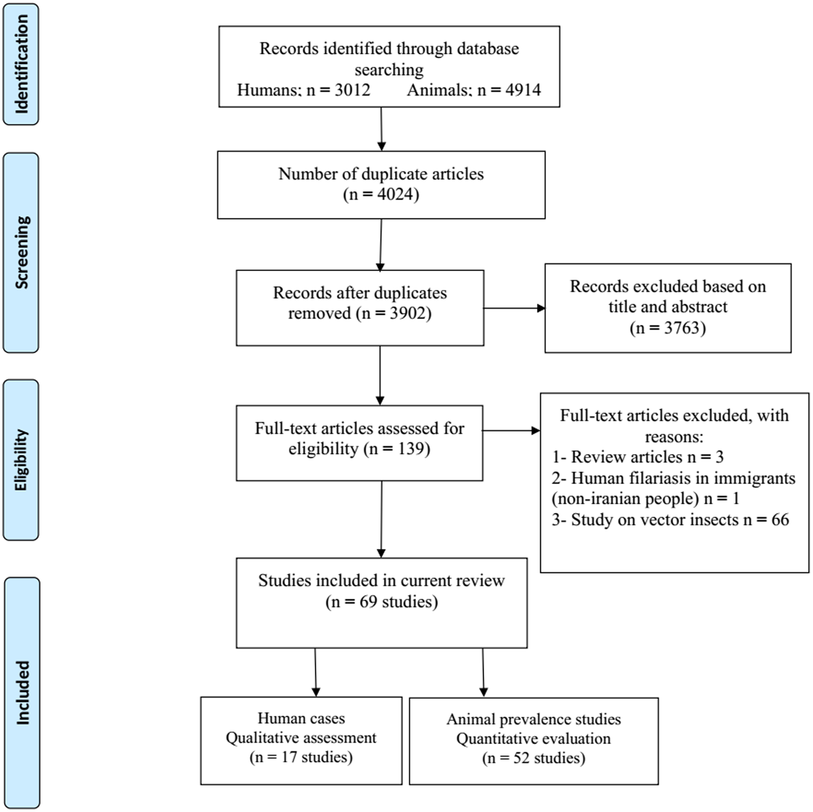
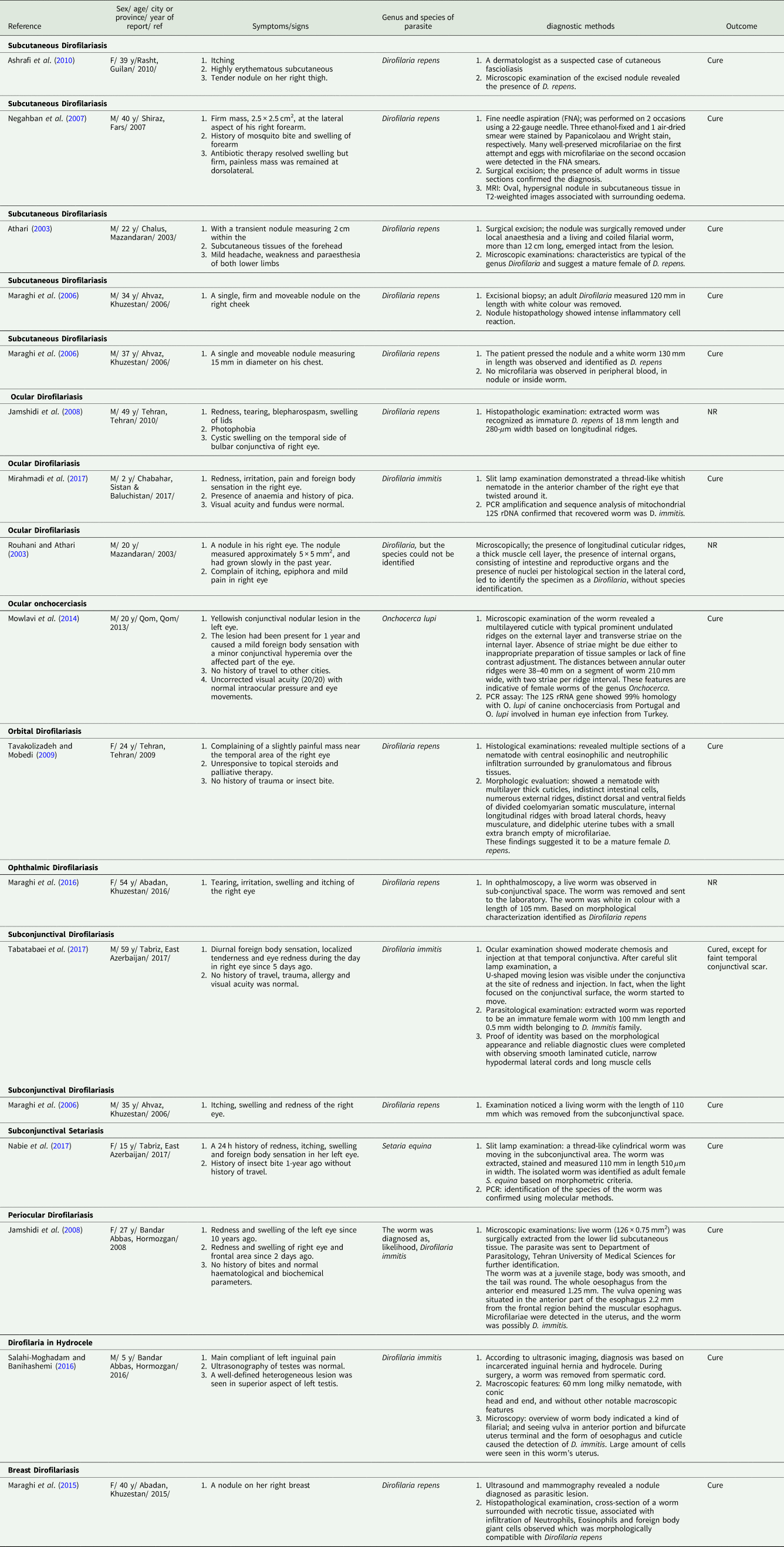
We analysed 3012 papers from database searching, while 2561 were irrelevant (based on their title/abstract, were review papers and/or study on a non-Iranian immigrant), 450 were excluded for duplication, and one article was omitted due to lack of proper diagnosis. Finally, 17 entries relevant to case reports of human filariasis in Iran met our inclusion criteria (Fig. 1). According to Table 1, the cases were reported from 9 provinces of Iran, mostly caused by D. repens. The routine diagnosis was based on microscopic identification of the worm and/or examination of histopathologic sections; only three studies used polymerase chain reaction (PCR) and sequencing methods for molecular evaluation. The right eye was the most parasitised organ (7 cases), followed by left eye (2 cases), as well as single cases in the lower eyelid, cheek, forehead, spermatic cord, breast, chest, forearm and thigh. Cases were reported more frequently in men than women (64.7% vs 35.29%), and among adults than children (82.35% vs 17.64).
PRISMA flow diagram describing included/excluded studies up to 1 December 2019 (Moher et al., Reference Moher, Liberati, Tetzlaff and Altman2009)

Characteristics of human filariasis case report studies in Iran up to 1 December 2019

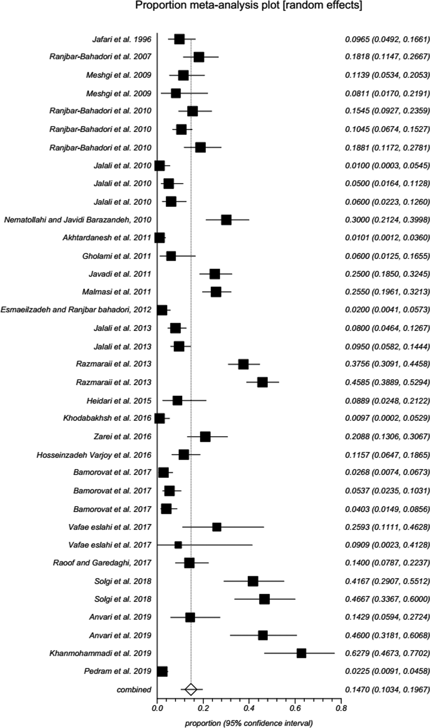
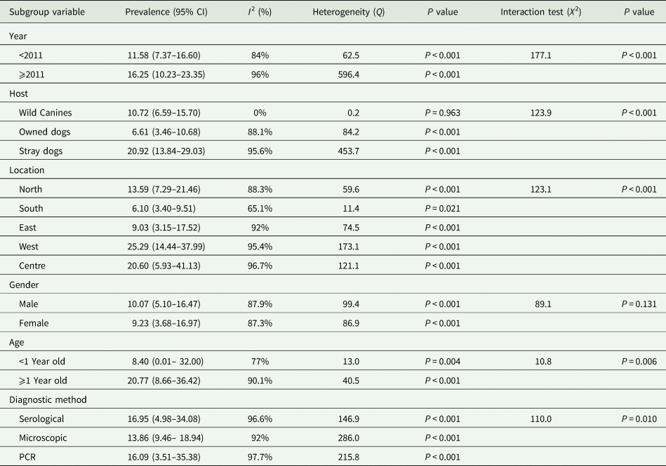
Information related to Filarioidea spp identified in various animal hosts (camel, cattle, small ruminants, dog, wild canids (jackals and red fox), horse, donkey, rodent, cat and pigeon) in Iran was illustrated as Supplementary Table 1. The systematic searching for original prevalence studies in animal hosts yielded 52 eligible papers based on our inclusion/exclusion criteria (Fig. 1). According to random-effects model meta-analysis, the pooled prevalence of D. immitis in canids of Iran was 14.69% (95% CI: 10.33–19.67) (Table 2; Supplementary Table 2; Fig. 2). Subgroup analysis was performed with summarized details in Table 3. Year-based analysis demonstrated a higher prevalence trend of D. immitis in published literature beyond 2011 [16.25% (95% CI: 10.23–23.35)] than those before 2011 [11.58% (95% CI: 7.37–16.60)] (P < 0.001). The higher prevalence was detected in free-ranging dogs (20.92%; 95% CI: 13.84–29.03), followed by wild canids (jackals and red fox) (10.72%; 95% CI: 6.59–15.70) and owned dogs (6.61%; 95% CI: 3.46–10.68) (P < 0.001). Geographical distribution showed the highest and lowest prevalence rates in western [25.29% (95% CI: 14.44–37.99)] and southern [6.10% (95% CI: 3.40–9.51)] parts of Iran, respectively (P < 0.001). Based on gender of dogs in Iran, D. immitis was more prevalent in males (10.07%; 95% CI: 5.10–16.47) than females (9.23%; 95% CI: 3.68–16.97) (P = 0.131). The heartworm was more prevalent in animals equal or more than 1-year old (20.77%; 95% CI: 8.66–36.42) than those younger (8.40%; 95% CI: 0.01– 32.00) (P = 0.006). According to the diagnostic method, the pooled prevalence of D. immitis in canids of Iran was as follows: microscopic 13.86% (95% CI: 9.46– 18.94), PCR 16.09% (95% CI: 3.51–35.38) and serology 16.95% (95% CI: 4.98–34.08) (P = 0.01).
Forest plot of the prevalence of D. immitis in canids of Iran up to 1 December 2019. A square is appointed to each individual study with a horizontal line as confidence intervals and the area of each square is proportional to the study's weight in the meta-analysis. Also, a diamond is assigned to the meta-analysed measure of effect. A vertical line representing no effect is also plotted. If the confidence intervals for individual studies overlap with this line, it demonstrates that at the given level of confidence their effect sizes do not differ from no effect for the individual study.

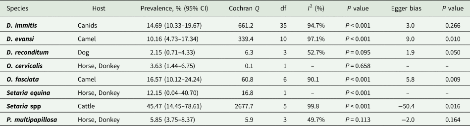
Prevalence, publication bias, and heterogeneity of Dirofilaria immitis, Dipetalonema spp, Onchocera spp, Setaria spp and Parafilaria multipapillosa in animal hosts in Iran up to 1 December 2019

Prevalence of Dirofilaria immitis in canines according to year, host, location, gender, age and diagnostic method in Iran up to 1 December 2019

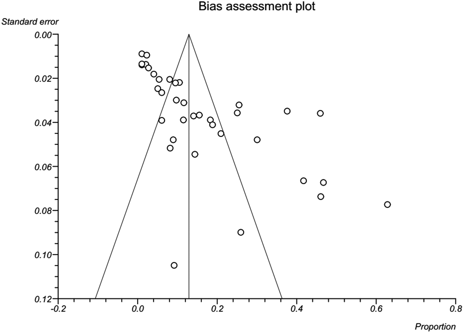
Results revealed a strong significant heterogeneity (Q = 661.2, df = 35, I 2 = 94.7%, P < 0.001) among the selected studies. Subgroup analysis revealed that there were statistically significant differences between the overall prevalence of D. immitis in canids of Iran and year (X 2 = 177.1, P < 0.001), host (X 2 = 123.9, P < 0.001), location (X 2 = 123.1, P < 0.001), age (X 2 = 10.8, P = 0.006) and diagnostic method (X 2 = 110.0, P = 0.010). Publication bias was checked by Egger's regression test, showed that it may not have a substantial impact on total prevalence estimate (Egger; bias: 3.0, P = 0.266) (Fig. 3).
A bias assessment plot from Egger for the prevalence of D. immitis in canids of Iran up to 1 December 2019. In the absence of publication bias, it assumes that studies with high precision will be plotted near the average, and studies with low precision will be spread evenly on both sides of the average, creating a roughly funnel-shaped distribution. Deviation from this shape can indicate publication bias.

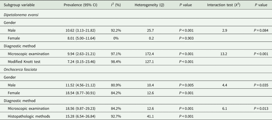
Based on the random-effects model meta-analysis, the pooled prevalence, publication bias and heterogeneity of other filarioids in Iran are depicted in Table 2. Furthermore, Table 4 demonstrates the subgroup analysis of Dipetalonema evansi (D. evansi) and Onchocerca fasciata (O. fasciata) in camels of Iran (Supplementary Figs 1–7).
Subgroup analysis of Dipetalonema evansi and Onchocerca fasciata in camels of Iran up to 1 December 2019

Discussion
Nowadays, the complex interaction between humans and animals has subjected to revolutionary changes in several aspects including human behaviour, demographics, land use and environment changes (Otranto and Deplazes, Reference Otranto and Deplazes2019). This also leads to an unprecedented encounter between humans and infectious agents and the problem of the emergence of infectious zoonotic diseases. Regarding filariasis, this occurs in those areas of the world where insect vectors inhabit, particularly in developing countries, inflicting significant threat to human and animal health (Thompson et al., Reference Thompson, Lymbery and Smith2010). With respect to the importance of this issue and increased number of human cases in recent years, we sought to extend our knowledge on poorly-known filarial infections in the human and examined animals in a vast Middle Eastern country, Iran.
Regarding human cases, most reports were from Khuzestan province, southwestern Iran. This territory is located in the vicinity of the Persian Gulf and possesses favourable milieu such as abundant dams and lagoons as well as tropical temperatures throughout the year, required for the colonization of blood-sucking arthropod vectors (Hamidinia et al., Reference Hamidinia, Maraghi, Azimi, Ai and Shirian2016). The eyes or the conjunctiva are a frequent site of choice for filarioids (Otranto and Eberhard, Reference Otranto and Eberhard2011), as we also found most Iranian cases in right and left eyes, respectively. Dirofilariasis due to Dirofilaria spp. were the most abundant filarial infection in humans in Iran. Despite remarkable seroprevalence of antibodies to Dirofilaria in human societies of endemic areas (Simon et al., Reference Simon, Muro, Cordero and Martin1991; Espinoza et al., Reference Espinoza, Cordero, Muro, Lorente and Simón1993; Vieira et al., Reference Vieira, Vélez, Montoya, Agudelo, Alvarez, Genchi and Simon1998; Tasić-Otašević et al., Reference Tasić-Otašević, Gabrielli, Tasić, MiladinovićTasić, Kostić, Ignjatović, Dragonjić, Milošević, Arsić-Arsenijević and Cancrini2014), human dirofilariasis is underdiagnosed, though the substantial increase in cases has been recorded worldwide mainly as subcutaneous/ocular form (Simón et al., Reference Simón, Siles-Lucas, Morchón, González-Miguel, Mellado, Carretón and Montoya-Alonso2012). In the current review, it was realized that D. repens was the most isolated species, commonly from subcutaneous tissue and right eye. Iranian cases with D. immitis had an eye infection as well as an interesting case removed from the spermatic cord in relation to testicular hydrocele (Salahi-Moghadam and Banihashemi, Reference Salahi-Moghadam and Banihashemi2016). There was only a single case of intraocular involvement (anterior chamber/vitreous body) among Iranian dirofilariasis cases (Mirahmadi et al., Reference Mirahmadi, Maleki, Hasanzadeh, Ahoo, Mobedi and Rostami2017), similar to some cases from other countries including Turkey and Brazil (Gungel et al., Reference Gungel, Kara, Pinarci, Albayrak, Baylancicek and Uysal2009; Otranto et al., Reference Otranto, Diniz, Dantas-Torres, Casiraghi, de Almeida, de Almeida, dos Santos, Furtado, de Almeida Sobrinho and Bain2011a). Cases of human pulmonary dirofilariasis, which has been predominantly reported from Japan and USA (Simón et al., Reference Simón, Siles-Lucas, Morchón, González-Miguel, Mellado, Carretón and Montoya-Alonso2012), were not detected in Iran, which could be due to lack of available serological assessment, though clinicians should be alerted to it (Khedri et al., Reference Khedri, Radfar, Borji, Azizzadeh and Akhtardanesh2014). In an international scale, human infections by D. repens are prevalent in Eurasia region, with the highest incidences of subcutaneous/ocular dirofilariasis occurring in Europe particularly in Russia and Italy (Simón et al., Reference Simón, Siles-Lucas, Morchón, González-Miguel, Mellado, Carretón and Montoya-Alonso2012). In Asia, Sri Lanka and India are predominant regarding subcutaneous/ocular dirofilariasis (Simón et al., Reference Simón, González-Miguel, Diosdado, Gómez, Morchón and Kartashev2017). Subcutaneous nodules due to D. repens usually emerge over a period of weeks or months, with rigid, elastic solidity (Simón et al., Reference Simón, Siles-Lucas, Morchón, González-Miguel, Mellado, Carretón and Montoya-Alonso2012). Approximately, 30–35% of D. repens-related infections involve ocular sites, entailing considerable consequences such as floaters, damaged vision, glaucoma, crystalline lens, the opacity of the vitreous humor and blepharedema (Pampiglione and Rivasi, Reference Pampiglione and Rivasi2000; Stringfellow et al., Reference Stringfellow, Francis, Coroneo and Walker2002). Altogether, the spread of human dirofilariasis due to D. repens may be the result of vector habitat outreach, impaired immune responses to subcutaneous parasites and inadequate diagnosis and therapy in the primary hosts, i.e. dogs (Otranto and Deplazes, Reference Otranto and Deplazes2019).
Among Iranian case reports, subconjunctival infection to Onchocerca lupi (O. lupi) in a 20-year-old male commuter inhabiting Qom province was interesting, since it was the first human case of Onchocerca infection in the country (Mowlavi et al., Reference Mowlavi, Farzbod, Kheirkhah, Mobedi, Bowman and Naddaf2014). Mowlavi et al. mentioned that the patient with multiple parasitic nodules in his eye may have got infected by blackflies in northern Tehran or Culicoides species in Qom suburbs (Mowlavi et al., Reference Mowlavi, Farzbod, Kheirkhah, Mobedi, Bowman and Naddaf2014). Onchocerca lupi was first described in a wolf (Canis lupus cubanensis) in Russia in 1967 (Rodonaja, Reference Rodonaja1967). Otranto et al., in 2011 confirmed the first certain case of ocular O. lupi in a human by morphological and molecular analysis in Turkey where there was no report of canine onchocerciasis before (Otranto et al., Reference Otranto, Sakru, Testini, Gürlü, Yakar, Lia, Dantas-Torres and Bain2011b, Reference Otranto, Dantas-Torres, Cebeci, Yeniad, Buyukbabani, Boral, Gustinelli, Mounir, Mutafchiev and Bain2012). Based on a published systematic review, there have been increasing reports of human infections with O. lupi, especially during last decade, raising the zoonotic potential of this nematode in Europe, Middle East and USA (Grácio et al., Reference Grácio, Richter, Komnenou and Grácio2015). Although ocular cases are more common, O. lupi has occasionally been recovered from the spinal cord or subcutaneous nodules of infected humans (Grácio et al., Reference Grácio, Richter, Komnenou and Grácio2015). Notwithstanding reported infections in dogs and cats from some European countries (Hermosilla et al., Reference Hermosilla, Hetzel, Bausch, Grübl and Bauer2005; Faísca et al., Reference Faísca, Morales-Hojas, Alves, Gomes, Botelho, Melo and Xufre2010; Maia et al., Reference Maia, Annoscia, Latrofa, Pereira, Giannelli, Pedroso and Otranto2015; Tudor et al., Reference Tudor, Turcitu, Mateescu, Dantas-Torres, Tudor, Bărbuceanu, Ciuca, Burcoveanu, Acatrinei and Rinaldi2016; Hodžić et al., Reference Hodžić, Hinney, König, Naucke, Duscher and Joachim2018) as well as USA and Canada (Labelle et al., Reference Labelle, Daniels, Dix and Labelle2011; Otranto et al., Reference Otranto, Giannelli, Latrofa, Dantas-Torres, Trumble, Chavkin, Kennard, Eberhard and Bowman2015b), there exist paucity of data about O. lupi prevalence in canid population of Iran and its potential blackfly (Simuliidae) or Culicoides vectors.
Setaria spp. are parasites of some herbivores, with adults causing fibrinous peritonitis and immature forms aberrantly migrating in accidental hosts (Ahmad and Srivastava, Reference Ahmad and Srivastava2007). A subconjunctival setariasis due to Setaria equina (S. equina) in the left eye of a 15-year-old girl was reported from northwestern Iran (Nabie et al., Reference Nabie, Spotin and Rouhani2017). So far, only few cases of human infection to adult Setaria spp. has been reported globally. Panaitescu et al., documented four human cases of subconjunctival S. labiatopapillosa infection in Romania with photophobia, swelling and tearing signs (Panaitescu et al., Reference Panaitescu, Preda, Bain and Vasile-Bugarin1999). Another report from Romania emphasized subconjunctival infection of an old man with Setaria sp. (Ţălu et al., Reference Ţălu, Ştefănuţ, Mihalca and Coroiu2012). Despite a wide array of Culicidae mosquito vectors for setariasis around the world, comprising Aedes, Anopheles, Armigeres, Culex and Mansonia (Azari-Hamidian et al., Reference Azari-Hamidian, Norouzi and Harbach2019), less is understood about the possible vectors in Iran. Azari-Hamidian et al., in 2009 did the only present survey and found Anopheles maculipennis mosquitoes in northwestern Iran infected to Setaria (Azari-Hamidian et al., Reference Azari-Hamidian, Yaghoobi-Ershadi, Javadian, Abai, Mobedi, Linton and Harbach2009).
Dirofilaria immitis, agent of cardiopulmonary dirofilariasis in canids, was the predominant species of filarioids among all examined animals in Iran. The highest detection rate was obtained by serology (16.95%; 95% CI = 3.51–35.38%) as convenient techniques for screening or field-based appraisal, while the lowest prevalence was determined using microscopy (13.86%; 95% CI = 9.46–18.94%). Serological methods are appropriate, especially for the diagnosis of amicrofilaremic infections (Simón et al., Reference Simón, Siles-Lucas, Morchón, González-Miguel, Mellado, Carretón and Montoya-Alonso2012). Geographically, both D. immitis and D. repens are sympatric in most territories (Simón et al., Reference Simón, González-Miguel, Diosdado, Gómez, Morchón and Kartashev2017), although the latter was only detected in a recent multiplex-PCR study in Iran with 26% prevalence in dogs (Pedram et al., Reference Pedram, Tabrizi, Hosseinzadeh, Pourmontaseri and Rakhshandehroo2019). Globally, several studies have reported the prevalence of canine D. immitis infection. In continental Portugal where heartworm is endemic, 4–9% prevalence was reported (Alho et al., Reference Alho, Meireles, Schnyder, Cardoso, Belo, Deplazes and de Carvalho2018). The prevalence in Turkey, neighbouring Iran, was zero to 18% (Köse and Erdoğan, Reference Köse and Erdoğan2012). In Greece, canine D. immitis prevalence ranged between 0.7% and 25% (Angelou et al., Reference Angelou, Gelasakis, Verde, Pantchev, Schaper, Chandrashekar and Papadopoulos2019; Diakou et al., Reference Diakou, Soubasis, Chochlios, Oikonomidis, Tselekis, Koutinas, Karaiosif, Psaralexi, Tsouloufi and Brellou2019). In Poland, Eastern Europe very low prevalence (<1%) was observed (Krämer et al., Reference Krämer, Schaper, Schunack, Połozowski, Piekarska, Szwedko, Jodies, Kowalska, Schüpbach and Pantchev2014). The highest canine heartworm prevalence was reported from Madeira Island with 40% (Genchi and Kramer, Reference Genchi and Kramer2019). Reports from the Far East countries are rare, with 2–15% and 18% in China (Liu et al., Reference Liu, Yang, He, Yang and Sun2013)and Thailand (Boonyapakorn et al., Reference Boonyapakorn, Srikitjakarn, Morakote and Hoerchner2008), respectively. Moreover, 4.7–29.5% prevalence rates were observed in India (Borthakur et al., Reference Borthakur, Deka, Islam, Sarma and Sarmah2015). A few studies have reported D. immitis and D. repens in African countries including Algeria (Tahir et al., Reference Tahir, Damene, Davoust and Parola2017), Tunisia (Rjeibi et al., Reference Rjeibi, Rouatbi, Mabrouk, Tabib, Rekik and Gharbi2017), Mozambique (Schwan and Durand, Reference Schwan and Durand2002) and Tanzania (Mukendi et al., Reference Mukendi, Kimbita, Mbanzulu, Maindo and Misinzo2016) with 1.4–14.5% prevalence rates. The highest prevalence of D. immitis in the Americas has been reported in US Eastern states, Caribbean Islands and some parts of Argentina and Brazil (20.4–74%) (Lee et al., Reference Lee, Montgomery, Theis, Blagburn and Eberhard2010; Little et al., Reference Little, Beall, Bowman, Chandrashekar and Stamaris2014; Barrett and Little, Reference Barrett and Little2016; Simón et al., Reference Simón, González-Miguel, Diosdado, Gómez, Morchón and Kartashev2017).
Based on the findings of this current review, the highest prevalence of D. immitis was observed in stray dogs (20.92%; 95% CI = 13.84–29.03%), whereas 10.72% (95% CI = 6.59–15.7%) and 6.61% (95% CI = 3.46–10.68%) prevalence rates were reported in wild canids (jackals and red fox) and owned dogs, respectively, in Iran. An increasing trend has been shown of heartworm infection among European populations of jackals (7.7–23.3%), foxes (3.7–35%) and raccoon dogs (31.1%) (Marconcini et al., Reference Marconcini, Magi, Macchioni and Sassetti1996; Cirovic et al., Reference Cirovic, Penezic, Pavlovic, Kulisic, Cosic, Burazerovic and Maletic2014; Simón et al., Reference Simón, González-Miguel, Diosdado, Gómez, Morchón and Kartashev2017). A recent study in Canada showed 4.8% prevalence of D. immitis in wild canids (Kotwa et al., Reference Kotwa, Jardine, Berke, Pearl, Mercer and Peregrine2019). Wild canids, directly or indirectly, possibly play a critical role in the maintenance and transmission of D. immitis (Simón et al., Reference Simón, González-Miguel, Diosdado, Gómez, Morchón and Kartashev2017). Distribution patterns of D. immitis in canids may be influenced by different ecosystems, such that foxes in agricultural regions of Europe were more infected than those foxes in semiarid or mountainous territories. High interactions among wildlife, pets and humans in suburban/agricultural areas could affect the transmission dynamics of dirofilariasis (Gortázar et al., Reference Gortázar, Castillo, Lucientes, Blanco, Arriolabengoa and Calvete1994; Marks and Bloomfield, Reference Marks and Bloomfield1998).
Based on our findings, D. immitis prevalence was prevalent in western (25.29%; 95% CI = 14.44–37.99%) and northern (13.59%; 95% CI = 7.29–21.46%) Iran. From a historical perspective, the first observation of the heartworm in Iran dates back to 1969 when Sadighian reported necropsy documentation of stray dogs in Caspian Sea littoral, northern Iran (Sadighian, Reference Sadighian1969). Climate and environment are important extrinsic factors for survival and development of vector mosquitoes and subsequent occurrence of dirofilariasis. Regarding the ectothermic nature of mosquitoes and their reliance on water supplies, climatic parameters including humidity/precipitation and temperature substantially impact their colonization, population density, diversity and activity (Simón et al., Reference Simón, González-Miguel, Diosdado, Gómez, Morchón and Kartashev2017). Also, from a parasitic standpoint, extrinsic incubation (8–20 days with 22–30°C temperatures) is influential for L3 larvae development (Simón et al., Reference Simón, Siles-Lucas, Morchón, González-Miguel, Mellado, Carretón and Montoya-Alonso2012). Such favourable circumstances are provided in the western and northern parts of Iran, where there exist huge water resources, irrigation systems and high precipitation rates annually. In contrast, low prevalence rates in eastern and southern parts of Iran are observed, where weak water supplies and low annual precipitation exist. An expedient exemplary of climate impact on the prevalence of dirofilariasis is represented in Grand Canary Island, where various altitudes possess different semitropical climates. Accordingly, D. immitis prevalence among canines of various zones differ significantly, from 30.4% in mild climate zone to 10% in the temperate cold climate zone (Montoya-Alonso et al., Reference Montoya-Alonso, Mellado, Carretón, Cabrera-Pedrero, Morchón and Simón2010). Although not significant, the prevalence in males was partly more than females in our review (10.07% vs 9.23%; P = 0.131), which is consistent with findings of other investigations (Reifur et al., Reference Reifur, Thomaz-Soccol and Montiani-Ferreira2004; Simsek et al., Reference Simsek, Utuk, Koroglu and Rishniw2008). Also, canids over 1-year old were over 2-fold more susceptible than younger (<1-year old) ones (20.77% vs 8.40%; P = 0.006), in agreement with another study in Brazil (Reifur et al., Reference Reifur, Thomaz-Soccol and Montiani-Ferreira2004). Adult animals had probably accumulated more exposure time to insect bites, thus had a higher prevalence of infection.
Besides canine Dirofilaria infection in Iran, we only found two studies regarding feline dirofilariasis in Ardabil and Khuzestan provinces. Heartworm infection is a subclinical condition in domestic/wild felids and only a few worms reach maturity; hence, there may be a limited number of blood microfilariae, which, in turn, substantially reduces the chance of transmission (Simón et al., Reference Simón, Siles-Lucas, Morchón, González-Miguel, Mellado, Carretón and Montoya-Alonso2012; Penezic et al., Reference Penezic, Selakovic, Pavlovic and Cirovic2014). Otranto et al. (Reference Otranto, Cantacessi, Dantas-Torres, Brianti, Pfeffer, Genchi, Guberti, Capelli and Deplazes2015a) also corroborate our findings, highlighting the lower contribution of felids in the epidemiology of heartworm disease (Otranto et al., Reference Otranto, Cantacessi, Dantas-Torres, Brianti, Pfeffer, Genchi, Guberti, Capelli and Deplazes2015a). In the USA, 3–19% prevalence ranges have been reported regarding feline dirofilariasis. Studies in Europe have shown a 7–27% (Italy) and 33% (Canary Islands) seroprevalence rates. In Japan, 2–5.2% of cats were seropositive for dirofilariasis (Simón et al., Reference Simón, Siles-Lucas, Morchón, González-Miguel, Mellado, Carretón and Montoya-Alonso2012).
Other known, but less frequent filarioid nematodes found in Iran are D. evansi (syn. Deraiophoronema evansi) in camels, and Acanthocheilonema reconditum (A. reconditum) in dogs. The camel parasite was isolated from 7 provinces with a total prevalence of 10.16% (95% CI = 4.73–17.34%). Male dromedaries were more parasitised than females (10.62% vs 8.01%), consistent with Mahran study (Mahran, Reference Mahran2004), although it was not significant (P = 0.084). A significant association was observed between the prevalence of D. evansi in Iranian camels and diagnostic method (X 2 = 13.2; P < 0.001). It seems that using a microscope examination is more appropriate for identifying D. evansi than the Knott test. One-humped camel (Camelus dromedarius) is the dominant species in Iran, with particular tolerance to the harsh desert environment and some pathogens, although D. evansi induces clinical disease (Sazmand and Joachim, Reference Sazmand and Joachim2017). Adult worms accumulate in large number in various affected organs, comprising spermatic cord, epididymis, testicles, heart and lungs. The acute disease could lead to emaciation, orchitis, heart failure, arteriosclerosis and nervous impairment (Oryan et al., Reference Oryan, Valinezhad and Bahrami2008). Our knowledge on this parasite is limited to prevalence studies in arid, semi-arid countries of the world, including Egypt, Nigeria, Saudia Arabia, Iran and India (Pathak and Chahabra, Reference Pathak and Chahabra2010; Sazmand et al., Reference Sazmand, Tafti, Hekmatimoghaddam and Moobedi2013; Egbe-Nwiyi et al., Reference Egbe-Nwiyi, Paul and Muhammed2016; El-Khabaz et al., Reference El-Khabaz, Abdel-Hakeem and Arfa2019). Globally, the estimated prevalence in adult camels was 2.5–4%, while it was 47.5% in less than 1-year-old camels (Muhammad and Athar, Reference Muhammad, Athar and Gahlot2000). A molecular study by PCR and sequencing methods in 2016 revealed paraphyly of D. evansi and D. gracile, which deserves further investigations (Sazmand et al., Reference Sazmand, Eigner, Mirzaei, Hekmatimoghaddam, Harl, Duscher, Fuehrer and Joachim2016). Acanthocheilonema reconditum living in canine subcutis and on muscle fascia develops mild parasitism in dogs with no major damages (Saari et al., Reference Saari, Nareaho and Nikander2019). Approximately, 2.15% (95% CI = 0.71–4.33%) of the dog population in Iran was reported to have this infection. In a multispecies survey in Romania on filarioid infections, A. reconditum DNA was only detected in a red fox (0.33%) (Ionică et al., Reference Ionică, Matei, D'Amico, Ababii, Daskalaki, Sándor, Enache, Gherman and Mihalca2017). Diagnosis is important only to differentiate their mfs from the life-threatening species, D. immitis (Otranto and Deplazes, Reference Otranto and Deplazes2019).
With respect to other filarioids infecting domestic livestock in Iran, we could only estimate the weighted prevalence of Parafilaria multipapillosa (P. multipapillosa) (referred to as Filaria haemorrhagica) in equids (5.85%; 95% CI: 3.75–8.37%), S. equina in equids (12.15%; 95% CI: 0.04–40.7%) and S. digitata in ruminants (45.47%; 95% CI: 14.45–78.61%). Several Onchocerca spp. parasitise livestock including, O. fasciata (camel; connective tissue, ligamentum nuchae) (16.57%; 95% CI: 10.12–24.24%), O. cervicalis (horse and donkey; cervical ligament) (3.63%; 95% CI: 1.44–6.57%) and O. reticulata (horse and donkey; connective tissue, flexor tendon) which are found in Iran. We found a statistically significant association between prevalence of O. fasciata in Iranian camels and gender (X 2 = 4.4: P = 0.035) and diagnostic method (X 2 = 6.1; P = 0.013). It seems that using microscopic examination may be more likely to detect O. fasciata than histopathologic methods. Prevalence studies are actually rare on P. multipapillosa, while there is more on P. bovicola of cattle (not found in Iran) (Bech-Nielsen et al., Reference Bech-Nielsen, Bornstein, Christensson, Wallgren, Zakrisson and Chirico1982; Solismaa et al., Reference Solismaa, Laaksonen, Nylund, Pitkanen, Airakorpi and Oksanen2008; Borgsteede et al., Reference Borgsteede, van Wuijckhuise, Peutz, Roumen and Kock2009). Setariosis is a benign infection and even high rates of microfilaraemia could be well tolerated (Hornok et al., Reference Hornok, Genchi, Bazzocchi, Fok and Farkas2007). Survey of filarial nematodes of 188 donkeys in Egypt showed a total infection rate of 86.7% mostly in males (86.73%), regarding O. cervicalis (82.98%), O. reticulata (4.26%), S. equina (36.17%) and P. multipapillosa (5.32%) (Radwan et al., Reference Radwan, Ahmed, Elakabawy, Ramadan and Elmadawy2016). In Hungary, 18 of 195 (9.2%) horses had mfs by Knott technique for S. equina (Hornok et al., Reference Hornok, Genchi, Bazzocchi, Fok and Farkas2007). In a Finnish study, 209 skin biopsies of cattle revealed 78 (37%) positive for Onchocerca sp. mfs (Solismaa et al., Reference Solismaa, Laaksonen, Nylund, Pitkanen, Airakorpi and Oksanen2008). Altogether, the above information obtained from current systematic review and meta-analysis represents that there is a gap in our knowledge and understanding in the field of filariasis in livestock that needs global collaboration for better realization of such arthropod-borne helminths.
Concluding remarks
As we stated in the current systematic review and meta-analysis, Dirofilaria spp. infections (D. repens cases in humans and D. immitis in canids) were the most frequently found species of all filarioid nematodes in Iran. Most cases have been reported from rainy provinces with favourable temperatures for vector hosts. Also, most cases were detected during the last two decades, which indicates increased prevalence, and/or improved diagnostics or awareness in the public and clinical understanding of Dirofilaria infections. In this context, there is an urgent need to nationwide epidemiological surveys, e.g. serodiagnosis to detect human pulmonary cases, as well as preventive therapy of, at least pet dogs. Regarding less evident, but existing filarioids, such as the interesting human case of O. lupi in Qom province and those being found in herbivores much large-scale studies in the context of the host-parasite-vector axis are recommended to be done in the future for better understanding of the epidemiology of these filarioid infections.
Supplementary material
The supplementary material for this article can be found at https://doi.org/10.1017/S003118202000058X.
Acknowledgements
The authors sincerely declare their appreciation of Professor Domenico Otranto (Head of Department of Veterinary Medicine, University of Bari, Italy) for his noteworthy comments on this manuscript.
Financial support
This research received no specific grant from any funding agency, commercial or not-for-profit sectors.
Conflict of interest
None.
Ethical standards
Not applicable.