Right valve of right horn of systemic venous sinus nearly divides the right atrium into two separate chambers in early embryonic period. The incomplete involution of the valve of systemic venous sinus results in variable anatomical formations.Reference Loukas, Sullivan, Tubbs, Weinhaus, Derderian and Hanna 1 We present a case of persistent valve of systemic venous sinus as an intermediate form between Chiari network and division of right atrium (cor triatriatum dexter).
Case report
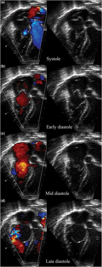
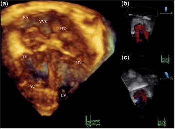
A 2-week-old Malay baby girl, weighing 3.2 kg, presented with mild cyanosis. She was delivered at 35 weeks of gestation with uneventful prenatal and perinatal history. She was well with unremarkable systemic review, though recorded an O2 saturation of 87% in room air. Electrocardiogram and chest radiograph were normal. Transthoracic echocardiography revealed redundant highly mobile membranous strands across the right atrium with attachments at the inferior caval vein to right atrium junction, near superior caval vein and near coronary sinus opening into right atrium (Fig 1). The aneurysmal redundant tissue prolapsed through the tricuspid valve into right ventricle during the diastolic phase causing moderate right ventricular inflow obstruction (Supplementary video S1). There was a right to left shunt across the patent oval foramen causing the cyanosis. Right ventricle, pulmonary and tricuspid valve size were normal. In addition, there was a small-sized patent arterial duct. The anatomical findings were confirmed on 3D echo and a diagnosis of persistent valve of systemic venous sinus (cor triatriatum dexter/Chiari network) was made (Fig 2, Supplementary video S2). In view of her mild cyanosis and only moderate obstruction, she was followed up conservatively. At 2 months of age, she was still active with O2 saturation of 80–86%. There was no involution of the redundant tissue, and surgical resection was performed. Per-operatively, the tissue mainly consisted of two membranes arising from terminal crest and eustachian valve, intertwining and inserting at coronary sinus opening rim dividing the right atrium into three compartments with communicating fenestrations avoiding severe right ventricular inflow obstruction (Supplementary figure S3). Patent oval foramen and three additional small secundum atrial septal defects were found and closed. The arterial duct was ligated. She had an uneventful course and was discharged on the 5th post-operative day with oxygen saturation 99% in room air.
Transthoracic two-dimensional echo with colour Doppler showing the redundant tissue from persistent valve of systemic venous sinus prolapsing into right ventricle causing moderate obstruction in the late diastolic phase. Multiple perforations are visible in the prolapsed structure.

Transthoracic 3D echocardiogram with colour Doppler showing multiple stands in the right atrium in systolic phase ( a ) and moderate right ventricular inflow obstruction during diastole ( b , c ). LV = left ventricle; MV = mitral valve; PFO = patent foramen ovale; RA = right atrium; RV = right ventricle; TV = tricuspid valve; VVS = valve of systemic venous sinus.

Discussion
The valves of right horn of systemic venous sinus are prominent structures within the right atrium during early embryonic period. These ridge-like invaginations redirect the blood from superior and inferior caval veins across the oval foramen into the left atrium. The right valve nearly divides the right atrium into two separate chambers. With the normal development of the heart, these structures involute between the 9th and 15th week of gestation. The left valve gets incorporated into atrial septal secundum. The right valve cranially involutes into terminal crest and caudally develops rudimentary eustachian valve at the inferior caval vein to right atrium junction and thebesian valve at coronary sinus to right atrium junction.Reference Smallhorn and Anderson 2
Involution may not be complete, resulting in a spectrum of anatomical presentations. Persistent valve of systemic venous sinus, in its simplest form as a prominent or giant eustachian valve, is a common finding. Chiari network represents a relatively more incomplete involution of the embryonic structure presenting in 2–4% of population.Reference Blasco, Torres, Domènech, Blanch, Herrera and Comas 3 Divided right atrium or cor triatriatum dexter is the most severe form with no or minimal involution of valve tissue.Reference Barrea, Rubay, Wagner and Ovaert 4
There are no strict criteria to distinguish between the three anatomical forms and reports overlap in the literature while describing these anomalies.Reference Martínez-Quintana, Rodríguez-González, Marrero-Santiago, Santana-Montesdeoca and López-Gude 5 Broadly categorising, the redundant tissue extending from eustachian valve with no attachment to interatrial septum is defined as prominent or giant eustachian valve. Multiple thin or thick strands arising from the eustachian valve, terminal crest or superior caval vein to right atrium junction, intercommunicating in a net-like structure terminating in thebesian valve is termed as Chiari network.Reference Aypar, Sert and Odabas 6 Thick membrane arising from same landmarks with no perforation dividing the right atrium into two separate chambers is termed as division of the right atrium (cor triatriatum dexter).Reference Barrea, Rubay, Wagner and Ovaert 4 The intermediate stages are difficult to classify objectively. As evident from our case, the membranes, strands, and intercommunications had characteristics of both Chiari network and division of right atrium. There have been previous reports of Chiari network causing moderate right ventricular inflow obstruction because of minimal perforation and non-obstructive division of the right atrium due to adequate perforations.Reference Blasco, Torres, Domènech, Blanch, Herrera and Comas 3
The physical presentation may also overlap. Prominent eustachian valve is essentially a benign tissue in the right atrium, but has been reported to provide focus for atrial pre-excitation, infective endocarditis, as well as thrombosis and paradoxical embolisation.Reference Loukas, Sullivan, Tubbs, Weinhaus, Derderian and Hanna 1 , Reference Bowers, Krimsky and Gradon 7 The same complications have been reported in the Chiari network in addition to right ventricular inflow obstruction, which in turn is the complication of division of right atrium.Reference Smallhorn and Anderson 2 Cyanosis in neonates has been reported with either forms.Reference Ko, Chen and Lin 8 The degree of right ventricular inflow obstruction determines the development of the right side of the heart.Reference Loukas, Sullivan, Tubbs, Weinhaus, Derderian and Hanna 1 , Reference Smallhorn and Anderson 2 In most severe cases, the tissue forms a windsock membrane across tricuspid valve causing severe right ventricular inflow obstruction.Reference Trento, Zuberbuhler, Anderson, Park and Siewers 9 In severely obstructive cases, bipartite right ventricle, pulmonary atresia, and even tricuspid atresia have been reported. Our case had a well-developed right heart with normal valves. Persistent arterial duct found in our case might be an incidental finding associated with prematurity as it has not been reported previously.
The management of persistent valve of systemic venous sinus depends on the degree of right ventricular inflow obstruction and cyanosis resulting from right to left shunt across patent oval foramen. Spontaneous involution of the Chiari network with moderate right ventricular inflow obstruction has been reported in early infancy.Reference Bendadi, van Tijn, Pistorius and Freund 10 In our case, we followed the patient conservatively but involution was not seen up to 2 months of age. Transcatheter resection of membrane has been reported in case of the Chiari network, although the presence of this redundant network in itself makes catheterisation difficult because of the risk of tissue entrapment in the catheter or devices for patent oval foramen and atrial septal defect occlusion.Reference Aydin, Gürol, Yilmazer and Dagdeviren 11 Surgical resection is the treatment of choice for persistent valve of systemic venous sinus with significant right ventricular inflow obstruction. There has been no report of recurrence following resection in the literature.Reference Loukas, Sullivan, Tubbs, Weinhaus, Derderian and Hanna 1 , Reference Bendadi, van Tijn, Pistorius and Freund 10
It is important to recognise persistent valve of systemic venous sinus, especially in neonates with cyanosis, as surgical resection is completely curative. Even in the absence of cyanosis or right ventricular inflow obstruction, special attention has to given due to possible complications, as well as challenging transcatheter interventions.Reference Loukas, Sullivan, Tubbs, Weinhaus, Derderian and Hanna 1
Acknowledgement
The authors acknowledge cardiovascular technologists Mohammad Rabani Bin Rosman and Nalinimalar Kumarasamy for their help in arranging the images.
Financial Support
This research received no specific grant from any funding agency, commercial or not-for-profit sectors.
Conflicts of Interest
None.
Ethical Standards
Being a brief report, the standard institutional protocols were used for the management of the child. There was no experimental drug or intervention involved, hence does not carry any ethical concerns.