INTRODUCTION
Toxoplasmosis is a worldwide disease caused by the protozoan Toxoplasma gondii, which affects both humans and warm-blooded animals (mammals and birds). Toxoplasmosis is a major food-borne illness, when raw and/or undercooked infected meat is consumed. In the veterinary field, toxoplasmosis in cats (Baril et al. Reference Baril, Ancelle, Goulet, Thulliez, Tirard-Fleury and Carme1999), chickens (Dubey et al. Reference Dubey, Laurin and Kwowk2016), sheep (Klun et al. Reference Klun, Djurkovic-Djakovic and Thulliez2007), pigs (Steinparzer et al. Reference Steinparzer, Reisp, Grunberger, Kofer, Schmoll and Sattler2015) and goat (Edelhofer and Prossinger, Reference Edelhofer and Prossinger2010), which are considered possible sources of infection to humans, is really critical.
In human infections, the presence or absence of specific antibodies is enough to diagnose the disease. However, in toxoplasmosis it is not enough to detect the specific antibodies, but also to estimate the time of infection, especially for pregnant women and immunocompromised patients. If a woman becomes infected during the gestation period, the consequences to fetus can be serious (Durlach et al. Reference Durlach, Kaufer, Carral, Freuler, Ceriotto, Rodriguez, Freilij, Altcheh, Vazquez, Corazza, Dalla Fontana, Arienti, Sturba, Gonzalez Ayala, Cecchini, Salomon, Nadal, Gutierrez and Guarnera2008; El-Awady et al. Reference El-Awady, Mahgoub, Naguib and Ismail2009). Early treatment of toxoplasmosis during pregnancy reduces transplacental transmission; however, anti-parasite drugs can be harmful for both the mother and the fetus. Diagnosis of acute toxoplasmosis is essential to avoid unnecessary treatment.
Toxoplasmosis is mainly diagnosed by serological methods, i.e. latex agglutination test (LAT). LAT reagents are especially useful to analyse large numbers of samples due to their simplicity, rapidness and low cost. LAT has been previously applied for detecting several diseases, such as leishmaniasis, Chagas'disease, leptospirosis and neosporosis (Cummins et al. Reference Cummins, Moody, Lalloo and Chiodini1994; Smits et al. Reference Smits, Chee, Eapen, Kuriakose, Sugathan, Gasem, Yersin, Sakasi, Lai-a-Fat, Hartskeerl, Liesdek, Abdoe, Goris and Gussenhoven2001; Gonzalez et al. Reference Gonzalez, Garcia, Vega, Marcipar, Meira and Gugliotta2010; Garcia et al. Reference Garcia, Gonzalez, Vega, Marcipar and Gugliotta2012, Reference Garcia, Gonzalez, Caudana, Vega, Marcipar and Gugliotta2013, Reference Garcia, Gonzalez, Marcipar, Vega and Gugliotta2014; Moraveji et al. Reference Moraveji, Hosseini, Moghaddara, Namavari and Eskandari2012).
In this context, the goal of this work is to obtain latex particles sensitized with antigen of T. gondii, which can act as immunoagglutination (IA) reagents to detect specific antibodies and discriminate whether toxoplasmosis was recently acquired or it is a past infection. By choosing antigens which has selective reactivity for the antibodies generated in the acute phase of the disease to sensitize latex particles, the resulting IA reagents would be able to discriminate the acute infection from the chronic one. The P22 (SAG2) protein has been preliminary evaluated by assessing the specific IgG levels, having shown to be very sensitive during the acute phase (Parmley et al. Reference Parmley, Sgarlato, Mark, Prince and Remington1992; Li et al. Reference Li, Maine, Suzuki, Araujo, Galvan, Remington and Parmley2000). The recombinant antigen used in this work correspond to the abbreviated region P22Ag bearing the main antigenic sites, as bioinformatically predicted through specific software (Costa et al. Reference Costa, Peretti, García, Peverengo, Gonzalez, Gugliotta, Dalla Fontana, Lagier and Marcipar2016). Latex–protein complexes (LPC) obtained by using the P22Ag and the homogenate of the parasite were evaluated in this work.
In this way 14 LPC including particles of different sizes, chemical functionality (carboxyl or epoxy groups) and varied densities of surface functional groups, were tested to determine which ones have improved levels of discrimination between control sera. Subsequently, selected LPC were tested against a panel of sera and their ability to detect acute phase antibodies anti-T. gondii was evaluated.
MATERIALS AND METHODS
Materials
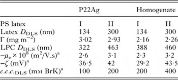
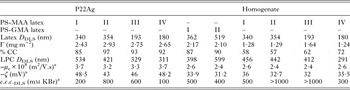
All the employed reagents were of analytical grade. Also, doubly deionized and distilled water was used. LPC were previously synthesized (Peretti et al. Reference Peretti, Costa, Gonzalez, Marcipar and Gugliotta2016) by physical adsorption (PA) of antigens onto polystyrene (PS) latexes (latexes PS I and PS II) and by covalent coupling (CC) of antigens with the external functional groups of carboxylated (PS-MAA I, PS-MAA II, PS-MAA III and PS-MAA IV) and epoxylated latexes (PS-GMA I and PS-GMA II). In the so-called sensitization process, either the acute phase recombinant protein of T. gondii P22Ag or the homogenate of the parasite (obtained from peritoneal exudates of infected mouse) were employed. Construction of expression plasmids and purification protocol of P22Ag is described by Costa et al. (Reference Costa, Peretti, García, Peverengo, Gonzalez, Gugliotta, Dalla Fontana, Lagier and Marcipar2016). The following characteristics were measured: average particle diameter (D DLS) of both the latexes and the LPC, by dynamic light scattering (DLS, Brookhaven Instruments Inc., Holtsville, USA) at 90°, Zeta potential (ζ) and electrophoretic mobility (μ e) employing a Zetasizer Nano (Malvern Instruments, Worcestershire, UK), and critical coagulation concentration (c.c.c.), at different concentrations of electrolyte (KBr), by DLS at pH 8. The amount of protein bound to the particle surface (Γ, mg m−2) and the percentage of protein covalently coupled with respect to the total bound protein (% CC) were also calculated. Characteristics of LPC obtained by PA and CC are shown in Tables 1 and 2, respectively (Peretti et al. Reference Peretti, Costa, Gonzalez, Marcipar and Gugliotta2016).
Final characteristics of the LPC obtained from polystyrene latexes PS I and PS II by physical adsorption of the antigens P22Ag and homogenate of T. gondii

a Determinated at pH 8.
Final characteristics of the LPC obtained from carboxylated latexes PS-MAA I, II, III and IV by covalent coupling of the antigens P22Ag or homogenate of T. gondii and epoxylated latexes PS-GMA I and II by covalent coupling of P22Ag
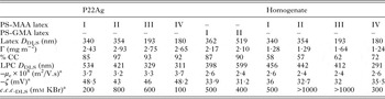
a Determinated at pH 8.
The Central Laboratory of the Province of Santa Fe (Argentina) provided 72 pregnant women serum samples classified in four groups. Serum samples were analysed by reference techniques following recommendations made by Durlach et al. (Reference Durlach, Kaufer, Carral, Freuler, Ceriotto, Rodriguez, Freilij, Altcheh, Vazquez, Corazza, Dalla Fontana, Arienti, Sturba, Gonzalez Ayala, Cecchini, Salomon, Nadal, Gutierrez and Guarnera2008) and classified according to their results. IgG and IgM were detected by indirect immunofluorescence assay and double-sandwich IgM enzyme-linked immunosorbent assay (ELISA, DS-IgM-ELISA, Radim, Pomezia, Italy), respectively. The ELISA kit, VIDAS-TOXO (bioMérieux, Marcy-l’Étoile, France), was used to assess IgG avidity index (AI) according to the manufacturer's instructions. Eighteen samples displayed negative reactions for both specific IgG and IgM antibodies (not infected group, NI). Eighteen showed positive reaction for specific IgG and negative for IgM and high AI (chronic, C). Thirty-six samples displayed positive reaction for specific IgG and IgM, and were analysed using IgG avidity test. Eighteen samples turned out to have low AI (LA, AI ⩽ 20%), and 18 samples displayed high AI (HA, AI ⩾ 30%). Serum samples were stored at −20 °C until tested.
Protein antigenicity evaluation by ELISA
Polystyrene microplates (GBO, Seattle, USA) were sensitized with 500 ng of P22Ag and homogenate (in carbonate buffer, pH 9·6) or about 20 µg of each LPC, which corresponds to approximately 500 ng of antigen (in borate buffer, pH 8) per well, and they were incubated over night at 4 °C. Then, microplates were washed with Tween in phosphate-buffered saline (PBS) at 0·01% (v/v) for three times, and a solution of skimmed milk in PBS at 5% (w/v) was employed during 1 h at 37 °C to block the free PS surfaces. Afterwards, sensitized microplates were incubated with a 1:100 dilution of the serum samples in 1% (w/v) skimmed milk in PBS. Then, microplates were washed thrice with Tween in PBS at 0·01%, and incubated with a 1:2000 dilution of the Peroxidase-conjugated goat anti-human IgG, Fcγ (Zymed, San Francisco, USA) in skimmed milk in PBS at 1% (v/v). The enzymatic reaction was developed using 100 µL of tetramethyl benzidine (Zymed) in H2O2 and stopped with 100 µL of 2 N H2SO4 (Peretti et al. Reference Peretti, Gonzalez, Marcipar and Gugliotta2014).
An ELx808 Absorbance Microplate Reader (BioTek Instruments, Winooski, USA) was employed to record ELISA results as optical density (OD) at 450 nm. All serum samples were evaluated by simultaneous determinations and the mean OD of these duplicates was calculated.
Immunoagglutination assays
LPC were conditioned before being employed in the agglutination test. First, they were centrifuged and redispersed in a pH 8 saline solution of bovine serum albumin (BSA). The effect of the ionic strength and the presence of glycine and/or polyethylene glycol 8000 (PEG) were studied, with the aim of reducing non-specific agglutinations during the immunoassays. BSA was used to block the hydrophobic zones of the particles, and glycine to reduce free carboxyl or epoxy groups present on the particles surface. In addition, PEG was employed to allow better exposure of the antigen epitopes bound to the particle surface.
For LAT, a dispersion containing the investigated LPC at a concentration corresponding to 1 unit of absorbance (A) was evaluated by turbidimetry, by measuring the time evolution of A at 570 nm. If during the period of analysis A remains unchanged, the LPC is considered stable. Then, 950 µL of the investigated LPC was mixed with 50 µL of serum. After 5 min of reaction, A was measured and the agglutination reaction was detected. The increment in A (ΔA) was determined by subtracting the absorbance of the LPC without serum to the absorbance measured for the sample (LPC + serum).
Statistical analysis
Graphics and statistical analysis were performed using GraphPad Prism version 6.00. Statistical significance was determined by ANOVA and Tukey's multiple comparisons post-test. Receiver operating characteristic (ROC) curves were constructed. The area under the ROC curve (AUC), the sensitivity, the specificity, the positive and the negative predictive values (PPV and NPV, respectively) were calculated. Toxoplasmosis prevalence in pregnant women was obtained from Carral et al. (Reference Carral, Kaufer, Durlach, Freuler, Olejnik, Nadal, Corazza, Pari, García, Córdoba, Rodriguez, Ceriotto and García2008). The AUC provides information about the discrimination between the analysed populations independently of the cut-off value. According to Swets and Pickett (Reference Swets and Pickett1982), if the AUC is between 0·5 and 0·7, the test discrimination is bad; if the AUC is between 0·7 and 0·9, the test may be useful in some cases; while AUC values >0·9 indicate a good diagnostic performance.
RESULTS AND DISCUSSION
Protein antigenicity evaluation by ELISA
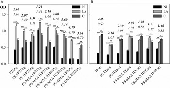
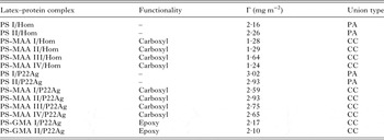
The acute phase recombinant protein P22Ag, the homogenate of T. gondii and the LPC obtained from them (Table 3), were first employed to sensitize PS microplates. Results are shown in Fig. 1.
ELISA results obtained for P22Ag (A), the homogenate (B) and the LPC produced by physical adsorption and chemical coupling of both antigens. Numbers on the bars indicate the ratio ODac/ODneg (bold) and the ratio ODac/ODch (non-bold). Error bars represent the range of duplicate samples. Statistical significance was determined by ANOVA and Tukey's multiple comparisons post-test. Statistically not different (ns: P > 0·05), statistically different (*: 0·01 < P < 0·05; **: 0·01 < P < 0·001; ***:0·001 < P < 0·0001; ****: P < 0·0001).

Latex–protein complexes analysed in ELISA and immunoagglutination assays

PA, physical adsorption; CC, covalent coupling.
The ratio between the OD obtained when the low avidity serum sample, and the OD for a not infected or a chronic serum was evaluated. When the recombinant protein P22Ag was used, the OD produced by the LA serum (ODLA) was greater than those obtained with the chronic (ODC) and the NI (ODNI) sera. These results correlate well with those reported by Costa et al. (Reference Costa, Peretti, García, Peverengo, Gonzalez, Gugliotta, Dalla Fontana, Lagier and Marcipar2016) for the same antigen and panel of sera. However, when employing the homogenate of T. gondii to sensitize the LPC, no significant differentiation between low avidity or chronic sera was observed. This was because the homogenate is a lysate of the total parasite, which includes non-specific antigens of the acute phase of the illness. The best performance was obtained with the complexes PS-GMA I/P22Ag (Γ = 2·17 mg m−2) and PS-MAA II/P22Ag (Γ = 2·93 mg m−2), which produced ODac/ODneg = 4·79 and 2·18, respectively; and ODac/ODch = 4·79 and 1·66, respectively. In all cases, the response obtained with the different LPC were similar to those obtained with the single antigen (Peretti et al. Reference Peretti, Gonzalez, Marcipar and Gugliotta2014), thus confirming that the antigen was not affected during the sensitization process, and consequently the produced LPC were able to recognize specific antibodies.
Immunoagglutination assays from control sera
LPC were studied in IA assays, under low ionic strength (20 mm) in the presence of glycine (0·1 m) and PEG 8000 (3% w/v). The reaction time is defined as the interval between the mixture of the serum with the LPC and the A reading, and it was fixed in 5 min (Garcia et al. Reference Garcia, Gonzalez, Marcipar, Vega and Gugliotta2014). Even though not shown, in all cases the best results, in the sense of maximizing discrimination of acute sera from negative and chronic ones, were obtained for latexes exhibiting higher values of c.c.c. and charge density. Furthermore, when comparing latexes of different sizes, it was noted that particles of about 350 nm of diameter produced better performance. The best results obtained with carboxylated complexes were from the latex PS-MAA II/P22Ag (Γ = 2·93 mg m−2), and, regarding the LPC derived from epoxylated latexes, only PS-GMA I/P22Ag (Γ = 2·17 mg m−2) achieved discrimination of acute serum from negative and chronic ones.
Immunoagglutination assays from a panel of sera
The performance of the LPC to discriminate the low avidity sera from the high avidity and the chronic ones was evaluated through ROC analysis (LA vs HA and LA vs C). Table 4 shows the values of AUC, cut-off, sensitivity, specificity NPV and PPV obtained for the three LPC evaluated.
Performance of carboxylated PS-MAA II/P22Ag or PS-MAA II/Hom and epoxylated PS-GMA I/P22Ag in immunoagglutination assays

NPV, negative predictive value; PPV, positive predictive value. Groups of sera: LA, low avidity; HA, high avidity and C, chronic.
The cut-off value was fixed in order to obtain 100% of sensitivity. Although the low specificities obtained, this criterion allows to be sure that all samples that are negative belong to pregnant women who are not attending a recent infection. When the prevalence of the disease is low, a negative result will allow the disease to be ruled out with greater safety, reflecting a high NPV (100%). On the contrary, a positive result will not allow confirming the diagnosis, resulting in a low PPV, and serum samples should be analysed with confirmatory techniques of high specificity.
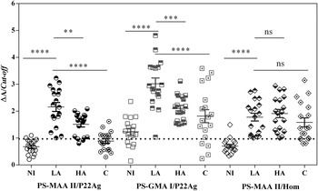
Figure 2 shows the ΔA/cut-off distributions of the three LPC evaluated. LPC obtained from homogenate of T. gondii showed similar ΔA values for the three groups of infected sera (LA, HA and C), because the heterogeneous homogenate is not specific for either the acute or the chronic sera, and it is not useful to differentiate stages of infection. Regarding the LPC derived from the recombinant protein P22Ag, lower ΔA C and higher ΔA LA values were observed, achieving better discrimination of low avidity sera. The mean ΔA LA for the carboxylated LPC PS-MAA II/P22Ag was 2·15 and it was statistically different to the mean ΔA NI (0·68), ΔA HA (1·50) and ΔA C (0·88). Discrimination of low avidity sera from high avidity sera was according to the performance of this antigen when it was evaluated by ELISA (Costa et al. Reference Costa, Peretti, García, Peverengo, Gonzalez, Gugliotta, Dalla Fontana, Lagier and Marcipar2016). With respect to the influence of chemical functionality of latex particles, the mean ΔA LA for the epoxylated LPC PS-GMA I/P22Ag was 3·02 and it was statistically different to the mean ΔA NI (1·23), ΔA HA (2·10) and ΔA C (1·81). Note that in the case of epoxylated LPC, various chronic sera were above the mean ΔA LA, which was not observed for the carboxylated complex. Even when the antigen conformation may be affected the performance of the epoxylated LPC, from the point of view of colloidal stability the lower μ e, ζ and c.c.c. values observed for PS-GMA I/P22Ag could explain the worst performance with respect to the carboxylated one. Moreover, nonspecific agglutination due to possible serum interferences could be produced, e.g. the presence of other proteins which could be adsorbed unspecifically on the hydrophobic surface of the epoxylated complexes.
Comparison of relative distributions (ΔA/cut-off) obtained for the carboxylated PS-MAA II/Hom and PS-MAA II/P22Ag and for the epoxylated PS-GMA I/P22Ag. Dashed line indicates ΔA/cut-off = 1. Horizontal lines indicate the mean and standard error of the mean (s.e.m.). Statistical significance was determined by ANOVA and Tukey's multiple comparisons post-test. Statistically not different (ns: P > 0·05), statistically different (**: 0·01 < P < 0·001; ***: 0·001 < P < 0·0001; ****: P < 0·0001).

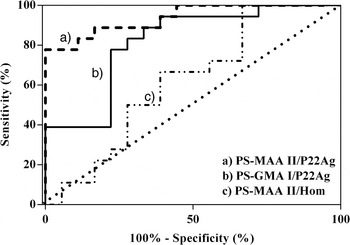
Figure 3 shows the ROC curves corresponding to the three LPC evaluated for LA vs C groups of sera. When PS-MAA II/P22Ag and PS-GMA I/P22Ag were evaluated, the AUC were 0·94 and 0·82, respectively (Table 4, LA vs C groups) indicating a better discrimination for the carboxylated LPC than for the epoxylated LPC. As expected, when the LPC obtained from the homogenate was tested, the AUC was 0·62.
ROC curves constructed between low avidity sera vs chronic sera for the three LPC studied.

Otherwise, the results shown for LPC obtained from the recombinant antigen and the carboxylated latex PS-MAA II allowed a good discrimination of the low avidity sera, and therefore it could be useful to rule out an early toxoplasmosis in pregnant.
Concluding remarks
P22Ag and the homogenate of the parasite linked to the particles were not practically affected during the sensitization by PA and CC.
The recombinant protein P22Ag were more reactive against low avidity sera, indicating that such antigen might be useful to detect acute phase antibodies.
The IA test based on latex particles with carboxyl functionality and the recombinant P22Ag produced an AUC of 0·94, a sensitivity of 100% and a NPV of 100% when comparing LA vs C groups of sera. Results are indicative of the ability of this reagent to rule out the acute infection, being necessary its evaluation with a larger panel of sera to confirm its applicability. In this way, the proposed test could be useful for the toxoplasmosis diagnosis in pregnant women, with the advantages of being cheap, rapid and easy to be implemented.
ACKNOWLEDGEMENTS
We are grateful to M. L. Dalla Fontana and C. Pastor for the provision of serum samples.
FINANCIAL SUPPORT
The authors would like to thank CONICET and Universidad Nacional del Litoral for their financial support.