The ubiquitous nontuberculous mycobacterium (NTM) constitutes a potential source of infectious disease in a broad range of animal species, including humans. Environmental studies have isolated NTM from natural and potable water sources worldwide. Water obtained from municipal treatment facilities, hospitals, and homes grew NTM in 10% to 95% of the samples in European and American surveys.Reference Phillips and von Reyn1
Rapidly growing nontuberculous mycobacteria (RGM), a subset of NTM, are defined as mycobacteria that grow within 7 days.Reference Runyon2, Reference Runyon3 RGM are capable of forming a protective biofilm that is important for their survival in a given environment. Among RGM, Mycobacterium mucogenicum, established as a separate species in 1995, is one of the most common organisms for central line–associated bloodstream infection (CLABSI).Reference Springer, Bottger, Kirschner and Wallace4Mycobacterium canariasense is a newly proposed RGM that is most closely related to Mycobacterium diernhofer.Reference Jimenez, Campos-Herrero, Garcia, Luquin, Herrera and Garcia5, Reference Campos-Herrero, Garcia, Figuerola, Suarez, Campo and Garcia6 We describe herein an outbreak of suspected CLABSI due to RGM including M. mucogenicum and M. canariasense during a 5-month period in a hematology-oncology ward at a Japanese tertiary care center. The investigations undertaken to identify clinical cases and sources of bloodstream infection are discussed together with the prevention and control measures implemented.
METHODS
Setting
An outbreak investigation was performed in the hematology ward at Tokyo Metropolitan Tama General Medical Center, a 789-bed tertiary care center in Tokyo, Japan. The hematology-oncology ward has 15 inpatient beds, including 7 transplant-unit beds (all single room accommodations with en-suite showers and toilets) where patients undergoing bone marrow transplantation are treated in protective isolation. The remaining 8 inpatient beds are located in an adjacent ward, which has 4-bedded rooms, communal showers, sinks, and toilets. The hematology-oncology ward performs approximately 20 new transplantation operations annually.
Case Finding
Cases were initially identified with the assistance of the microbiology laboratory as all patients showing a positive NTM blood culture result from October 15, 2011, through February 17, 2012. We then reviewed the medical charts of patients with an RGM bloodstream infection to determine whether the epidemiologic link was identical for all of the patients. Demographic characteristics, clinical data including the grade of graft-versus-host diseases,Reference Glucksberg, Storb and Fefer7 and microbiologic data were collected. No cases were detected before or afterward during a period of follow-up.
Standard Central Venous Catheter (CVC) Maintenance
The study institution’s standard protocol for CVC dressing care at the time of the outbreak included hand hygiene prior to changing the dressing, wearing nonsterile clean gloves when doing so, using povidone-iodine or a chlorhexidine-based antiseptic to disinfect the site, and covering the site with a sterile, transparent dressing. Frequency of dressing change was every 7 days if the dressing barriers were intact. However, dressing changes were performed at intervals of less than 7 days if there were signs of blood, precipitate, submersion of water after showering or bathing, or other defects.
Environmental Investigation
To investigate potential sources of infection within the environment, a total of 74 different water and environmental samples were obtained. Water samples were obtained from the room faucets, bathroom faucets, showers from each single and 4-bedded room, and faucets in the nursing station, whereas environmental samples were obtained from the showerheads in each single room, showerheads and bathtubs from the communal shower room, and disposable wet wipes in the hematology-oncology ward. The municipal water system supplied water to the patient care units, lavatories, toilets, and showers in the patient rooms. The potable water in the transplant unit had been sterilized both by chlorination and by purification using a hollow fiber membrane ultrafiltration filter. Prior to collecting the samples, the taps were run for 2 min to minimize contamination from tap spouts. The water was collected in a sterile 0.2-L container for samplingReference Livni, Yaniv and Samra8, Reference Ashraf, Swinker and Augustino9 then centrifuged at 3000 rpm for 20 minutes and cultured on 2% Ogawa medium (Serotec) for 4 weeks at 37°C.
Bacterial Identification
Blood cultures from patients with an RGM bloodstream infection were grown using an automated BacT/Alert 3D (Sysmex, bioMérieux). Colonized catheter tips were sent for microbiologic analysis and were processed by qualitative culture. Acid-fast staining was performed with the colonies grown on sheep’s blood. Species identification was performed using the standard method.Reference Hawkins, Qi, Warren and Stosor10 Isolates from the blood and the environmental/water cultures were sent to the Tokyo Metropolitan Institute of Public Health for final identification, after initial isolation by the Clinical Microbiology Laboratory at our hospital. To confirm the identity of the acid-fast bacteria from the blood cultures and environmental/water samples, the fragments encompassing the 16SrRNA gene were amplified by polymerase chain reaction and the amplified products were sequenced. Sequences were compared with those in the DNA Data Bank of Japan databases using blast analysis.Reference Cooksey, Jhung and Yakrus11–Reference Roth, Reischl and Streubel16 Pulsed-field gel electrophoresis of Xbal-digested genomic DNA was performed and banding patterns were compared visually.Reference Hawkins, Qi, Warren and Stosor10 In addition, amplification was performed using random amplified polymorphic DNA polymerase chain reaction. Similar isolates with the same banding pattern were assigned to the same random amplified polymorphic DNA type.Reference Hawkins, Qi, Warren and Stosor10, Reference Zhang, Rajagopalan, Brown and Wallace17
RESULTS
Epidemiology
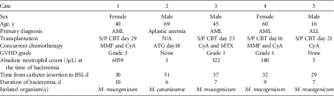
During the study period, a bloodstream infection due to RGM developed in 5 patients with a CVC (Table 1). The pathogens implicated in these cases were M. mucogenicum (n=4) and M. canariasense (n=1). All of the patients received a long-term CVC (i.e., Hickman catheter) inserted into the internal jugular vein. Four patients underwent cord blood transplantation for acute myeloid leukemia and acute lymphatic leukemia, and the remaining patient received antithymocyte globulin for aplastic anemia. None of the patients showed signs of disseminated NTM infection on the basis of examination and imaging studies. Long-term CVCs were immediately removed when RGM was identified in the blood culture. All of the patients defervesced immediately after catheter removal and were carefully monitored without antimicrobial therapy against RGM. All patients have had no evidence of relapses. In March 2012, the CVC management protocol and covering of the catheter-implantation site during bathing were also reinforced. No CVC-related mycobacterial bloodstream infections occurred during the following year.
Clinical Data for Patients with Central Line–Associated Bloodstream Infection Due to Rapidly Growing Non-Tuberculous Mycobacteria

Note. All patients had a Hickman catheter, and all patients were cured. ALL, acute lymphocytic leukemia; AML, acute myeloid leukemia; ATG, antithymocyte globulin; BSI, bloodstream infection; CBT, cord blood transplantation; CyA, cyclosporine; GVHD: graft-versus-host disease; MMF, mycophenolate mofetil; MTX, methotrexate; N/A, not applicable; S/P, status post. The grading of GVHD was based on the Glucksberg grade.
Description of the Catheter Care after Insertion
The frequency of dressing changes was documented in the nursing notes on the electronic patient records. Four of 5 patients required frequent changes at the catheter insertion site (every 2–5 days) for reasons that included submersion of the catheter site during showering or bathing. Only 1 patient (case patient 3) required a dressing change every 7 days, even without submersion of the catheter.
Microbiological Results
Clinical Samples
All of the patients had multiple blood cultures performed, and for each patient more than 1 blood culture result was positive for mycobacteria. The median time from catheter insertion to a positive blood culture was 32 days (range, 29–51 days). The median duration of bacteremia was 7 days (range, 6–10 days). Four of 5 catheter tip cultures (80%) showed mycobacterial growth.
Environmental/Water Samples
Water samples from different sites in the unit were cultured for NTM. NTM was not isolated from sterilized potable water. However, M. mucogenicum was isolated from shower water from 1 of the single rooms and a communal shower room. M. canariasense was also isolated from toilet water from 1 of the 4-bedded rooms. The remaining water and environmental cultures showed no mycobacterial growth.
Water chlorination in the main water tank at the hospital was measured daily. This hospital considers chlorine levels between 0.10 to 0.40 ppm to be adequate for maintaining sterility. During the outbreak, the chlorination level was kept at approximately 0.11 ppm.
Microbiologic Analysis
Typing by pulsed-field gel electrophoresis and random amplified polymorphic DNA revealed a genetic match between blood isolates of M. mucogenicum from patients 1, 3, and 4 and a shower water isolate of M. mucogenicum from the single rooms. A blood isolate of M. canariasense from patient 2 matched M. canariasense isolated from a toilet of the 4-bedded room. The other isolate of M. mucogenicum from patient 5 was found to have different patterns from that of patients 1, 3, and 4 by pulsed-field gel electrophoresis and random amplified polymorphic DNA typing. No organism genetically matched with that from patient 5 was isolated from the water or the environmental culture.
DISCUSSION
We experienced a cluster of RGM bloodstream infections in a hematology ward over a 5-month period. This outbreak was caused by 2 different clones of M. mucogenicum as well as M. canariasense. M. mucogenicum is commonly implicated in outbreaks of bloodstream infections resulting from contaminated hospital equipment and contaminated tap water, whereas M. canariasense is a rare species of RGM and the spectrum of clinical diseases attributable to it is not clearly understood.Reference Jimenez, Campos-Herrero, Garcia, Luquin, Herrera and Garcia5, Reference Campos-Herrero, Garcia, Figuerola, Suarez, Campo and Garcia6
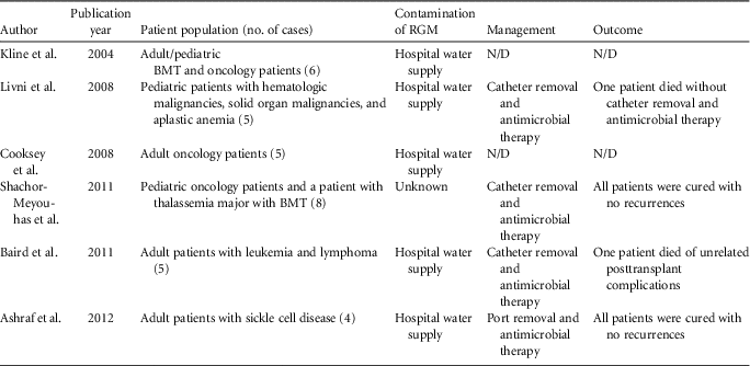
Although the incidence of CLABSI due to RGM, especially M. mucogenicum, in hematopoietic stem cell transplant recipients or patients on intense immunosuppressive agents, appears to be low, 6 outbreaks of bloodstream infections among such patients have been reported between 2004 and 2012 (although the outbreak reported in 2004 actually occurred in 1998; Table 2).Reference Livni, Yaniv and Samra8, Reference Ashraf, Swinker and Augustino9, Reference Cooksey, Jhung and Yakrus11, Reference Kline, Cameron and Streifel18–Reference Shachor-Meyouhas, Sprecher, Eluk, Ben-Barak and Kassis20
Outbreaks of Central Line–Associated Bloodstream Infection Due to Rapidly Growing Non-Tuberculous Mycobacteria
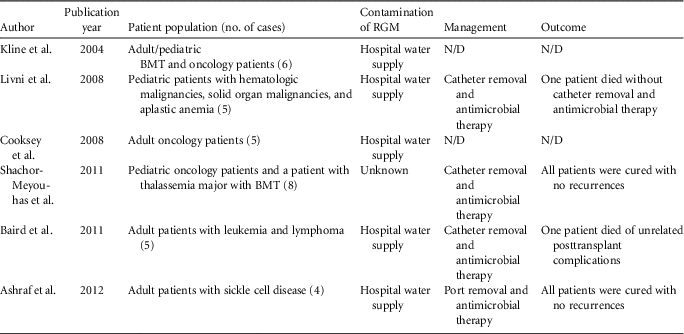
Note. BMT, bone marrow transplant; N/D, not documented; RGM, rapid growing mycobacteria.
In this outbreak, 4 of 5 patients received dressing changes at the catheter site owing to submersion of the catheter during showering. Moreover, the result of the environment/water culture and type matching of isolates from the blood cultures with isolates from the environmental/water cultures indicated that the origin of these organisms was the tap water supply. As with previous instances,Reference Livni, Yaniv and Samra8, Reference Ashraf, Swinker and Augustino9, Reference Baird, Taori, Dave, Willocks, Roddie and Hanson19 we considered the portal of entry for both of these organisms into the bloodstream in this outbreak was likely to be the submersion of CVC during bathing, showering, or toileting. Enhancing the CVC dressing prior to bathing or showering is crucial. The latest US guideline regarding prevention of catheter-related bloodstream infections also noted that submerging a catheter in water should be avoided and an impermeable covering should be applied to the catheter during showering.Reference O'Grady, Alexander and Burns21
The present study also highlighted the treatment strategies for CLABSI due to RGM. Given the lack of high-quality evidence, ideal treatment strategies remain unclear. In the outbreak discussed here, all of the patients defervesced after prompt catheter removal following the detection of the bloodstream infection, and all were carefully observed without antimicrobial therapy. Follow-up blood cultures revealed no bacterial growth, and there was no clinical evidence of recurrence. In most case series or outbreaks of NTM bloodstream infection, catheter removal was the most important step to achieving a successful outcome although many patients in these studies also received antimicrobial agents against NTM.Reference Gaviria, Garcia, Garrido, Corey and Boeckh22–Reference Holland, Chen, Chew and Gilbert26 Single case series also demonstrated successful management of RGM CLABSI by prompt catheter removal without antimicrobial therapy.Reference Rodgers, Mortensen, Blecker-Shelly, Fisher and Long27 Furthermore, removal of the catheter was associated with a significant decrease in the rate of bacteremia relapse.Reference Raad, Vartivarian, Khan and Bodey24 These findings correlate with the pathogenicity of RGM species, specifically with reference to their tendency to form a microbial biofilm matrix on the surface of the indwelling catheters.
In summary, we experienced a cluster of CLABSI due to RGM among patients with hematologic disorders. Since hospital tap water can be contaminated even with adequate chlorination, it is imperative that precise instructions for infection control in patients with an indwelling vascular catheter should be provided to the patients themselves, the healthcare workers, and family members/friends who may be providing care, especially when patients shower or bathe.
Acknowledgments
We thank Tokyo Metropolitan Institute of Public Health for performing the microbiologic investigation.
Financial support. H.H. reports that he is funded by Tokyo Metropolitan Clinical Research Funding (grant H260502001).
Potential conflicts of interest. All authors report no conflicts of interest relevant to this article.