
Introduction
Neural prostheses provide a means to understand and influence neurological injury and disease through bi-directional communication. Peripheral nerve injuries have a detrimental impact on military personnel and veterans as the greatest source of long-term disability including limb loss from combat-related injuries. Specifically, peripheral nerve recording and stimulation have the potential to influence neurological injury through bi-directional communication using peripheral nerve devices. Extraneural peripheral nerve electrodes are commonly used for peripheral nerve injury; unfortunately, extraneural devices have poor selectivity and signal resolution.[Reference Grill and Mortimer1] This trade-off motivates the use of intraneural devices. However, the long-term performance of intraneural devices is limited. An immunological foreign body response (FBR) to implanted devices limits the efficacy of chronic stimulation and recording capabilities over time due to the acute response of macrophages and subsequent glial sheath formation leading to encapsulation around the implant which blocks the signal from nervous tissue to device electrodes.[Reference Lago, Ceballos, Rodríguez, Stieglitz and Navarro2–Reference de la Oliva, Navarro and del Valle4] There have been a variety of approaches to mitigate the FBR and enhance device function including the implementation of intrinsically conducting polymers (CPs) at the device–tissue interface to improve recording and stimulation of neural devices. An intriguing option for deployment of the CPs is in situ polymerization of the conductive polymer. Specifically, here we demonstrate that poly(3,4-ethylenedioxythiophene) (PEDOT) interfaces can be created in situ, resulting in a less invasive intraneural electrode for intrafascicular targeting.
Previously, PEDOT has been studied for neural interfacing applications for microelectrodes and biomedical devices.[Reference Abidian and Martin5–Reference Yang, Kim, Hendricks, Leach, Northey and Martin10] PEDOT exhibits high chemical stability and conductivity and significantly improves the electrochemical properties of devices, specifically decreasing impedance magnitude over all frequencies.[Reference Ludwig, Uram, Yang, Martin and Kipke11, Reference Wilks, Woolley, Ouyang, Martin and Otto12] PEDOT has been applied to neural devices in the central nervous system and has been shown to enhance signal quality, charge storage capacity (CSC), and charge transport for electrical stimulation and recording.[Reference Wilks, Richardson-Burns, Hendricks, Martin and Otto9, Reference Ludwig, Uram, Yang, Martin and Kipke11, Reference Cui and Zhou13–Reference Venkatraman, Hendricks, King, Sereno, Richardson-Burns, Martin and Carmena16] PEDOT has also been implemented for other biomedical applications due to its ease of synthesis, ability to entrap and release bioactive molecules and drugs[Reference Alba, Du, Catt, Kozai and Cui17–Reference Chikar, Hendricks, Richardson-Burns, Raphael, Pfingst and Martin19], and ability to be modified and tailored for specific applications.[Reference Bhagwat, Kiick and Martin20–Reference Guimard, Gomez and Schmidt22]
In situ polymerization of PEDOT has shown great potential in vitro, in tissue slice culture, and in vivo for recording and stimulation.[Reference Wilks, Woolley, Ouyang, Martin and Otto12, Reference Ouyang, Shaw, Kuo, Griffin and Martin23–Reference Richardson-Burns, Hendricks and Martin25] There have been few studies using PEDOT in the peripheral nervous system (PNS)[Reference Egeland, Urbanchek, Peramo, Richardson-Burns, Martin, Kipke, Kuzon and Cederna26, Reference Urbanchek, Kung, Frost, Martin, Larkin, Wollstein and Cederna27] and none known by the authors using in situ polymerization methods of PEDOT in the PNS. This work demonstrates a method of in situ polymerization of an injectable PEDOT electrode for peripheral nerve interfacing to develop a less invasive ionic and electron charge transport pathway between the device and surrounding neural tissue to improve long-term functionality and performance of bi-directional interfaces.
Experimental methods
In situ electrochemical polymerization
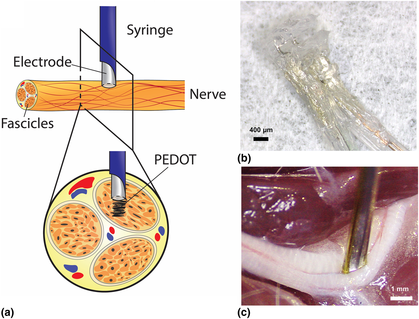
Polymerization was quantified initially in an agarose gel and subsequently in excised rat sciatic nerves to assess deposition. A monomer solution was made by mixing 0.01 M 3,4-ethylenedioxythiophene (EDOT) (97%, Sigma Aldrich, St. Louis, MO, USA) in 1X phosphate buffered saline (PBS) (pH 7.4) with 0.1 wt% poly(4-sodium styrene sulfonate) (PSS, M n ~ 70,000 g/mol, Sigma Aldrich, St. Louis, MO, USA). In situ polymerization was initially performed in a 0.5 wt% agarose gel (0.5 wt% agarose in 0.01 M EDOT:PSS) to determine the relationship between deposition time and PEDOT size. Intraneural PEDOT polymerization was performed in excised rat sciatic nerves using 100 µm diameter platinum iridium (PtIr) microwire to assess electrodeposition with small diameter electrodes. Polymerization was also assessed using a custom designed Plastics One guide-electrode device (C316I-MS303/2/GDE/ELECT 31GA, Plastics One, Roanoke, VA, USA) consisting of a 31-gauge cannula with two 200 µm diameter stainless steel (SS) electrodes. Figure 1(a) depicts the insertion and injection of monomer for electrochemical polymerization. The epineurium was separated from the nerve for facile insertion of the electrode into nerve and the cuff array was subsequently placed around the nerve shown in Figs. 1(b) and 1(c).[Reference Tong, Murbach, Subramanian, Chhatre, Delgado, Martin, Otto, Romero-Ortega and Johnson28] Once inserted into the nerve, 6–10 µL of EDOT:PSS was injected at a constant rate of 0.2 µL/min using a Quintessential Stereotaxic Injector (Stoelting QSI™, Wood Dale, IL, USA). While injecting the monomer solution, constant voltage (potentiostatic) polymerization was performed using a three-electrode cell configuration using an Autolab PGSTAT128N (Metrohm Autolab B.V., Utrecht, The Netherlands) at 2 V with the PtIr microwire or Plastics One SS device as the working electrode (WE), SS as the counter electrode (CE), and an Ag|AgCl glass body electrode (Thermo Fisher Scientific) as the reference electrode (RE).
(a) Schematic of delivery and in situ polymerization of PEDOT with a Plastics One guide-electrode device in peripheral nerve. (b) 3D printed silicone cuff electrode arrays. (c) A Plastics One guide-cannula device with an injection port and 200 µm SS electrodes for simultaneous injection and polymerization of PEDOT.

Electrochemical impedance spectroscopy
Electrochemical impedance spectroscopy (EIS) measurements were recorded prior to and after polymerization of PEDOT using an Autolab PGSTAT128N with a three-electrode cell configuration. EIS is commonly performed to assess the device–tissue interface. EIS measurements were collected by applying a voltage sine wave between the WE and Ag|AgCl RE while measuring the current magnitude and phase over a 0.01–10 kHz frequency range, using a 15-sine 10 mV RMS excitation voltage. Pt wire was used as the CE.
Cyclic voltammetry measurements
Cyclic voltammetry (CV) measurements were performed using an Autolab PGSTAT128N by sweeping the voltage of the WE at a constant rate to assess the resultant current flow between the WE and CE. The voltage range applied for SS was −0.6 to +0.8 V and for PEDOT was −0.8 to +0.6 V to remain within the limit to prevent hydrolysis reduction and oxidation reactions. Fast sweep rates (5 V/s) were performed to assess static equilibrium and determine the CSC of the bare devices and devices with PEDOT. From the measurements, the peaks represent the reduction and oxidation reactions occurring at the interface, and the peak amplitudes represent the number of charge carriers present. The cathodal CSC (Q cap) was determined by integrating the cathodal current density and dividing by the sweep rate and surface area of the electrode.
Tissue processing, histology, and microscopy
After polymerization, nerves were fixed in 4% paraformaldehyde (PFA) (95%, Sigma Aldrich, St. Louis, MO, USA) for 24 h, washed with 1X PBS for 24 h, and cryoprotected in 30% sucrose for at least 24 h, all at 4 °C. To analyze polymerization in tissue, nerves were frozen in 2-methyl butane at −40 °C for 30 s before slicing. Tissue was embedded with Optimal Cutting Temperature solution (Sakura Finetek, Alphen aan den Rijn, The Netherlands) and horizontally sectioned into 20 µm thick slices (Leica CM 1950, Buffalo Grove, IL, USA). Tissue sections were then washed with 1X PBS, allowed to dry, and stained for cell nuclei with 4’,6-diamidino-2-phenylindole (DAPI) (H-100, Vector Lab, Burlingame, CA, USA). Stained tissue slices were imaged using fluorescence and light microscopy (Leica DMi 8, Buffalo Grove, IL, USA) at 10× magnification using a 405 and 488 nm excitation filter.
Select samples were whole-mount collected and prepared for imaging via CLARITY to evaluate PEDOT in whole nerve samples and complement the classic histology techniques.[Reference Chung, Wallace, Kim, Kalyanasundaram, Andalman, Davidson, Mirzabekov, Zalocusky, Mattis, Denisin, Pak, Berstein, Ramakrishnan, Grosenick, Gradinaru and Deisseroth29] CLARITY removes opaque lipids from tissue, resulting in a clear or transparent sample ideal for imaging by laser light sheet microscopy. Nerves were fixed in 4% PFA for 24 h, incubated in a hydrogel solution at 4 °C for 3 days, and polymerized at 37 °C for 3.5 h to crosslink the hydrogel. The sample was then placed in a clearing solution containing sodium dodecyl sulfate (>85%, Tokyo Chemical Industry, Tokyo, Japan) for 3 months to clear lipids from the tissue. Once optically clear, the tissue was washed overnight in 1X PBS at 37 °C and incubated in a series of 2,2’-thiodiethanol (TDE, Sigma Aldrich, St. Louis, MO, USA) solutions for 1 h per dilution (20, 47, and 63% TDE in 1X PBS) at 37 °C with agitation for refractive index correction to ~1.45. Laser light sheet microscopy (Zeiss Z.1 Light sheet, Germany) was used to assess large cleared samples three-dimensionally for the presence of polymerized PEDOT in nerve. PEDOT fluorescence, visible in the GFP spectrum, was acquired using a 488 nm laser and 5× magnification.
Statistics
Mean 1 kHz impedance magnitude, Q cap, and binned frequency ranges were evaluated for statistical significance using Student's t-tests (P < 0.01). Error bars for all data were calculated using the standard error of the mean from n = 4 bare devices and devices with PEDOT for impedance and phase and n = 6 bare devices and devices with PEDOT for CV measurements. The approximate area of each electrode was 31,400 µm2.
Results, analysis, and discussion
Initially, custom-made 3D printed cuff electrodes were used to polymerize PEDOT in an attempt to direct polymerization through nerve.[Reference Tong, Murbach, Subramanian, Chhatre, Delgado, Martin, Otto, Romero-Ortega and Johnson28] Due to the robust and heterogeneous epineurium, this method proved difficult to electrodeposit PEDOT through nerve at low current thresholds. Therefore, the in situ deposition method through syringe injection was employed to simultaneously inject and polymerize a PEDOT electrode within peripheral nerve in addition to placement of 3D printed cuff electrodes around the nerve. Impedance magnitude, phase angle, CV measurements, and histology of nerve slices were analyzed before and after polymerization using both 3D printed cuff arrays and injectable in situ PEDOT methods. Using the injectable in situ polymerization method, intraneural polymerization was observed in excised rat sciatic nerves through electrochemical impedance and phase measurements, fluorescence microscopy, and laser light sheet microscopy.
Electrochemical impedance spectroscopy
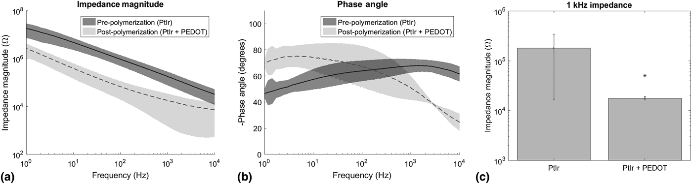
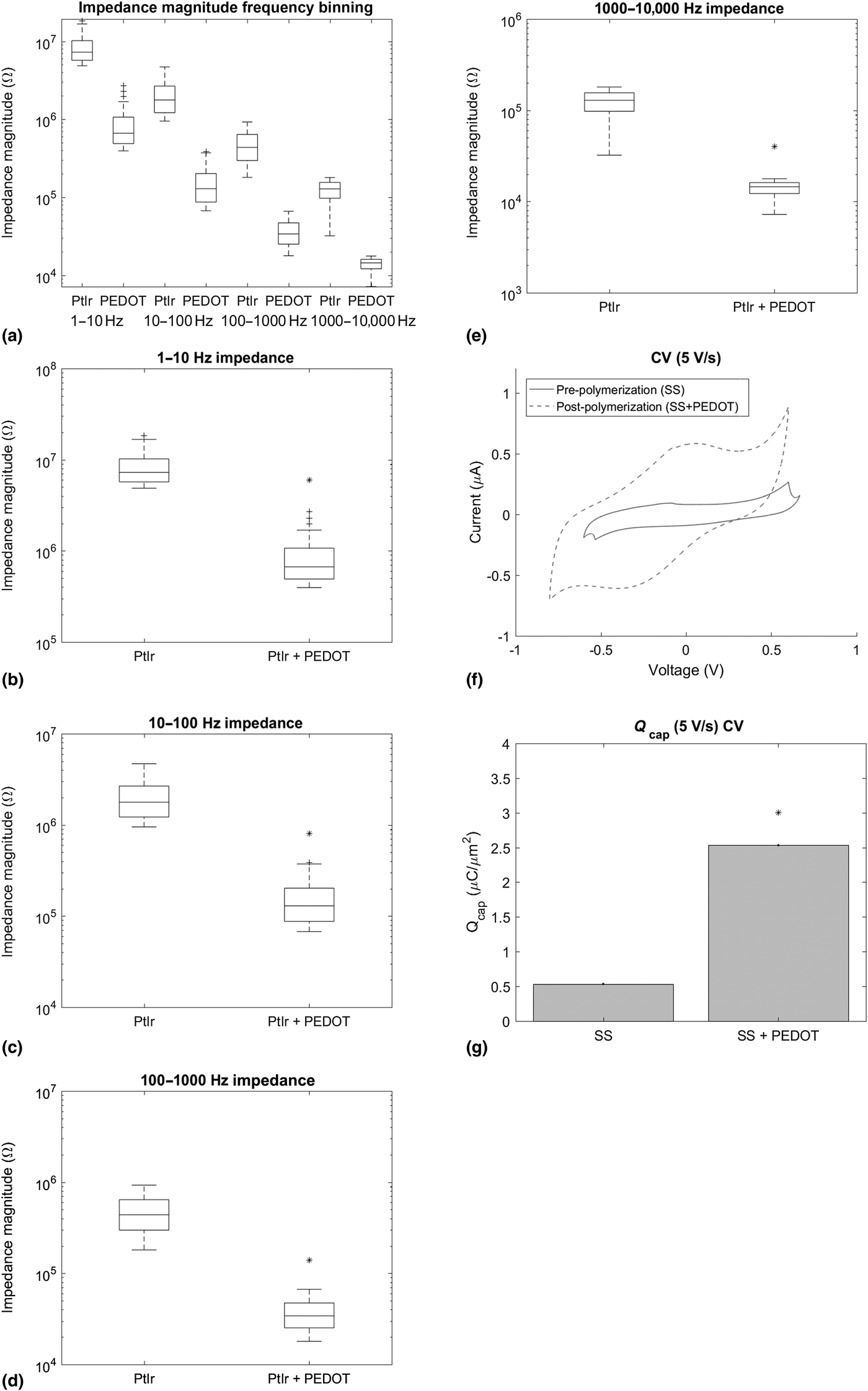
After polymerization of PEDOT in nerve, the impedance magnitude decreased over all frequencies measured as compared with bare PtIr electrodes as shown in Fig. 2(a). The drop in impedance is associated with the increase in electrochemical surface area and porosity of PEDOT that can likely increase detection sensitivity to neural activity. Lower impedance magnitude is associated with better signal quality, higher signal-to-noise ratios for improved recordings, and lower power required for stimulation. Phase values near 0 are indicative of a highly resistive interface, while phase values near −90 represent a largely capacitive interface. Above 100 Hz, PEDOT possessed a lower phase than bare electrodes [Fig. 2(b)], characteristic of a more resistive interface. Impedance and phase measurements were filtered using a Savitsky-Golay filter to smooth noise, especially evident around 60 Hz. At 1 kHz frequency, injectable PEDOT electrodes had a significantly lower mean impedance magnitude of 17.8 kΩ than the 1 kHz impedance magnitude of 179.7 kΩ for bare electrodes, as shown in Fig. 2(c). The 1 kHz impedance magnitude was analyzed for the recording of neural devices because this value has relevance in biological systems as the fundamental frequency of an action potential. Frequency ranges were binned and assessed for stimulation applications demonstrated in Figs. 3(a)–(e). This data exhibited a significant decrease in impedance magnitude for all frequency ranges for electrodes with PEDOT. Significantly lower impedance magnitude over all frequencies measured demonstrates lower power required for stimulation at lower current densities with PEDOT, which has been shown previously.[Reference Wilks, Richardson-Burns, Hendricks, Martin and Otto9, Reference Wilks, Woolley, Ouyang, Martin and Otto12]
Electrochemical impedance spectroscopy (EIS) measurements. (a) Impedance magnitude pre-polymerization (PtIr) and post-polymerization (PtIr + PEDOT) between 1 and 10,000 Hz. (b) Pre-polymerization (PtIr) and post-polymerization (PtIr + PEDOT) phase angle measurements. (c) 1 kHz impedance magnitude of PtIr and PtIr + PEDOT demonstrates a significant decrease in impedance of PEDOT electrodes.

EIS and cyclic voltammetry measurements. (a) PtIr electrodes and PtIr + PEDOT electrodes mean impedance magnitude of binned frequency ranges scaled logarithmically. (b) 1–10 Hz, (c) 10–100 Hz, (d) 100–1000 Hz, and (e) 1000–10,000 Hz binned frequency range impedance magnitudes of bare PtIr electrodes pre-polymerization compared with PtIr + PEDOT electrodes post-polymerization. Electrodeposited PEDOT impedances are significantly lower than the impedance magnitude of the bare electrodes across all frequency ranges. (f) Cyclic voltammetry measurements of SS devices and devices with electrodeposited PEDOT. Fast (5 V/s) sweep rates of PEDOT electrodes demonstrate increased cathodal charge storage capacity (Q cap) compared with SS devices. (g) Mean Q cap calculations show significantly greater charge available at the device–tissue interface with PEDOT electrodes (P < 0.01).

CV measurements
CV measurements were performed using Plastics One SS electrodes and Plastics One SS electrodes with electrodeposited PEDOT to provide information on the CSC of devices alone and devices with PEDOT. A fast sweep rate (5 V/s) was applied to assess information at the electrode tip, whereas slower sweep rates provide information of the electrode tip and shaft with electrolyte penetration underneath the insulation.[Reference Cogan30] PEDOT electrodes have a significantly greater mean Q cap of 2.53 µC/μm2 compared with bare SS electrodes with a mean Q cap of 0.53 µC/μm2 represented in Figs. 3(f) and 3(g) for fast sweep rates (5 V/s) demonstrating the increased charge available. The greater Q cap of PEDOT electrodes may be useful for physiologically relevant electrical stimulation compared with bare electrodes. Lower power applied for stimulation minimizes the potential formation of reactive species in tissue and enhances device longevity. While Plastics One devices facilitate intraneural polymerization, they are not ideal for electrophysiological stimulation. The combined use of Plastics One devices and 3D printed cuff arrays for polymerization and stimulation will be assessed in future studies as a chronic neural interface. Previously, the 3D printed cuff arrays have been fabricated with electrodeposited PEDOT designed for peripheral nerve stimulation.[Reference Tong, Murbach, Subramanian, Chhatre, Delgado, Martin, Otto, Romero-Ortega and Johnson28]
Tissue processing, histology, and microscopy
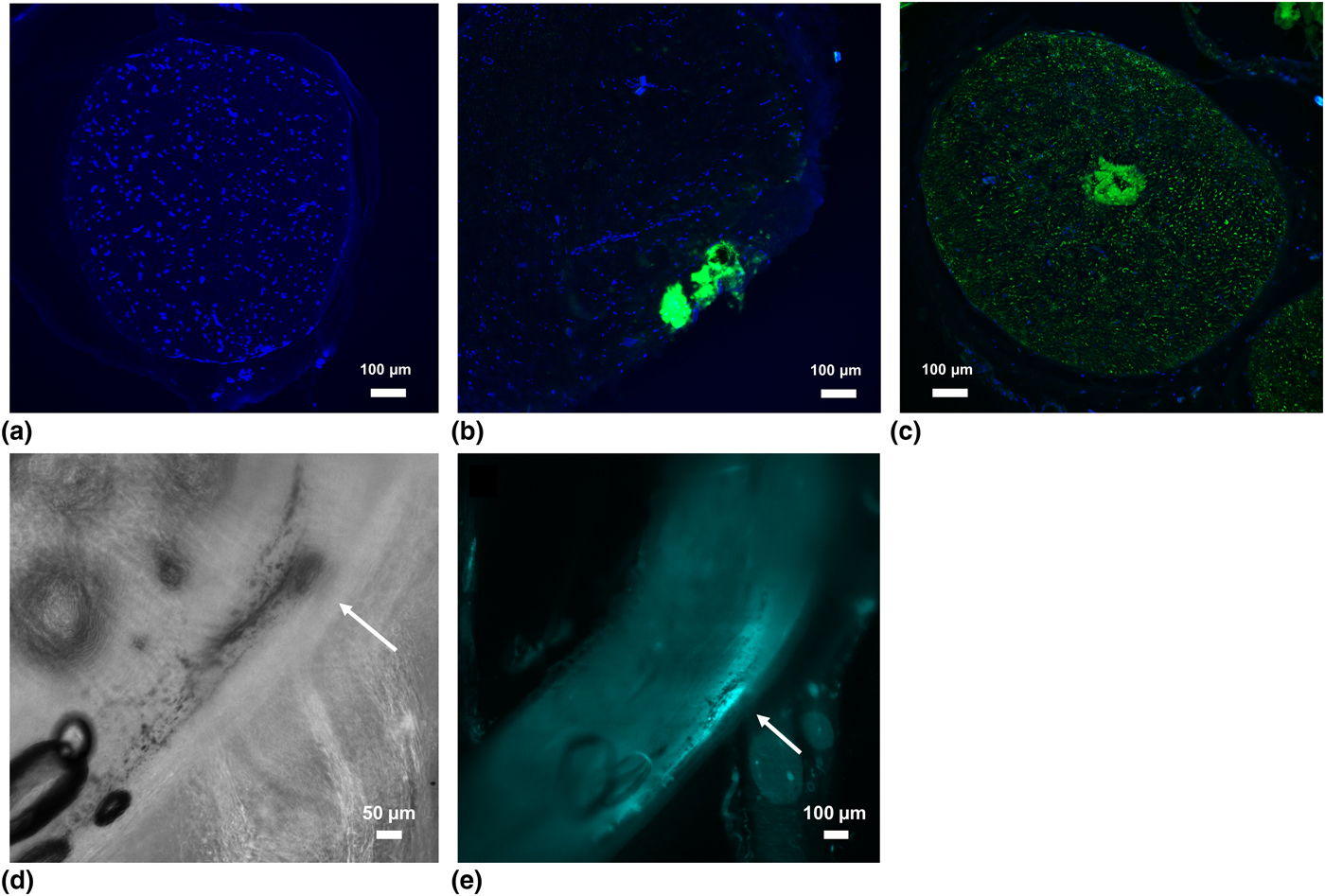
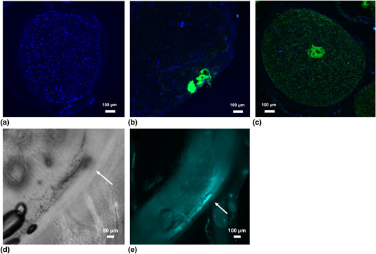
Polymerization was initially assessed in 2D cryosectioned nerve slices. Figure 4(a) represents a control sciatic nerve stained with DAPI for cell nuclei without PEDOT, and Fig. 4(b) depicts a nerve slice containing PEDOT that was polymerized using 3D printed nerve cuffs alone for shallow intraneural polymerization. PEDOT was imaged by fluorescence microscopy due to its intrinsic fluorescence with a 488 nm laser.[Reference Wilks, Woolley, Ouyang, Martin and Otto12] Precise intraneural polymerization of PEDOT deep within fascicles was achieved by in situ injection and polymerization methods demonstrated by the decreased impedance magnitude post-polymerization in Figs. 2(a) and 3(a)–3(e) as well as fluorescence spectroscopy in 2D nerve slices shown in Fig. 4(c). The nerve in Fig. 4(c) expresses green fluorescent protein (GFP) background due to its acquisition from a transgenic rat expressing GFP ubiquitously.
Histology and microscopy of tissue slices with (a) a control nerve without PEDOT, (b) intraneural PEDOT polymerization using 3D printed cuff arrays, and (c) intraneural PEDOT polymerization by in situ injection and polymerization method with a 3D printed cuff array. Scale bars are 100 µm. (d–e) Laser light sheet microscopy images of whole peripheral nerve samples demonstrating intraneural in situ PEDOT polymerization. (d) Bright field image of PEDOT polymerized in rat sciatic nerve. (e) PEDOT fluorescence in nerve. The precise location of electrodeposition is depicted by white arrows.

Laser light sheet microscopy reveals intraneural PEDOT polymerization in whole tissue samples shown in Figs. 4(d) and 4(e). Figure 4(d) represents a bright field image of PEDOT polymerized in rat sciatic nerve, and the same location of PEDOT fluorescence was imaged with a 488 nm laser demonstrating intraneural PEDOT polymerization in Fig. 4(e). PEDOT deposition is depicted by white arrows in Figs. 4(d) and 4(e). Rat sciatic nerves imaged by laser light sheet microscopy were fixed and cleared prior to polymerization due to tissue damage of PEDOT nerves caused by active electrophoretic clearing techniques. Polymerization in fixed tissue is believed to have different mechanical and structural properties than fresh tissue, which may affect diffusion and deposition of PEDOT in nerve. The mechanical integrity of in situ deposited PEDOT must be studied and characterized further to assess chronic stability in peripheral nerve.
Conclusions
We have demonstrated that PEDOT can be locally electrodeposited within individual peripheral nerves using an electrode and cannula delivery system. The in situ electrochemical PEDOT polymerization resulted in lower impedance magnitude over all frequencies measured and significantly decreased impedance magnitude for all frequencies for stimulation and 1 kHz impedance for recording applications. PEDOT deposition resulted in greater Q cap for stimulation compared with bare Plastics One SS electrodes. Histology and microscopy confirm intraneural polymerization from PEDOT autofluorescence in nerve sections as well as laser light sheet microscopy of whole nerve samples. Long-term stability and in vivo assessment in the PNS is required for further investigation of injectable PEDOT electrodes for peripheral nerve stimulation and recording.
Acknowledgments
This work was sponsored by the Defense Advanced Research Projects Agency (DARPA) Biological Technologies Office (BTO) Targeted Neural Plasticity (TNT) program under the auspices of Drs. Doug Weber and Tristan McClure-Begley through the DARPA Contracts Management Office: No. HR0011-17-2-0019 and the National Science Foundation (NSF CMMI—1739318).