Introduction
Sialolithiasis accounts for 50 per cent of major salivary gland disease.Reference Epker1 In the UK, the incidence of symptomatic salivary calculi is 59 cases per million per annum.Reference Escudier and McGurk2 Eighty per cent of salivary calculi are found in the submandibular gland, 5–10 per cent in the parotid gland, and 0–5 per cent in the sublingual and minor salivary glands.Reference Bodner3
Previously, the treatment of choice for calculi lying in the proximal Stenson's duct or the gland has been parotidectomy, which carries a 3–7 per cent risk of facial nerve injury.Reference Gallo, Berloco, Bruschini and McGurk4 Reluctance to proceed with surgery, due to fear of facial nerve damage, delays definitive treatment and leads to stone enlargement and fibrosis of the gland.
In 1991, Katz used a 0.8 mm flexible endoscope with a dormia basket for diagnosis and extraction of calculi, using a blind technique.Reference Katz5 Lithotripsy was introduced in 1989Reference Iro, Nitsche, Schneider and Ell6 for the treatment of salivary gland stones. It has since become the ‘gold standard’, especially in mainland Europe and North America.
The first documented case of interventional sialendoscopy was performed by Konigsberger et al. in 1990,Reference Konigsberger, Feyh, Goetz, Schilling and Kastenbauer7 using an endoscopically controlled, electrohydraulic, intracorporeal shock wave lithotripsy technique. Nahleili and Baruchin reported the maximum number of cases, in 1999.Reference Nahlieli and Baruchin8
Lithotripsy uses two methods of shock wave application: extracorporeal shock wave lithotripsy and endoscopically controlled intracorporeal shock wave lithotripsy. Continuous endoscopic monitoring during therapy allows direct application of the shock wave to the stone under direct vision and avoids injury to the duct.
Extracorporeal lithotripsy for parotid duct stone removal is used in the UK, but intracorporeal shock wave lithotripsy has not found favour thus far.Reference McGurk, Escudier, Thomas and Brown9 We here report our initial experience with the use of endoscopically assisted intracorporeal shock wave lithotripsy in the treatment of a parotid duct stone.
Case report
A 57-year-old woman presented with a three-year history of a recurrent swelling of her left parotid gland. The initial episodes had had an infective presentation. She had previously been admitted to hospital with septicaemia due to bacterial sialadenitis and had required intravenous antibiotics. Subsequently, in the past year, this swelling appeared after each meal, and it was painful and restricted the patient's mouth-opening.
The patient presented to the out-patient clinic in one of the quiescent phases. Locally, there was not much to suggest a chronic, recurrent parotid condition. Intra-oral examination revealed clear fluid draining from the duct opening, which appeared to be almost normal despite recurrent infective episodes. There was no calculus on a bimanual palpation.
A plain X-ray showed a calcific duct opacity lying superficially in the parotid gland. Multiple unsuccessful attempts were made to cannulate the duct for a sialography.
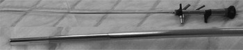
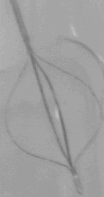
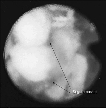
An interventional, diagnostic sialendoscopy was performed under general anaesthesia, using a lacrimal probe to dilate the opening of Stenson's duct. We used a double-channelled, offset, semi-rigid Olympus 7 F ureteroscope (Olympus, Keymed, Southend, UK; Figure 1). This endoscope had an outer diameter of 2.4 mm. The 3.2 F working channel was used to insert the electrokinetic lithotripter probe and a Segura basket (Figure 2). The 2 F port was used to irrigate the duct during lithotripsy with isotonic saline (with a filling pressure of 100 cm of water), which exited via the 3.2 F channel and allowed efficient clearance of debris.
Semi-rigid ureteroscope: full length view, with close up view of tip.

The Segura basket: note the outwardly bowed, flat wire spring strips. The device was originally described for removing stones from the kidneys, ureter and biliary duct.

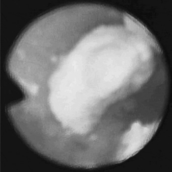
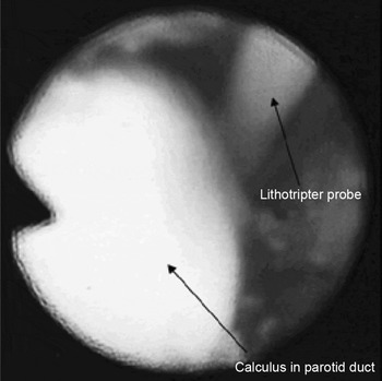
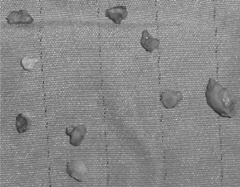
The semi-rigid ureteroscope was advanced until the stone was visualised. The stone itself measured 4 mm (Figure 3) and was positioned 4 cm from the duct opening. Fragmentation of the stone was achieved using the 3 F electrokinetic lithotripter probe (Figure 4) inserted through the 3.2 F working channel. The stone fragments were extracted using the Segura basket (Figure 5).
Endoscopic view of the sialolith in situ.

Endoscopic view showing application of lithotripter probe to the surface of the parotid duct calculus.

Endoscopic view showing fragmented sialolith held within the Segura basket during retrieval.

Following complete evacuation of all the fragmented stone debris (Figure 6), the ureteroscope was advanced further to rinse the duct. It was also used to confirm the integrity of the proximal duct.
Extracted fragments of the parotid duct calculus.

The procedure lasted 45 minutes and was done under cover of oral antibiotics and corticosteroids. Post-operative hydrotherapy to ensure continued irrigation of the parotid ductal system was encouraged, via sour stimulus and gentle massage to the area.
The post-operative period was uneventful, and the patient was asymptomatic after 18 months of follow up. Audiometric assessment did not reveal any hearing loss due to the therapy.
Discussion
Currently, over 3000 patients per year are treated by the UK National Health Service for sialadenitis or sialolithiasis.Reference Escudier and McGurk2 In earlier years, the standard management for these cases would have been parotidectomy.
The present options for management of salivary duct calculi include dormia basket retrieval of stones, lithotripsy or transoral surgical removal of duct stones.
Lithotripsy is now the gold standard for treatment of all parotid stones, apart from those that are small enough and amenable to basket retrieval.Reference McGurk, Escudier, Thomas and Brown9
Interventional sialendoscopy was first used for salivary gland stones in 1991.Reference Katz5 This procedure allows extraction of sialoliths with or without prior fragmentation and avoids the need for open surgery.Reference Marchai, Dulguerov, Becker, Barki, Disant and Lehmann10
Endoscopes capable of being introduced into the ductal system of the parotid and submandibular salivary glands may be flexible, rigid or semi-rigid. The semi-rigid endoscope combines the acceptable properties of both the rigid and flexible types and as a result is probably the instrument of choice.Reference Marchai, Dulguerov, Becker, Barki, Disant and Lehmann10
Zenk et al. reported Stensen's duct diameters of between 0.5 and 1.5 mm in 25 human cadavers.Reference Zenk, Hosenmann and Iro11 Papillotomy of Stenson's duct is rarely necessary – dilatation with a lacrimal probe is usually sufficient.
Since 1990, several techniques have been developed to fragment sialoliths. Extracorporeal lithotripsy was popularised by Iro et al. in 1992.Reference Iro, Schneider, Fodra, Waitz, Nitsche and Heinritz12 Intracorporeal procedures such as laser,Reference Ito and Baba13 electrohydraulicReference Konigsberger, Feyh, Goetz and Kasenbauer14 and pneumoblasticReference Iro and Zenk15 lithotripsy have been reported to achieve similar ends.
In principle, Electrokinetic and pneumoblastic lithotripsy are similar. While in a pneumoblastic lithotripsy it is the pneumatic energy that pushes a projectile against the end of a metal probe (impactor) to affect it's to and fro motion, this action of the impactor is achieved by electromagnetic energy in the EKL.
Electrokinetic lithotripsy has been found to be safe, inexpensive and reliable in the treatment of lower ureteric stones.Reference Keeley, Pillai, Smith, Chrisofos and Tolley16 However, its use for the treatment of salivary stones has not previously been reported. Stones lying free in the duct can be trapped in a basket and fragmented; this makes extraction safer than attempting to remove the stones in toto. The trapped stone is fragmented until the basket can be withdrawn without force.
The size of a parotid sialolith is probably the most important factor in predicting the success of interventional sialendoscopy.Reference Marchai, Dulguerov, Becker, Barki, Disant and Lehmann10 For stones smaller than 3 mm, the success rate of this technique is 35 per cent. With the addition of fragmentation, the success rate for larger stones increases to 72 per cent.Reference Marchai, Dulguerov, Becker, Barki, Disant and Lehmann10
External lithotripsy usually requires several sessions, at intervals of a few weeks. As no sialendoscopy has been described along with this technique, fragmented stones are left in the ductal system in anticipation of spontaneous excretion.
The diagnostic benefits offered by sialendoscopy are beyond those of the conventional methods of radiography, sialography and ultrasonography. Sialendoscopy accurately identifies, under direct vision, radiolucent stones, polyps, stenoses, mucous plugs, foreign bodies and anatomical abnormalities.
• Symptomatic salivary stones in the middle or proximal parotid duct are conventionally treated by gland excision. This approach is associated with a significant risk of facial nerve paralysis
• Electrokinetic lithotripsy is normally used for the treatment of lower ureteric stones. This paper describes its application in a case of parotid calculus
• Continuous, endoscopically monitored electrokinetic lithotripsy with good irrigation gives a well illuminated field and absolute delivery of energy to the target. It avoids side effects caused by the impact of the shock wave on the parotid duct and adjacent anatomical structures, thereby making it a safer procedure
At present, interventional sialendoscopy is limited by the diameter of the endoscope. Stones lying proximally in the duct, and a fibrosed canal wall with a reduced diameter, are the other restrictions of this otherwise excellent technique.
Conclusion
Interventional sialendoscopy allows the extraction and/or fragmentation of the majority of sialoliths and reduces the need for salivary gland excision. Continuous, endoscopically monitored electrokinetic lithotripsy avoids the side effects of shock wave application to the surface of the stone. It also allows fragmentation of stones, making removal safer and easier. Despite its apparent simplicity, intracorporeal electrokinetic lithotripsy is a technically challenging procedure. However, the authors are of the opinion that this procedure can be safely performed once learnt, allowing its benefits to be offered to all patients.