Introduction
Cutaneous-leishmaniasis manifestations encompass a disease complex characterized as tegumentary leishmaniasis (TL). Briefly, the distinctive clinical forms of American tegumentary leishmaniasis (ATL) can be classified as follows: (i) localized cutaneous leishmaniasis (LCL) with ulcerative skin lesions developing at the site of the sandfly bite; (ii) mucosal leishmaniasis showing destructive mucosal inflammation and (iii) diffuse cutaneous leishmaniasis (DCL) with multiple non-ulcerative nodules (Reithinger et al. Reference Reithinger, Dujardin, Louzir, Pirmez, Alexander and Brooker2007). In addition to these typical clinical manifestations, some studies have reported cutaneous-leishmaniasis cases that do not fit any of the well-characterized clinical forms of the disease. These cases are known as atypical ATL (Lescure et al. Reference Lescure, Bonnard, Chandenier, Schmit and Douadi2002; Baptista et al. Reference Baptista, Schubach, Madeira, Leal, Pires, Oliveira, Conceição-Silva, Rosalino, Salgueiro and Pacheco2009; Guimarães et al. Reference Guimarães, Machado, Lago, Morgan, Schriefer, Bacellar and Carvalho2009; Guimarães et al. Reference Guimarães, Queiroz, Silva, Silva, Magalhães, Lago, Machado, Bacellar, Wilson, Beverley, Carvalho and Schriefer2016). The presence of unusual lesions instead of the typical ulcers characterizes atypical ATL (Meireles et al. Reference Meireles, Maia, Soares, Teodoro, Gadelha, da Silva and de Lima2017). Patients may exhibit verrucous, crusted, vegetative, lupoid or other lesions.
The clinical manifestations of ATL and its response to treatment are partly influenced by the Leishmania species and/or strain involved. The parasite's contribution to the broad spectrum of clinical manifestations of cutaneous leishmaniasis has been demonstrated by studies showing correlations between genetic variants of Leishmania and different clinical forms (Queiroz et al. Reference Queiroz, Sousa, Heine, Cardoso, Guimarães, Machado, Carvalho, Riley, Wilson and Schriefer2012; Oliveira et al. Reference Oliveira, Valete-Rosalino, Pacheco, Costa, Schubach and Pacheco2013). Some studies of intra-specific genetic variability in subgenus Leishmania (Viannia) and some other related to L. (V.) braziliensis have been published (Nolder et al. Reference Nolder, Roncal, Davies, Llanos-Cuentas and Miles2007; Oddone et al. Reference Oddone, Schweynoch, Schönian, de Sousa, Cupolillo, Espinosa, Arevalo, Noyes, Mauricio and Kuhls2009; Rougeron et al. Reference Rougeron, Meeûs, Hide, Waleckx, Bermudez, Arevalo, Llanos-Cuentas, Dujardin, De Doncker, Le Ray, Ayala and Bañuls2009, Reference Rougeron, De Meeus, Hide, Waleckx, Dereure, Arevalo, Llanos-Cuentas and Banuls2010; Kuhls et al. Reference Kuhls, Cupolillo, Silva, Schweynoch, Boité, Melo, Mauricio, Miles, Wirth and Schönian2013), although this issue still deserves further investigation.
The present study analyses 95 DNA samples of L. (V.) braziliensis collected from ATL patients in the Xakriabá Indigenous Reserve, Minas Gerais state, Brazil. This region is particularly interesting because the patients exhibit a variety of clinical forms of ATL and because diverse reservoir and vector species are present (Quaresma et al. Reference Quaresma, Rêgo, Botelho, da Silva, Moura-Júnior, Teixeira-Neto, Madeira, Carvalho, Paglia, Melo and Gontijo2011; Rêgo et al. Reference Rêgo, Rugani, Shimabukuro, Tonelli, Quaresma and Gontijo2015). Molecular characterization of the parasites confirmed the presence of genetic diversity among the L. (V.) braziliensis samples and demonstrated that this diversity was correlated with the different clinical manifestations of ATL in the surveyed patients.
Material and methods
Study area and population
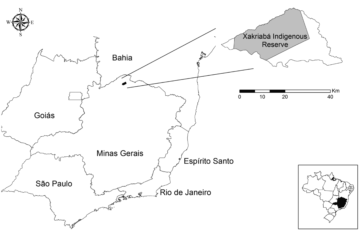
The Xakriabá Indigenous Reserve is located in the municipality of São João das Missões (latitude 14°53′01″ south and longitude 44°05′26″ west), in northern Minas Gerais, Brazil (Fig. 1). The reserve covers 78% of the entire municipality and has a population of 8380 inhabitants (Quaresma et al. Reference Quaresma, Rêgo, Botelho, da Silva, Moura-Júnior, Teixeira-Neto, Madeira, Carvalho, Paglia, Melo and Gontijo2011). The region predominantly displays Brazilian savannah (cerrado) vegetation mixed with areas of caatinga (scrubland). Lutzomyia (Nyssomyia) intermedia is the main vectors transmitting L. (V.) braziliensis in Xakriabá area. The leishmaniasis reference center of the Januária city serves as reference for the diagnosis and treatment of ATL in the region. The prevailing economic activities of the residents are subsistence agriculture and livestock raising, usually carried out by the male population, while the female is engaged in activities domestic.
Location of study area. The municipality of São João das Missões is located in the northern area of Minas Gerais State, Brazil. The Xakriabá Indigenous Reserve, where the study was performed, is indicated.

Patients and samples
A total of 164 people resident in the Imbaúbas village at Xakriabá Indigenous Reserve participated in a population survey study between July and October 2008. Among these, 151 subjects performed all stages of the study: personal interview, clinical examinations, immunological, parasitological and diagnostic methods. Furthermore, all persons in the Indigenous Reserve with ATL-suggestive skin lesions from June 2008 to August 2010 were actively sought and invited to participate in the study. Those with clinical symptoms of ATL were referred for sample collection by biopsy and/or lesion-aspiration procedures used for conventional parasitological and molecular diagnosis. Ten ATL cases were diagnosed from the survey study (July–October 2008) and 77 patients were found from active search over the years 2008–2010.
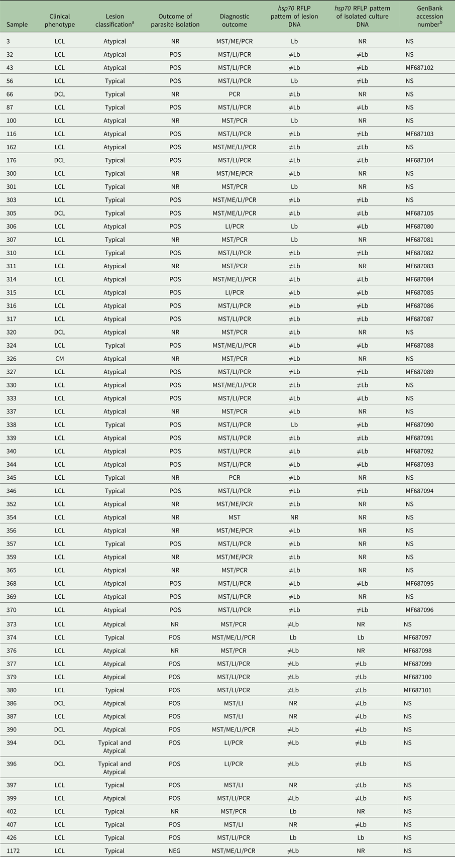
In total, 87 patients with cutaneous lesions were evaluated in the study of clinical and genetic analysis. Several diagnostic methods were used to detect the ATL cases, such as: Montenegro Skin Test (MST), microscopic examination of wound's imprinting in the glass slide searching for amastigotes forms (ME), Leishmania promastigotes isolation (LI) and hsp70-PCR. Seventy-three patients had some positive parasitological examination (parasitological confirmation = ME- and/or LI- and/or PCR-positive) and 14 patients were detected based on immunological diagnosis (MST-positive).
It was impossible to get parasite DNA from the samples of 27 patients, because they were diagnosed only by MST and/or ME, or because the isolation in culture did not progress to obtain satisfactory amount of promastigote cells (n = 13). Therefore, the molecular analyses were performed on 95 samples (56 DNA samples isolated directly from the biopsy fragments plus 39 DNA samples extracted from cultured cells) collected from 60 patients. It means that duplicate DNA analysis was performed in some patients (n = 35). In this study, all possible sources of DNA for each patient were used to increase the success of the next molecular experiments.
Disease definition
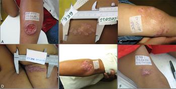
The patients’ lesions were classified taking into account the morphological aspects, number and localization of wounds. The particularities of endemic area and previous literature reports (Guimarães et al. Reference Guimarães, Queiroz, Silva, Silva, Magalhães, Lago, Machado, Bacellar, Wilson, Beverley, Carvalho and Schriefer2016; Meireles et al. Reference Meireles, Maia, Soares, Teodoro, Gadelha, da Silva and de Lima2017) were also considered. The classic initial clinical sign of the cutaneous leishmaniasis is the appearance of small papules and an erythematous nodule, which may be single or multiple, usually located in an exposed region of the tegument where, after a few months, it develops into ulcers. Typical lesions were characterized like circular or oval ulcers with an erythematous, infiltrated, firm, reddish and granular base and a well-delimited, elevated border. Atypical lesions were defined by the presence of unusual cutaneous wounds and classified as crusted, vegetative lesion, papules, verrucous and ulcerated plaques due to L. (V.) braziliensis (Fig. 2).
Examples of typical (A) and atypical lesions (B–F) of ATL patients in the Xakriabá Indigenous Reserve. (A) Typical lesions: circular or oval ulcers with an erythematous, infiltrated, firm, reddish and granular base and a well-delimited, elevated border. (B through F) Different types of atypical lesions: B – plaque, C – papules, D – verrucous, E – crusted and F – ulcerated plaques.

Promastigotes isolates and Leishmania species identification
The aspirates and lesion fragments were added to tubes containing NNN medium (Novy and McNeal, Reference Novy and McNeal1903; Nicolle, Reference Nicolle1908) enriched with liquid Schneider's medium and incubated at 25 ± 1 °C. The cultures were examined weekly and were considered positive when Leishmania promastigotes were observed. When no flagellate forms were observed in a tube after 6 weeks of incubation, the culture was considered negative. The isolated samples were cryopreserved and deposited at the strain collection of the IRR Leishmaniasis Laboratory for further characterization by isoenzyme electrophoresis (MLEE – multilocus enzyme electrophoresis) and other molecular methods.
Identification of the cultured promastigotes using MLEE was performed by the Leishmania Isolation, Culture and Typing Service of the Leishmania Collection of the Oswaldo Cruz Institute (Coleção de Leishmania do Instituto Oswaldo Cruz – CLIOC) according to the protocol described by Cupolillo et al. (Cupolillo et al. Reference Cupolillo, Grimaldi and Momen1994). Four enzymatic loci were used: G6PDH – glucose-6-phosphate dehydrogenase (Enzyme Commission Number 4.2.1.3), 6PGDH – 6-phosphogluconate dehydrogenase (E.C. 1.1.1.43), PGM – phosphoglucomutase (E.C. 1.4.1.9) and IDH – isocitrate dehydrogenase (E.C. 1.1.1.42).
hsp70 PCR-restriction-fragment length polymorphism
The PCR reaction was performed using the primers HSP70for, 5′-GAC GGT GCC TGC CTA CTT CAA-3′, and HSP70rev, 5′-CCG CCC ATG CTC TGG TAC ATC-3′ (Garcia et al. Reference Garcia, Kindt, Bermudez, Llanos-Cuentas, De Doncker, Arevalo, Tintaya and Dujardin2004), which amplified a 1300-bp fragment of the hsp70 gene. The PCR reaction was prepared to a final volume of 25 µL using 1.5 mm MgCl2, 200 µ m dNTP mix (New England, USA), 5.0% dimethyl sulfoxide (Invitrogen, USA), 1.5 units of platinum Taq DNA polymerase (Invitrogen, USA), 0.4 pmol of sense primer and 0.4 pmol of antisense primer (IDT prodimol, Brazil). The DNA amplification was performed in a thermal cycler using 35 cycles of denaturation at 94 °C for 30 s, primer-specific annealing at 61 °C for 1 min and extension at 72 °C for 3 min, followed by one final extension at 72 °C for 10 min. The amplification products were visualized on 2% agarose gels stained with ethidium bromide (10 µg mL−1). Samples showing the 1300-bp-specific band were digested with the HaeIII enzyme to analyse their restriction-fragment length polymorphism (RFLP). The digestion reaction was prepared using a final volume of 15 µL, containing 1 µL of HaeIII (New England Biolabs) (10 U µL−1), 1.5 µL of enzyme buffer and 12.5 µL of the PCR product. This mixture was incubated at 37 °C for 2 hours. The restriction profiles were compared with those obtained from HaeIII digestion of the PCR products from the reference strains L. (L.) amazonensis (IFLA/BR/1967/PH8), L. (V.) braziliensis (MHOM/BR/1975/M2903), L. (L.) infantum (MHOM/BR/1974/PP75) and L. (V.) guyanensis (MHOM/BR/1975/M4147).
hsp70 Gene sequencing and phylogenetic analysis
The 1300-bp hsp70 gene fragment was sequenced to identify the parasite species and to analyse the nucleotide polymorphisms and possible variants strains of the L. (V.) braziliensis. To prepare the samples for sequencing, the PCR products obtained from amplifications with primers HSP70for and HSP70rev were purified using the QIAquick PCR purification kit (Qiagen) according to the manufacturer's instructions. The sequencing reactions were performed using the BigDyeTerminator v3.1 Cycle Sequencing Kit and the DNA sequencing was conducted in the ABI 3730 automated DNA sequencer at Fiocruz sequencing facilities. The hsp70 sequences from other L. (V.) species were detected using the Basic Local Alignment Search Tool (BLAST) algorithm and downloaded from GeneBank, NCBI, National Institute of Health, USA (http://www.ncbi.nlm.nih.gov). All hsp70 sequences were used in the sequence alignment analyses conducted using the Molecular Evolutionary Genetics Analysis package version 6.0 (MEGA 6.0) (Tamura et al. Reference Tamura, Stecher, Peterson, Filipski and Kumar2013).
The de novo assembly of the hsp70 sanger sequences were performed using the Phred, Phrap and Consed pipeline (PMID: 9521922, PMID: 9521923), and the alignment and sequence editing was performed using MEGA 6.0 (Tamura et al. Reference Tamura, Stecher, Peterson, Filipski and Kumar2013). Sequence reads of another species from Trypanosomatidae family [Leptomonas collosoma, Phytomonas serpens (strain 9T), Crithidia fasciculata, Herpetomonas samuelpessoai (strain HSP)] as well as Leishmania species [L. (V.) braziliensis – MHOM/BR/1975/M2904, L. (V.) peruviana – MHOM/PE/1990/HB86, L. (V.) panamensis – MHOM/PA/1994/PSC-1, L. (V.) shawi – MCEB/BR/1984, L. (V.) naiffi – MDAS/BR/1979/M5533, L. (V.) lainsoni – MHOM/BR/1981/M6426] were included and applied to the same workflow as sequences generated by this study. The samples of another tripanosomatids species were kindly provided by the ‘Oswaldo Cruz Foundation, Protozoa Collection (Coleção de Protozoários – Fundação Oswaldo Cruz)’ with the exception of C. fasciculata (code number: CFAC1_300044700.1), whose sequences were taken from the TriTrypDB® (Aslett et al. Reference Aslett, Aurrecoechea, Berriman, Brestelli, Brunk, Carrington, Depledge, Fischer, Gajria, Gao, Gardner, Gingle, Grant, Harb, Heiges, Hertz-Fowler, Houston, Innamorato, Iodice, Kissinger, Kraemer, Li, Logan, Miller, Mitra, Myler, Nayak, Pennington, Phan, Pinney, Ramasamy, Rogers, Roos, Ross, Sivam, Smith, Srinivasamoorthy, Stoeckert, Subramanian, Thibodeau, Tivey, Treatman, Velarde and Wang2010; Downing et al. Reference Downing, Imamura, Decuypere, Clark, Coombs, Cotton, Hilley, de Doncker, Maes, Mottram, Quail, Rijal, Sanders, Schönian, Stark, Sundar, Vanaerschot, Hertz-Fowler, Dujardin and Berriman2011; Rogers et al. Reference Rogers, Downing, Smith, Imamura, Sanders, Svobodova, Volf, Berriman, Cotton and Smith2014). All sequences produced in this study were submitted and are now available in GenBank® (Table 1).
DNA samples of Leishmania (Viannia) braziliensis analysed in this study

LCL, localized cutaneous leishmaniasis; DCL, disseminated cutaneous leishmaniasis; CM, cutaneous–mucosal; ME, microscopic examination; LI – Leishmania promastigotes isolation; MST, Montenegro Skin Test; PCR, hsp70-PCR; NEG, negative result; POS, positive result; Lb, restriction profile indistinguishable to L. braziliensis (MHOM/BR/1975/M2903); ≠Lb, L. braziliensis (MHOM/BR/1975/M2903) variant profile; NR, not realized; NS, trains that were not sequenced.
a Please see Fig. 2.
b hsp7 Sequence GenBank accession number.
To select the best nucleotide substitution model for the phylogenetic analysis, the 40 hsp70 nucleotide aligned sequences were submitted to Jmodeltest 2.0. The tree reconstruction was performed by PhyML 3.0 (PMID: 20525638) using the Maximum Likelihood method based on the Tamura–Nei nucleotide substitution model (TN93) and a discrete γ distribution of 0.46, previously predicted as the best model by Jmodeltest and using 1000 bootstrap replicates to evaluate the robustness of the nodes. The final tree was rooted with the L. collosoma sequence generated by our group since it is the most distinct known related species used in this analysis.
Statistical analysis
A univariate analysis was conducted using the χ 2 test, t-test and odds ratio (OR) to compare patients with typical ulcerated lesions vs atypical lesions and patients with different L. (V.) braziliensis hsp70 restriction-fragment profiles. The variables tested in the univariate analysis were: (1) demographic (gender, age, skin colour reported, weight/height, educational level, occupation, monthly income, village of residence, residence time in the Xakriabá reserve); (2) habits (alcoholism, medications for continuous use); (3) presence of comorbidities (tuberculosis, Chagas disease, sexually transmitted diseases, pneumonia, hypertension, diabetes, varicose veins); (4) clinical examination (search for lesions suggestive of ATL); (5) past infections (already had leishmaniasis, report of treatment desertion, alternative treatment – medicinal plants, number and description of the lesions, time of healing); (6) characteristics of current injuries (type, number, site of lesions, appearance – active, epithelialized, partially epithelialized, size, self-medication, presence of bacterial infection, injury time).
Variables with significant P values in the univariate analysis (using a significance level of 0.20) were selected for inclusion in multivariate models. The significance level of 0.20 was used for a preliminary selection of variables potentially associated with the outcome (for a selection of variables possibly predictors of the outcome), when the univariate analysis was done. The use of more conservative P values is a helpful strategy for coping with potential confounders. All the variables that reached this statistical significance were included in the multivariate logistic regression analysis: (1) alternative treatment – medicinal plants (P = 0.05); (2) drug treatment (P = 0.11); (3) lesion diameter (P = 0.07); (4) size of MST induration (P = 0.08); (5) L. braziliensis genetic profile (variant – ≠Lb – or indistinguishable – Lb, P = 0.06).
The adjusted OR obtained in the multivariate analysis showed the strength of the outcome association when the significance level of 0.05 was used. Using logistic regression with a significance level of 5%, the adjusted OR was calculated to evaluate the independent effect of each studied variable on the different outcomes observed (parasitological confirmation, lesion type and L. (V.) braziliensis hsp70 genetic profile).
Ethical considerations
This study was conducted in accordance with Resolution 196/96 of the National Health Council of the Brazilian Ministry of Health (Conselho Nacional de Saúde do Ministério da Saúde, Brazil), which regulates research on human subjects, and with the approval of the IRR Research Ethics Committee in Human Research and the National Committee for Research Ethics (Comissão Nacional de Ética em Pesquisa – CONEP) (process 355/2008). All adult subjects and a parent or guardian of any child participant provided informed consent written on their behalf. Permission to enter the indigenous land was provided by the National Indian Foundation (Fundação Nacional do Índio – FUNAI) (149/CGEP/08).
Results
Clinical features of cutaneous-leishmaniasis patients
Thirteen clinical cases were identified from 151 subjects evaluated in the population survey, which represents a prevalence of 8.6% (95% CI 4.9–13.9) in the Imbaúbas village.
Considering the 13 clinical cases diagnosed in population survey added to the cases actively located, a total of 87 ATL patients were included in this study. The age and gender distribution was similar in the two groups. Most of the 87 ATL clinical cases were patients with atypical lesions (n = 60; 69%); only 25 patients (29%) had typical ulcers. Two patients with disseminated leshmaniasis showed both typical and atypical lesions simultaneously. Seventy-three patients had some positive parasitological examination (parasitological confirmation) and 61 of these presented DNA samples to perform the molecular assays (Table 1). Among the 73 parasitological confirmed cases, 24 (33%) had typical lesions, while 47 (67%) had atypical lesions. Atypical lesions could be classified as plaque, papules, verrucous, crusted and ulcerated plaques (Fig. 2).
Leishmania species identification
All samples analysed by MLEE showed electromorphic profiles similar to that of the L. (V.) braziliensis reference strain (MHOM/BR/1975/M2903). No isoenzymatic variants were observed between parasites isolated from patients with typical and atypical clinical manifestations.
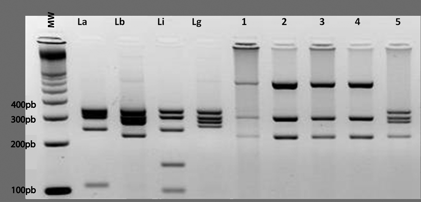
A total of 95 DNA samples were evaluated (39 extracted from isolates and 56 from lesion fragments). When the DNA extracted from cultured parasites was screened by hsp70 PCR-RFLP, the restriction profiles of most samples differed from those expected for L. (V.) braziliensis and the other Leishmania species tested (Fig. 3). Of 39 isolates evaluated, two showed a restriction profile corresponding to L. (V.) braziliensis (Lb), while 37 showed L. braziliensis variant profile (≠Lb). In addition, the PCR-RFLP analyses of DNA extracted directly from lesion fragments (56 samples) yielded ten (16.3%) samples with profiles identical to that of L. (V.) braziliensis and 46 with L. (V.) braziliensis variant profiles. Three subjects had lesions in which both parasites profiles were found. The hsp70 PCR-RFLP profiles of samples from these subjects differed from that of L. (V.) braziliensis when DNA extracted from cultured promastigotes was amplified but were indistinguishable to L. (V.) braziliensis when DNA extracted directly from lesion fragments was used. Two of these patients had typical ulcers, while one had an atypical lesion.
Representative PCR-RFLP profiles obtained by amplifying the hsp70 gene from DNA extracted from cultured samples and digesting the products with HaeIII. Molecular weight (MW) – 100 bp, La – Leishmania amazonensis reference strain (IFLA/BR/67/PH8), Lb – L. (Viannia) braziliensis reference strain (MHOM/BR/75/M2903), Li – L. (L.) infantum reference strain (MHOM/BR/74/PP75) and Lg – L. (V.) guyanensis reference strain (MHOM/BR/75/M4147). Lanes 1–5 – DNA samples from promastigotes isolated from the lesions of patients in the Xakriabá Indigenous Reserve, Minas Gerais state, Brazil. Lanes 1–4 – samples have a distinct profile from any of the reference strains shown. Lane 5 – sample has a profile indistinguishable from the L. braziliensis reference strain.

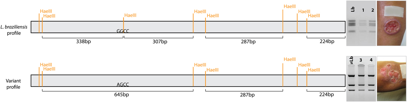
The 1300-bp hsp70 gene fragment was sequenced from samples of two different groups of study DNA samples, selected based on PCR-RFLP data (n = 26 patients), to validate the observed patterns of molecular genotypes, and to further confirm the parasite species identification. All sequences aligned with that of the L. (V.) braziliensis reference strains [L. (V.) braziliensis M2904 and M2903] and had similarity and identity values of 98–99%, but nucleotide polymorphisms were detected at some sites. Two groups were observed based on the genetic sequences patterns and they were coincident with the two PCR-RFLP profile types. One group of samples showed a mutation whose eliminates a site recognized by the restriction enzyme HaeIII (338 + 307 = 645 bp). In addition, the other group that exhibited Lb profile showed the preserved restriction sites, confirming the findings of the RFLP (Fig. 4). So, in summary, the results of RFLP analysis and sequencing of hsp 70 fragment show that there are two different genetic profiles of parasites circulating in this endemic area.
Genetic polymorphisms of the Leishmania (Viannia) braziliensis were correlated with clinical manifestations. (A) Leishmania braziliensis profile: represents the sequence and restriction pattern indistinguishable of the L. (V.) braziliensis: Lb – reference strain (M2903), 1 – patient number 374, 2 – patient number 426 – this profile is associated with patients whose had typical lesions. (B) Variant profile: represents the sequence and restriction pattern that presents: ≠Lb – mutation on the HaeIII restriction site (G > A) in comparison with L. (V.) braziliensis reference strain (M2903), 3 – patient number 306, 4 – patient number 332. Variant profile represents the sequence and restriction pattern that presents mutation on the HaeIII restriction site (G > A) in comparison with L. (V.) braziliensis reference strain (M2903); this profile is associated with patients whose had atypical lesions (logistic regression, P < 0.05, OR = 4.8, 95% CI 1.0–22.0).

Consensus sequences for hsp70 gene were obtained through forward and reverse strands assembly using STADEN Package soft-177 ware (MRC-LMB, UK). Taking into account comparison of the our sequences from ATL patients with those another L. (V.) species was possible recognized some polymorphisms which allowed distinguish of the L. (V.) guyanensis, L. (V.) panamensis, L. (V.) shawi, L. (V.) naiffi and L. (V.) lainsoni. Besides, the analysis of L. (V.) braziliensis complex strains evidenced the presence of two main haplotypes according to comparing with the reference strains (M2903 and M2904).
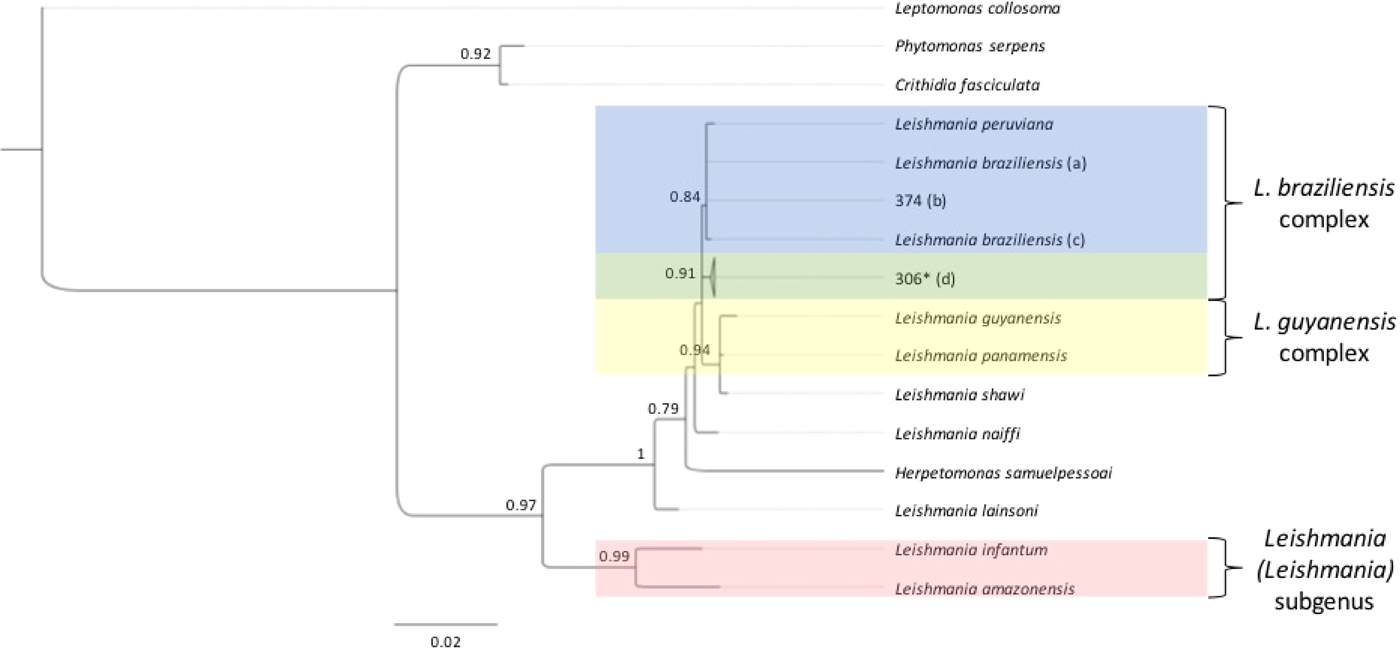
Phylogenetic tree was constructed for hsp70 gene fragment including all the 26 consensus sequences from the ATL patient's samples and comparing to other Leishmania and trypanosomatids species (Fig. 5). The Maximum Likelihood method based on the Tamura–Nei model provided strong statistical support for all nodes and showed high bootstrap support both at specie–complexes and species and strains level organization. The resulting tree supports a common origin of the patients parasites together with L. (V.) braziliensis (M2903 and M2904), clearly indicating that they belong to this species. Furthermore, we identified dendrogram branches within the L. braziliensis complex cluster that showed high bootstrap values among species and isolates due to the high number of samples and species analysed.
Maximum Likelihood phylogenetic of the hsp70 sequences. The tree was constructed using the Maximum Likelihood method based on the Tamura–Nei model (TN93), predicted as the best model by the program Jmodeltest. A discrete γ distribution was used to model evolutionary rate differences among sites (+G, parameter = 0.46). The analysis involved 40 nucleotide sequences, all sequences with 99% redundancy were collapsed in the * branches. All positions containing gaps and missing data were eliminated. The branches values are related to the bootstrap percentage. The bootstrap value used in this construction was 1000. Reference strain's sequences used to generate this dendrogram: Leishmania (Viannia) peruviana MHOM/PE/1990/HB86; L. (V.) guyanensis MHOM/BR/1975/M4147; L. (V.) panamensis MHOM/PA/1994/PSC-1; L. (V.) shawi MCEB/BR/1984/M8408; L. (V.) naiffi MDAS/BR/1979/M5533; L. (V.) lainsoni; Leptomonas collosoma; Phytomonas serpens (strain 9T); Crithidia fasciculate; Herpetomonas samuelpessoai (strain HSP). (a) Leishmania (V.) braziliensis MHOM/BR/1975/M2903; (b) L. (V.) braziliensis sample's patient 374; (c) L. (V.) braziliensis MHOM/BR/1975/M2904; (d) L. (V.) braziliensis sample's patients: 306, 43, 116, 176, 305, 307, 310, 311, 314, 315, 316, 317, 324, 327, 338, 339, 340, 344, 346, 368, 370, 376, 377, 379, 380.

Leishmania (V.) braziliensis genetic polymorphisms was correlated with typical and atypical clinical manifestations
Among the 73 parasitological confirmed cases, 24 (32.9%) had typical lesions, while 47 (67.1%) had atypical lesions. To assess whether the molecular profiles obtained directly from lesion-fragment samples were associated with the clinical characteristics of the confirmed cases, ten patients with profiles indistinguishable to that of L. (V.) braziliensis (Lb) were compared with 46 patients with L. (V.) braziliensis variant profile. After adjustment using multivariate logistic regression, the only variable associated with the L. (V.) braziliensis variant profile was the lesion type (OR = 4.8, 95% CI 1.0–22.0). The probability of having atypical lesions was almost five times higher among patients carrying parasites with the Lb hsp70 variant profile than among those with the Lb profile. In other words, the only feature associated with atypical lesions was a hsp70 gene-restriction profile after HaeIII digestion distinct to that of L. (V.) braziliensis reference strain (Fig. 4).
Discussion
Leishmania (V.) braziliensis is the most important cause of TL in several geographical regions in Brazil. The genetic variation of the parasite in this study was assessed based on the hsp70 gene-restriction profiles (in which mutations eliminate enzyme-recognition sites, thus altering the profile) and on their hsp70 gene sequencing and phylogenetic analysis. The comparison of our results with previously published studies (Garcia et al. Reference Garcia, Kindt, Bermudez, Llanos-Cuentas, De Doncker, Arevalo, Tintaya and Dujardin2004; da Silva et al. Reference da Silva, de Sousa, da Graça, Porrozzi and Cupolillo2010) showed that the PCR-RFLP variant pattern found in this study is different from those produced for other Leishmania species already tested using this methodology. Besides, two genetic variations of the L. (V.) braziliensis parasites were observed based on the hsp70 gene sequencing and phylogenetic analysis. These results indicate that there are two distinct strains of this parasite circulating among infected human hosts in the Xakriabá Reserve. This result suggests the existence of important variability within L. (V.) braziliensis that could be related to the distinct geographical location of these samples and a potential ancient dispersion of this species in Brazil.
Leishmania (V.) parasites are characterized by great genetic diversity, and the described species vary widely in their degrees of variability. Intra-specific polymorphisms are highly frequent in L. (V.) braziliensis (Boite et al. Reference Boite, Mauricio, Miles and Cupolillo2012; Kuhls et al. Reference Kuhls, Cupolillo, Silva, Schweynoch, Boité, Melo, Mauricio, Miles, Wirth and Schönian2013; Herrera et al. Reference Herrera, Hernández, Ayala, Flórez, Teherán and Ramírez2017). The results of this study and other reports highlight the use of techniques that can both identify the parasite species and differentiate genetically distinct variant strains of the same species. Such identifications are especially important in endemic areas where there are more than one species of Leishmania, also in studies that aim to differentiate cases of relapse and reinfection.
An important finding of our study is the large number of isolates that showed the L. (V.) braziliensis variant genotype. The phylogenetic analysis allowed to observe a potential population structure of L. braziliensis complex in the studied region, emphasizing that the majority of samples collected from ATL patients presented a variant genetic profile (represented in Fig. 5 by the branch of the sample 306) in accordance to PCR-RFLP previous data and in correlation with atypical clinical manifestations. Thus, the diversity found in L. braziliensis isolates from the Xakriabá community appears to be associated with host–parasite interactions. Indigenous populations may present distinct genetic backgrounds, which could be, in part, involved in the surprisingly high frequency of atypical cutaneous-leishmaniasis presentations detected. Although L. braziliensis strains have been increasingly shown as one major determinant of ATL outcome, its effect on the host may be somewhat modulated by the host genetics itself. For example, host genetics might influence the susceptibility of the human beings to specific strains of the parasite.
Regarding the clinical appearance of the lesions, most of the cases (70%) presented atypical lesions instead of the typical ulceration commonly reported in patients with L. (V.) braziliensis-caused ATL. The severity and the clinical presentation of leishmaniasis depend on many factors, including species of Leishmania involved, the number of parasites inoculated, the site of inoculum, and the nutritional status and immune competence of the host (Mitropoulos et al. Reference Mitropoulos, Konidas and Durkin-Konidas2009). A previous study of our team evaluated characteristics of the immune response of some patients with ATL in the Xacriabá community (Costa-Silva et al. Reference Costa-Silva, Gomes, Martins-Filho, Rodrigues-Silva, Freire, Quaresma, Pascoal-Xavier, Mendes, Serakides, Zauli, Campi-Azevedo, Melo, Gontijo, Peruhype-Magalhães and Teixeira-Carvalho2014). Our findings suggest that differential gene expression of cytokines and chemokines found in skin lesions from CL patients is associated with time evolution of lesions.
Other studies have reported the occurrence of atypical lesions in cutaneous leishmaniasis (Lescure et al. Reference Lescure, Bonnard, Chandenier, Schmit and Douadi2002; Convit et al. Reference Convit, Ulrich, Pérez, Hung, Castillo, Rojas, Viquez, Araya and Lima2005; Baptista et al. Reference Baptista, Schubach, Madeira, Leal, Pires, Oliveira, Conceição-Silva, Rosalino, Salgueiro and Pacheco2009; Guimarães et al. Reference Guimarães, Queiroz, Silva, Silva, Magalhães, Lago, Machado, Bacellar, Wilson, Beverley, Carvalho and Schriefer2016); however, these unusual ATL manifestations occurred less frequently in the areas where these studies were performed than in our study area. The two patients with both typical ulcers and atypical lesions are an interesting observation. It is important to emphasize that only the typical lesions of these patients were evaluated, but the presence of two types of clinical manifestations in the same patient may suggest that the clinical outcome is influenced by the parasite genotype in addition to the host's clinical status. Another interesting finding that should be discussed was the demonstration of three subjects with lesions in which different genetic profiles were found on a single lesion analysed (one DNA extraction and just one biopsy).
Furthermore, we found an important association between the two L. (V.) braziliensis variants and the observed clinical manifestations. Parasites with profiles identical to those of the L. (V.) braziliensis reference strains were associated with typical ulcerative lesions, while parasites with the variant profile were associated with atypical lesions. However, we cannot determine whether the variant parasite strain was responsible for the atypical clinical manifestations or whether the variant strain simply reflects genetic divergence between two distinct parasite populations. The observation that L. (V.) braziliensis variants are associated with different lesion types emphasizes the importance of further investigations on the relationship between clinical manifestations and genetic diversity in L. (V.) braziliensis. Further studies should focus on different genetic targets of the parasite and different molecular markers. Inter- and intraspecific variations have been demonstrated in Leishmania and more specifically in the subgenus Viannia (Schriefer et al. Reference Schriefer, Schriefer, Góes-Neto, Guimarães, Carvalho, Almeida, Machado, Lessa, de Jesus, Riley and Carvalho2004, Reference Schriefer, Guimarães, Machado, Lessa, Lessa, Lago, Ritt, Góes-Neto, Schriefer, Riley and Carvalho2009; Nolder et al. Reference Nolder, Roncal, Davies, Llanos-Cuentas and Miles2007; Pereira et al. Reference Pereira, Silva, Castilhos, Mastrantonio, Souza, Romão, Rezende, Pena, Beletti and Souza2009). However, the consequences of this ‘new’ type of interaction (variant parasite–host) are not yet well known. To what extent do differences in parasite genotypes influence virulence and pathogenicity and therefore the disease prognosis and response to treatment? Do variant parasites require different therapeutic approaches? Could variant strains be responsible for recidivans? Are variant parasites more resistant to chemotherapy? As yet, these questions lack definitive answers. Our results indicate the need for further research related to parasitic diseases in which knowledge of the parasite's genetic characteristics and its host interactions will enhance our understanding of these diseases in humans. Therefore, surveillance efforts and control activities in the studied region should consider the presence of different strains and clinical presentations to improve the diagnosis and treatment of the infection in human subjects.
List of genbank accession numbers
The sequences generated by our team were added to the database GenBank® (http://www.ncbi.nlm.nih.gov/genbank) and the accession numbers of the L. braziliensis collected from ATL patients can be viewed in Table 1. The accession numbers of the L. collosoma, P. serpens (strain 9T) and H. samuelpessoai (strain HSP) are listed, respectively: MF6807, MF687108 and MF687106.
Acknowledgements
The authors thank the inhabitants of the Xacriabá Indigenous Reserve for contributing to this study.
Financial support
Financial support was given by the National Council for Scientific and Technological Development (CNPq – grant number 479408/2010-6), the Minas Gerais Research Foundation (FAPEMIG grant number APQ-00289-10), Oswaldo Cruz Foundation (FIOCRUZ grant number 40356/2008-2), European Community LeishEpiNetSA (grant number 015407). C.M.F. Gontijo and C.F.A.Brito are researchers’ fellow from CNPq.