Background
Despite wider provision of organized stroke unit care and increasing use of reperfusion therapies that limit the extent of brain injury in the acute phase of stroke, a high proportion of patients remain permanently disabled. Even with optimal acute care, over 50% of stroke survivors have residual disability that restricts their independence.1 Conventional physical therapies can facilitate recovery of function and prevent maladaptive responses, but at present few interventions have evidence of sustained superiority over endogenous recovery processes, and some are applicable only to very specific deficits (for example, constraint-induced movement therapy for upper limb functionReference Wolf, Winstein, Miller, Thompson, Taub, Uswatte, Morris, Blanton, Nichols-Larsen and Clark2). Novel therapeutic approaches that have restorative or regenerative potential and that can be applied to the entire range of neurological deficits following stroke are therefore needed.
Stem cells are self-renewing cells that have the potential to differentiate into a range of specialized adult cells and offer an attractive possible approach to regeneration within the central nervous system (CNS) – a biological niche that was until recently thought to be devoid of significant endogenous regenerative potential. The broad range of cell types that are included under this general heading leads to much public and medical confusion, fuelled by a tendency for media commentaries to imply the imminence of revolutionary therapeutic changes. Clinical studies within properly regulated frameworks remain few, and all include only small numbers of subjects.
It is important to remember that stem cells represent only one therapeutic possibility – and one that has many practical, and some ethical, hurdles compared with alternatives that include small molecules acting on conventional neurotransmitter systems (e.g. dopamine agonists, amphetamines and other noradrenergic stimulants), antibodies directed against neural growth blocking molecules (e.g. anti-MAG), novel neurotransmitter systems (e.g. sigma site agonists such as SA4503), growth factors (e.g. granulocyte colony stimulating factor, G-CSF), and physical stimulation (e.g. transcranial magnetic stimulation).Reference Cramer3 Judging the likely strategy or strategies that will prove to be clinically useful at the present time is an exercise in crystal ball gazing.
Search methods
Background knowledge was supplemented by a PubMed search using the terms ‘stem cell’ AND ‘stroke’ AND ‘human.’ Reference lists of papers were also reviewed and content of recent meetings on the theme taken into account.
Translation of stem cell therapies
Stem cell approaches present a series of additional and unique issues for translation to clinical use beyond those of conventional small molecule drug therapies. However, they enter a field with a difficult background in general.
Safety and efficacy testing is undertaken in animal model systems where focal brain ischaemia is induced, usually by means of middle cerebral artery occlusion (MCAO) in rats. Cells may be derived from the same species as used for the ischaemia model, but are more often xenografts from other species, predominantly humans. Xenografting raises issues that include the routine use of immunosuppressant treatment, and its potential interactions with the cell graft, as well as difficulty in extrapolating cell engraftment and survival data to potential human use. In the field of stroke, there is ongoing debate about the failure to translate neuroprotective strategies that are highly effective in predominantly rodent models of focal ischaemia but almost uniformly ineffective or harmful in the clinic, and concern that the model systems may themselves inadequately represent the clinical situation.Reference Macleod, O'Collins, Howells and Donnan4 There is particular concern over potential bias introduced when randomization, treatment allocation concealment and blinding of outcome measures are not rigorously undertaken.Reference Macleod, O'Collins, Howells and Donnan4, Reference Crossley, Sena, Goehler, Horn, van der and Bath5 Publication bias has also been highlighted as a major factor that skews the literature towards over-optimistic assessment of likely effect sizes. Not all published animal studies using stem cell implantation have identified benefit,Reference Bliss, Andres and Steinberg6 and there are inconsistent findings between different laboratories for the same cell type in similar models.
Acute treatments can be assessed by tissue-based criteria (such as volume of early brain infarction), but in order to assess regenerative therapies, longer term testing involving histological, electrophysiological and behavioural testing is necessary. Behavioural testing in particular is undertaken as a surrogate for functional recovery after stroke. Rodents do not exhibit hemiplegia for any significant length of time after recovery from MCAO and a range of behavioural tests that seek to discriminate on the basis of more subtle aspects of neurological function have been developed.Reference Hicks, Schallert and Jolkkonen7 Whether changed performance on behavioural testing reflects adaptation rather than tissue regeneration, which tests are most sensitive to change, or whether there are differences depending on time point after MCAO, are the subject of debate. Certainly there are differences in the apparent sensitivity of different tests.Reference Hicks, Schallert and Jolkkonen7
Sources of stem cells
Stem cells can be derived from a variety of sources, from the early embryo through to adult tissues, and exhibit varying degrees of pluripotency depending on their source, and also their environment. Embryonic stem cells have the capacity to self-replicate indefinitely in their undifferentiated state and to differentiate into any cell lineage: properties shared by the more recently developed induced pluripotent stem cells. The differentiation of ‘adult’ stem cells, in contrast, is more restricted.
The clinical development of cells as therapy presents challenges that differ from drug treatments, or devices, and which vary according to the source of cells and any subsequent – often genetic – manipulation that is undertaken. Medicines regulatory systems for handling cell therapies are developing approaches, but at present have only extremely limited experience.
Since cell culture is fundamental to the expansion of stem cell numbers regardless of their origin, cells are inevitably exposed to media that include human or animal blood products or cells. Transmission of infectious agents (including non-human viruses or prions) therefore is a concern. Retention of cell characteristics through multiple passages in culture is also clearly required, as well as confirmation of karyotypic and genetic stability.Reference Puceat and Ballis8 These present significant technical hurdles in bringing cells to the clinic in the quantities required for clinical studies.
Embryonic stem cells (eSCs) are pluripotent cells derived from the inner wall of the blastocyst. Use of human embryonic material to obtain eSCs presents particular ethical issues that are frequently confused in media reports with other types of stem cell.
Murine eSCs grafted into the contralateral hemisphere after MCAO in rats migrate to the site of infarctionReference Hoehn, Küstermann, Blunk, Wiedermann, Trapp, Wecker, Föcking, Arnold, Hescheler, Fleischmann, Schwindt and Bührle9, Reference Hayashi, Takagi, Fukuda, Imazato, Nishimura, Fujimoto, Takahashi, Hashimoto and Nozaki10 but risk of tumour, particularly teratoma or teratocarcinoma, formation is a concern that has been raised in model systems.Reference Erdö, Bührle, Blunk, Hoehn, Xia, Fleischmann, Föcking, Küstermann, Kolossov, Hescheler, Hossmann and Trapp11 To add further complexity, a mouse stroke study found that teratoma risk was absent in xenografts but present in allografts.Reference Erdö, Bührle, Blunk, Hoehn, Xia, Fleischmann, Föcking, Küstermann, Kolossov, Hescheler, Hossmann and Trapp11 Clinical use of undifferentiated eSCs is therefore unlikely, and most studies have developed lineage-restricted cell lines such as neuronal precursors derived from eSCs by biochemical manipulation, for example, oligodendrocyte progenitor cells (the GRNOPC1 cell line) now entering a phase I clinical trial in thoracic spinal cord injury.Reference Keirstead, Nistor, Bernal, Totoiu, Cloutier, Sharp and Steward12
Induced pluripotent stem cells (ipSCs) potentially avoid ethical and practical issues associated with eSCs as these are produced from an individual's own somatic cells by ‘reprogramming’ to induce pluripotency. This was first achieved in mice by retroviral insertion of four transcription factor genes that regulate cell differentiation,Reference Okita, Ichisaka and Yamanaka13 and subsequently replicated in cells from other species including humans.Reference Yu, Vodyanik, Smuga-Otto, Antosiewicz-Bourget, Frane, Tian, Nie, Jonsdottir, Ruotti, Stewart, Slukvin and Thomson14 Recent developments include the introduction of relevant genes without the use of viral vectorsReference Okita, Nakagawa, Hyenjong, Ichisaka and Yamanaka15 or without transgenes. However, the reported methods are slow (several weeks), have very low efficiency for production of ipSCs,Reference Leeper, Hunter and Cooke16 and there are concerns about tumorigenicity both with respect to persistent undifferentiated cellsReference Miura, Okada, Aoi, Okada, Takahashi, Okita, Nakagawa, Koyanagi, Tanabe, Ohnuki, Ogawa, Ikeda, Okano and Yamanaka17 and the additional growth factor genes used, even when potentially oncogenic retrovirus delivery is avoided. Careful selection of cell populations from derived ipSCs appears to be necessary to mitigate this risk. There are a limited number of studies in animal disease models to date, including Parkinson's disease in rats and spinal cord injury in mice;Reference Tsuji, Miura, Okada, Fujiyoshi, Mukaino, Nagoshi, Kitamura, Kumagai, Nishino, Tomisato, Higashi, Nagai, Katoh, Kohda, Matsuzaki, Yuzaki, Ikeda, Toyama, Nakamura, Yamanaka and Okano18 in the latter, grafted ipSC-derived neural stem cells delivered 9 days after injury differentiated into functioning neurons and improved function, without any evidence of teratoma formation over a 24 week follow-up.Reference Tsuji, Miura, Okada, Fujiyoshi, Mukaino, Nagoshi, Kitamura, Kumagai, Nishino, Tomisato, Higashi, Nagai, Katoh, Kohda, Matsuzaki, Yuzaki, Ikeda, Toyama, Nakamura, Yamanaka and Okano18
Neural stem cells have more restricted differentiation, into neurones and glia, and may be derived from foetal tissue, cultured eSCs or ipSCs that exhibit neural differentiation markers and are induced to develop along neural lines by treatment with mediators such as retinoic acid, or tumour-derived neuronal tissues, including immortal teratocarcinoma-derived cells (e.g. NT2N and hNT cells).Reference Bacigaluppi, Pluchino, Martino, Kilic and Hermann19, Reference Borlongan, Tajima, Trojanowski, Lee and Sanberg20 Human foetal cortex cells have been immortalized by insertion of transcription factor genes including v-mycReference Cacci, Villa, Parmar, Cavallaro, Mandahl, Lindvall, Martinez-Serrano and Kokaia21 and c-myc.Reference Pollock, Stroemer, Patel, Stevanato, Hope, Miljan, Dong, Hodges, Price and Sinden22 The potential for adult somatic stem cells to differentiate into neural precursors has also been explored, with evidence that adult dermal cells can be induced to produce neural cells by culturing in a medium containing astrocyte-derived material.Reference Joannides, Gaughwin, Schwiening, Majed, Sterling, Compston and Chandran23 Animal studies with a number of NSC lines have been undertaken, with evidence of cell survival and in some studies improvement in behavioural outcomes after focal ischaemic injury.Reference Bacigaluppi, Pluchino, Martino, Kilic and Hermann19, Reference Borlongan, Tajima, Trojanowski, Lee and Sanberg20, Reference Pollock, Stroemer, Patel, Stevanato, Hope, Miljan, Dong, Hodges, Price and Sinden22, Reference Modo, Stroemer, Tang, Patel and Hodges24–Reference Wei, Cui, Snider, Rivkin, Yu, Lee, Adams, Gottlieb, Johnson, Yu and Choi28
The majority of NSC studies have used intraparenchymal delivery of cells,Reference Bacigaluppi, Pluchino, Martino, Kilic and Hermann19 with fewer studies of intravascular administration.Reference Chu, Kim, Park, Jeong, Park, Jung, Lee, Kang, Lee, Park, Kim and Roh25, Reference Jiang, Zhang, Ding, Silver, Zhang, Meng, Lu, Pourabdillah-Nejed-D, Wang, Savant-Bhonsale, Li, Bagher-Ebadian, Hu, Arbab, Vanguri, Ewing, Ledbetter and Chopp27 The time of cell implantation after focal ischaemia has ranged from 24 hours to 1 month.
In studies of NSCs derived from various sources, cells have migrated to the region of ischaemia after implantation in brain parenchyma (including contralesional striatum),Reference Modo, Stroemer, Tang, Patel and Hodges24, Reference Veizovic, Beech, Stroemer, Watson and Hodges29–Reference Kelly, Bliss, Shah, Sun, Ma, Foo, Masel, Yenari, Weissman, Uchida, Palmer and Steinberg31 or after intravenous injection,Reference Chu, Kim, Park, Jeong, Park, Jung, Lee, Kang, Lee, Park, Kim and Roh25, Reference Chu, Kim, Chae, Jeong, Kang, Jung, Kim, Kim, Kang, Kim and Yoon32 in most but not all studies. Cell migration has been documented both by histology and in vivo using magnetic resonance imaging (MRI).Reference Modo, Beech, Meade, Williams and Price33–Reference Sykova and Jendelova35 Initial cell proliferation and subsequent survival of grafted cells, and differentiation into electrically active neurones and glia, has been seen 4–12 weeks after acute (24 hours post-ischaemia)Reference Hayashi, Takagi, Fukuda, Imazato, Nishimura, Fujimoto, Takahashi, Hashimoto and Nozaki10, Reference Chu, Kim, Chae, Jeong, Kang, Jung, Kim, Kim, Kang, Kim and Yoon32, Reference Bühnemann, Scholz, Bernreuther, Malik, Braun, Schachner, Reymann and Dihné36 or 7 days post-ischaemiaReference Kelly, Bliss, Shah, Sun, Ma, Foo, Masel, Yenari, Weissman, Uchida, Palmer and Steinberg31 administration, and some human NSCs have been observed to survive in rat brain 540 days after IV injection.Reference Chu, Kim, Park, Jeong, Park, Jung, Lee, Kang, Lee, Park, Kim and Roh25 After IV administration, most peri-infarct human NSCs had differentiated into glia, with around 10% having neuronal characteristics,Reference Chu, Kim, Chae, Jeong, Kang, Jung, Kim, Kim, Kang, Kim and Yoon32 but others report predominantly neuronal differentiation of intrastriatal human NSCs.Reference Darsalia, Kallur and Kokaia37
In one study, intraparenchymal injection of human NSCs was performed 7 days after MCAO in three Cynomolgus monkeys, the only study of non-human primates reported.Reference Roitberg, Mangubat, Chen, Sugaya, Thulborn, Kordower, Pawar, Konecny and Emborg38 Cells survived up to 105 days post-implantation, with the estimated proportion between 3 and 30% and no clear relationship to time of histological examination. However, most cells were evident in the vicinity of the needle tracks, and few had migrated to the peri-infarct area. Also, few cells expressed neuronal phenotypes.
Different cell types may differ in their effects on behavioural outcome (e.g. hNT cells compared with foetal rodent striatal cells),Reference Borlongan, Tajima, Trojanowski, Lee and Sanberg39 and in one study were further improved by combination treatment with IV human NSCs and vascular endothelial growth factor (VEGF), presumed to be mediated by enhanced angiogenesis.Reference Chu, Park, Lee, Jung, Ko, Kang, Sinn, Lee, Kim, Kim and Roh40
Although most studies have routinely administered immunosuppressant drug treatments, one study found no difference in host immune response to intraparenchymal implantation of human cells in a rat model in the presence or absence of immunosuppressant drugs.Reference Modo, Rezaie, Heuschling, Patel, Male and Hodges41
Mesencyhmal stem cells (MSCs) are non-haematopoietic stem cells that are present in adult bone marrow (also commonly referred to as bone marrow stromal cells as they are derived from the same niche as cells that support haematopoietic cells), and represent a multipotent cell population with therapeutic potential that can be obtained fairly readily for clinical use. As their name indicates, MSCs can differentiate into mesodermal cell lineages including adipocytes, chondroblasts, fibroblasts and osteocytes, but in vitro and in vivo they may also differentiate into cells expressing markers of ectodermal tissue (including neurones) and also endodermal tissue (hepatocytes),Reference Prockop42 depending upon the local environment. The nomenclature of bone marrow-derived cells has been confused as cells of multiple types can be obtained from this source, not all of which have properties that are truly those of stem cells. Consensus criteria for nomenclature and definition of MSCs were published in 2005–2006 to address the difficulties in interpreting the literature. Studies before that time require careful review to ensure that the cell type corresponds to what would now be classed as MSCs. Criteria are based on behaviour in culture (plastic adherence), expression of specific cell surface markers (CD105, CD73 and CD90) and lack of expression of some other markers (CD45, CD34, CD14 or CD11b, CD79a or CD19, and HLA class II), and multipotent differentiation potential.Reference Dominici, Le Blanc, Mueller, Slaper-Cortenbach, Marini, Krause, Deans, Keating, Prockop and Horwitz43
When injected into the brain in a rat focal ischaemia model, adult rat MSCs express neuronal and glial markers,Reference Li, Chen and Chopp44 and human MSCs also exhibit neural phenotypes.Reference Zhao, Duan, Reyes, Keene, Verfaillie and Low45 As for neural stem cells, MSCs migrate to the ischaemic brain regionReference Eglitis, Dawson, Park and Mouradian46 after intravascular (intracarotidReference Li, Chen, Wang, Lu and Chopp47 or intravenous) administration, and systemic administration improves functional outcomes.Reference Chen, Li, Wang, Zhang, Lu, Lu and Chopp48 Functional improvement is evident despite only a small percentage of injected cells reaching the brain,Reference Chen, Li, Wang, Zhang, Lu, Lu and Chopp48 the great majority being cleared rapidly and appearing in lungs, spleen and kidneys after systemic administration.Reference Detante, Moisan, Dimastromatteo, Richard, Riou, Grillon, Barbier, Desruet, De Fraipont, Segebarth, Jaillard, Hommel, Ghezzi and Remy49 Acutely injected MSCs persist in the brain 1 year after stroke, but by then have been cleared from systemic organs.Reference Shen, Li, Chen, Cui, Zhang, Kapke, Lu, Savant-Bhonsale and Chopp50 Several potential mechanisms of benefit include production of neurotrophic factors, stimulation of angiogenesis and neurogenesis, immunomodulation, and stimulation of endogenous stem cell mobilization.Reference Shen, Li, Chen, Cui, Zhang, Kapke, Lu, Savant-Bhonsale and Chopp50–Reference Li, Chen, Chen, Wang, Gautam, Xu, Katakowski, Zhang, Lu, Janakiraman and Chopp56 Whether migration to the brain in sufficient numbers for a therapeutic effect is dependent on blood–brain barrier disruption in the subacute phase of ischaemic stroke, or persists for longer is not clearly established, although it is probable that cells can also cross an intact blood–brain barrier. The majority of animal studies administered MSCs within a week of ischaemia, and the longest interval reported is 1 month.Reference Shen, Li, Chen, Zacharek, Gao, Kapke, Lu, Raginski, Vanguri, Smith and Chopp57 Many of the outcome studies established reduction in infarct volume as an effect, in keeping with a ‘neuroprotective’ effect. This may have a bearing on the design of clinical studies with MSCs.
Bone marrow offers the possibility of autologous cell transplantation, with obvious advantages in avoiding rejection (and thus immunosuppressant drug treatment) and circumventing ethical concerns and practical barriers to availability that are inherent in other cell sources. However, the number of cells that can be obtained by marrow aspiration alone is significantly smaller than doses that are likely to have therapeutic effects, and there is evidence from animal models that functional recovery after stroke is dose dependent.Reference Chen, Li, Wang, Zhang, Lu, Lu and Chopp48 It is therefore necessary to expand cells in culture ex vivo, and then administer cultured cells after sufficient numbers have been obtained. This process introduces inevitable delay in preparation of cells for clinical use, of the order of several weeks.Reference Bang, Lee, Lee and Lee58 Infusion of autologous cells is not without potential problems, particularly in a patient population with manifest circulatory disease who are often elderly, and there are reports of ischaemic stroke complicating infusion of stored MSCs.Reference Hentschke, Hentschke, Hummel, Salwender, Braumann and Stang59 However, it appears that MSCs are not a target for immune rejection even when administered allogeneically, probably due to reduced expression of major histocompatability complex (MHC) antigens and T-cell stimulatory molecules, and they may therefore represent a source of adult-derived stem cells that may be suitable for therapeutic use as allografts.
Bone marrow mononuclear cells include other progenitor cell populations that may have therapeutic value in ischaemic disease including stroke. The mixed mononuclear cell population includes cells expressing the endothelial progenitor surface marker CD34 (CD34+ cells, also referred to as endothelial progenitor cells) that promote angiogenesis. Therapeutic angiogenesis has been attempted by infusion of bone marrow mononuclear cells in a number of clinical studies of subacute myocardial infarctionReference Strauer, Brehm, Zeus, Köstering, Hernandez, Sorg, Kögler and Wernet60, Reference Yousef, Schannwell, Kostering, Zeus, Brehm and Strauer61 or peripheral arterial disease,Reference Tateishi-Yuyama, Matsubara, Murohara, Ikeda, Shintani, Masaki, Amano, Kishimoto, Yoshimoto, Akashi, Shimada, Iwasaka and Imaizumi62 cells being delivered by intracoronary, intramyocardial, or intravenous delivery. Safety data are not directly applicable to stroke trials as small volumes of bone marrow-derived cells were generally delivered without ex vivo culture expansion to numbers equivalent to those used for stroke.
Multipotent adult progenitor cells (MAPCs) are a population of marrow cells with broad differentiation potentialReference Jiang, Jahagirdar, Reinhardt, Schwartz, Keene, Ortiz-Gonzalez, Reyes, Lenvik, Lund, Blackstad, Du, Aldrich, Lisberg, Low, Largaespada and Verfaillie63, Reference Kovacsovics-Bankowski, Mauch, Raber, Streeter, Deans, Maziarz and Van't Hof64 that can be culture-expanded to quantities suitable for commercial use.Reference Boozer, Lehman, Lakshmipathy, Love, Raber, Maitra, Deans, Rao and Ting65 MAPCs have immunomodulatory properties that make them potentially suitable as allografts without the need for immunosuppressant drugs. MAPCs are found in the hippocampus and are associated with improved functional outcomes in neonatal hypoxia–ischaemia models when given intravenously.Reference Yasuhara, Hara, Maki, Mays, Deans, Hess, Carroll and Borlongan66
Other peripheral blood stem cells
Bone marrow contributes a small number of circulating stem cells in peripheral blood in adults. Reduction in numbers of CD34+ endothelial progenitor cells (EPCs)Reference Asahara, Murohara, Sullivan, Silver, van der Zee, Li, Witzenbichler, Schatteman and Isner67 appears to correlate with severity (and possibly pathological subtype) of stroke in observational clinical studies.Reference Chu, Jung, Lee, Park, Sinn, Kim, Kim, Kim, Kim, Song, Kim, Lee and Roh68 Infusion of human EPCs in a mouse transient focal ischaemia model was associated with migration of cells to the peri-infarct zone, reduced infarct volume, improved behavioural recovery, and evidence of angiogenesis in the ischaemic region.Reference Fan, Shen, Frenzel, Zhu, Ye, Liu, Chen, Su, Young and Yang69 Ischaemia in any organ, including stroke, stimulates release of CD34+ cells into peripheral blood from bone marrow,Reference Dunac, Frelin, Popolo-Blondeau, Chatel, Mahagne and Philip70–Reference Paczkowska, Larysz, Rzeuski, Karbicka, Jałowiński, Kornacewicz-Jach, Ratajczak and Machaliński72 and migration of cells to the site of ischaemia, and CD34+ cell release can be further stimulated by administration of the polypepetide growth factor granulocyte-colony stimulating factor (G-CSF),Reference Yanqing, Yu-Min, Jian, Bao-Guo and Chuan-Zhen73 which is at least one of the endogenous factors that stimulate stem cell release and migration in response to tissue injury. Cells with other surface markers have also been considered to be EPCs but are probably not true endothelial progenitors; however, they may contribute to angiogenesis by release of trophic factors to support true EPCs.Reference Leeper, Hunter and Cooke16
Umbilical cord blood contains a small proportion of haematopoietic stem cells (estimated to be around 2%) and also other mononuclear cells that express non-haematopoietic stem cell markersReference Xiao, Nan, Motooka and Low74 that can be induced to express neural markers. Acute administration of human umbilical cord blood mononuclear cells was found to improve behavioural recovery indices in rodent focal ischaemia,Reference Xiao, Nan, Motooka and Low74–Reference Borlongan, Hadman, Sanberg and Sanberg76 and, in further studies, to have a dose-dependent benefit on behavioural outcomes and infarct size,Reference Vendrame, Cassady, Newcomb, Butler, Pennypacker, Zigova, Sanberg, Sanberg and Willing77 with possible superiority of IV administration over striatal implantation.Reference Willing, Lixian, Milliken, Poulos, Zigova, Song, Hart, Sanchez-Ramos and Sanberg78 However, others found no behavioural benefits of acute IV administration in a transient MCAO model.Reference Mäkinen, Kekarainen, Nystedt, Liimatainen, Huhtala, Närvänen, Laine and Jolkkonen79 Rodent studies demonstrate reduction in infarct size that is not dependent on entry of cells to the CNS.Reference Borlongan, Hadman, Sanberg and Sanberg76
Xenografts
Cells derived from porcine foetal tissue have been used in a small clinical study,Reference Savitz, Dinsmore, Wu, Henderson, Stieg and Caplan80 but there are major logistical and safety issues with the use of xenografts. Clinical studies to date have predominantly been in neurodegenerative diseases.
Genetic modification
A number of genetic modifications to stem cells have been tested in model systems. The majority of these involve insertion of trophic factor genes in order to enhance the delivery of specific molecules thought to be involved in neurogenesis or angiogenesis, including brain-derived neurotrophic factor (BDNF),Reference Kurozumi, Nakamura, Tamiya, Kawano, Kobune, Hirai, Uchida, Sasaki, Ito, Kato, Honmou, Houkin, Date and Hamada81 placental growth factor,Reference Liu, Honmou, Harada, Nakamura, Houkin, Hamada and Kocsis82 G-CSF, or VEGF.Reference Lee, Kim, Park and Kim83 Others have included the anti-apoptotic gene bcl-2.Reference Wei, Cui, Snider, Rivkin, Yu, Lee, Adams, Gottlieb, Johnson, Yu and Choi28 The introduction of novel genes, particularly growth promoters, raises additional safety issues for clinical studies since it is necessary to ensure that no adverse tumorigenic effects arise in the modified stem cells. The use of a growth factor gene to conditionally immortalize cells has been exploited, for example, in the CTX0E03 cell line that is in clinical trials at present, and has been used previously in other neural cell lines.Reference Gray, Grigoryan, Virley, Patel, Sinden and Hodges84
Endogenous neural stem cells
The brain's response to stroke includes reorganization of neural circuits that is thought to underpin spontaneous natural recovery of function. This is not simply functional plasticity, but also involves structural plasticity with changes in neuronal connections following ischaemia including dendritic and axonal sprouting and growth.Reference Benowitz and Carmichael85, Reference Murphy and Corbett86 In addition to these changes in the architecture of existing cells, recovery also involves the generation of new neurones from pools of neural progenitor cells (neural stem cells) that reside in the adult brain in the subventricular zone (SVZ) and the dentate gyrus.Reference Morshead, Reynolds, Craig, McBurney, Staines, Morassutti, Weiss and van der Kooy87 The discovery of these stem cells contradicted the traditional teaching that the adult CNS lacked any capacity to regenerate. Neurogenesis in the SVZ and cortex is increased after focal ischaemia in animal models of strokeReference Zhang, Zhang, Zhang and Chopp88, Reference Arvidsson, Collin, Kirik, Kokaia and Lindvall89 due to recruitment of new neural stem cells,Reference Zhang, Zhang, Wang, Wang, Gousev, Zhang, Ho, Morshead and Chopp52 and SVZ-derived neuroblasts migrate to the site of ischaemic injury.Reference Zhang, Zhang, Wang, Wang, Gousev, Zhang, Ho, Morshead and Chopp52, Reference Arvidsson, Collin, Kirik, Kokaia and Lindvall89 Basal levels of neurogenesis are reduced with advancing age in rats, but can be induced by stroke to a similar degree as in young animals.Reference Darsalia, Heldmann, Lindvall and Kokaia90 At least in rats, neurogenesis persists for several months after stroke.Reference Thored, Arvidsson, Cacci, Ahlenius, Kallur, Darsalia, Ekdahl, Kokaia and Lindvall91 There is evidence of neurogenesis near brain infarct in some cases of stroke in humans, including one very elderly subject aged 84 years at the time of death 1 week following stroke.Reference Minger, Ekonomou, Carta, Chinoy, Perry and Ballard92
While stimulation of endogenous neural stem cells may represent a therapeutic goal in itself, it is possible that the mode of action of exogenous stem cells may include stimulation of this process by trophic factors released by therapeutically administered cells.
Mechanisms of stem cell effects
Several stem cell lines that have been administered within 48 hours of focal ischaemia reduce infarct volume.Reference Bacigaluppi, Pluchino, Martino, Kilic and Hermann19 The presumed mechanisms include the release of trophic factors with neuroprotective properties, or modulation of the inflammatory response to tissue injury. The time window required for this type of protocol probably precludes any clinical use of autologous cell therapies, but may be feasible for allogeneic treatments with neural or mesenchymal stem cells if these can be developed as an ‘off the shelf’ treatment. Systemic delivery of cells would be advantageous, and perhaps essential, in this setting where a patient's clinical state is unlikely to allow delivery by any procedure more invasive than an intra-arterial approach.
Later administration of cells has targeted time periods between several days to 4 weeks after stroke in rodents, when regenerative mechanisms are potentially involved.
Assumptions that repair would result primarily from survival and differentiation of engrafted cells at the site of injury are almost certainly simplistic, and there is limited evidence for replacement of functioning neurones by NSCs or other cell types.Reference Bliss, Andres and Steinberg6 While cells administered systemically or at remote brain sites can migrate to the peri-infarct regionReference Modo, Mellodew, Cash, Fraser, Meade, Price and Williams34 (although as noted above, some studies have found no significant migration) and have been confirmed to survive and differentiate into cells expressing neuronal and glial surface markers, only small numbers of cells survive in the longer term, and the great majority are eliminated rapidly. Some surviving cells have electrophysiological properties of neurones and establish synapses with host cells,Reference Daadi, Li, Arac, Grueter, Sofilos, Malenka, Wu and Steinberg93 but whether this structural effect is sufficient to explain behavioural recovery has been questioned. Despite uncertainty over the possible contribution of cell replacement, dose-dependent therapeutic effects have been seen after delayed administration of NSCs,Reference Pollock, Stroemer, Patel, Stevanato, Hope, Miljan, Dong, Hodges, Price and Sinden22 and there is evidence that stem cells promote endogenous repair processes including neurogenesis,Reference Stroemer, Patel, Hope, Oliveira, Pollock and Sinden94 dendritic sprouting, axonal regeneration, and angiogenesis, again probably through the release of trophic factors (VEGF and thrombospondins being possible molecular mediators of several key processes).Reference Liauw, Hoang, Choi, Eroglu, Choi, Sun, Percy, Wildman-Tobriner, Bliss, Guzman, Barres and Steinberg95
Marrow-derived MSCs have significant immunomodulatory properties including suppression of T-cell responses and suppression of pro-inflammatory cytokine production by allogeneically administered cells that have led to their development as therapeutic agents for graft versus host disease in haematological use.Reference Ting, Mays, Frey, Hof, Medicetty and Deans96 This immunomodulatory activity may also represent part of the therapeutic effect. The immune-privileged status of MSCs may be exploited to avoid the need for immunosuppressant drug treatment in MSC allografts, for example with commercially developed MAPCs. Reduced immune response has also been seen with human MSCs administered in cancer or neurological diseases.
The potential for stem cells administered therapeutically to directly form axons and relevant synaptic connections that ‘rewire’ the brain may additionally be expected to have limitations based on anatomy: it is improbable that the very large malacic regions after large volume infarction would be amenable to substantial recovery of function, in contrast to smaller volumes of neuronal or axonal loss. Experimental findings where stem cells have been engrafted within a matrix or scaffold that provides a milieu for cell survivalReference Bible, Chau, Alexander, Price, Shakesheff and Modo97 suggest that in future this might also be a possibility for clinical studies, but present strategies deliver only small volumes of cells either systemically or by direct intraparenchymal injection, which makes restoration of large volumes of tissue loss unrealistic.
Clinical trials with stem cells
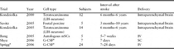
Completed clinical trials are summarized in Table 1.
Published human studies of cell treatments for stroke

*Refers to trials that administered G-CSF, for which stem cell mobilization represents only one possible moder of action.
Autologous stem cell treatment
Bang and colleaguesReference Bang, Lee, Lee and Lee58 derived MSCs from bone marrow aspirate obtained 1 week after stroke onset, expanded the cell population by ex vivo culture, and reintroduced the cells intravenously 1–2 months after stroke in a randomized, controlled clinical study involving 30 subjects (5 receiving MSC infusion and 25 controls). Patients were aged 30–75 years with MCA territory ischaemic stroke causing significant neurological impairment (NIH stroke scale score of 7 or more at day 7 after stroke onset). The dose of 1 × 10⁸ cells expressing the CD105 marker was predefined as one assumed equivalent to an effective dose in animal studies, and this dose was obtained after a mean of 31 days culture. Cells were delivered in two divided doses 4–5 weeks and again 7–9 weeks after stroke, with no adverse effects related to treatment. Compared with the 25 control subjects, indices of neurological deficit and disability were slightly better at 3, 6 and 12 months, but, unsurprisingly given the small number of subjects, there were no significant differences. A reduction in ventricular dilatation on follow-up brain magnetic resonance imaging in the MSC group was observed.
Honmou and colleagues have reported successful use of autologous serum for culture of autologous MSCs derived from bone marrow in five patients with stroke,Reference Honmou, Houkin, Matsunaga, Niitsu, Ishiai, Waxman and Kocsis98 and reinfusion of the cells 1–4 months after stroke onset, with full details of the study yet to be reported. The potential advantages of autologous culture include lack of several major sources of potential infection and greater speed of cell growth.
Further clinical trials with IV or intra-arterially delivered MSCs and other bone marrow-derived cell populations such as CD34+ cells are ongoing or planned.
Small clinical trialsReference Shyu, Lin, Lee, Liu and Li99, Reference Sprigg, Bath, Zhao, Willmot, Gray, Walker, Dennis and Russell100 involving 10 and 36 patients, respectively, and a recently completed larger recent trial, administered G-CSF to subacute stroke patients. Amongst other potential mechanisms, G-CSF mobilizes endothelial progenitor CD34+ cells from marrow, and a dose-dependent increase in circulating CD34+ cell counts was seen.Reference Sprigg, Bath, Zhao, Willmot, Gray, Walker, Dennis and Russell100 Safety concerns include the cell proliferative effect of G-CSF on haematopoietic cells, with potential for prothrombotic circulatory complications such as have been encountered in its use for treatment of haematological disease. While white cell counts rose in a similarly dose-dependent manner, no vascular complications were reported in stroke trials.
Multipotent adult progenitor cells are a commercially prepared cell population of mixed bone marrow origin (‘MultiStem’) that are planned to enter clinical trials in non-stroke patients. Potential development for stroke treatment is being kept as a possibility.
Stem cell allografts
Neural cells derived from an immortalized human teratocarcinoma cell line (NT2N cells, ‘LBS neurons’) had improved behavioural outcomes when implanted 1 month after MCAO in rats.Reference Borlongan, Tajima, Trojanowski, Lee and Sanberg20 This cell line was subsequently administered by stereotaxic injection to the putamen of human subjects in two small safety studies in the USA, with survival of cells documented in one individual who later died and had a post-mortem.Reference Kondziolka, Wechsler, Goldstein, Meltzer, Thulborn, Gebel, Jannetta, DeCesare, Elder, McGrogan, Reitman and Bynum101–Reference Nelson, Kondziolka, Wechsler, Goldstein, Gebel, DeCesare, Elder, Zhang, Jacobs, McGrogan, Lee and Trojanowski103 In the first study,Reference Kondziolka, Wechsler, Goldstein, Meltzer, Thulborn, Gebel, Jannetta, DeCesare, Elder, McGrogan, Reitman and Bynum101 2 or 6 million cells were implanted in 12 patients who had suffered an ischaemic stroke an average of 27 months earlier, predominantly involving the basal ganglia alone (8/12 subjects). All subjects were given oral ciclosporin for 8 weeks after surgery. No cell-related adverse events were reported and an improvement in motor function between implantation and 6-month follow-up was seen in half of the subjects by one of the clinical scales used for evaluation (although not evident on other clinical scales), after being stable over at least 1 month pre-operatively. An increase in 18F-fluorodeoxyglucose uptake on brain positron emission tomography scans at 6 month follow-up was reported in just over half of the subjects.
In a subsequent phase 2 open-label study conducted at two US centres,Reference Kondziolka, Steinberg, Wechsler, Meltzer, Elder, Gebel, Decesare, Jovin, Zafonte, Lebowitz, Flickinger, Tong, Marks, Jamieson, Luu, Bell-Stephens and Teraoka102 doses of 5 or 10 million cells were delivered intraparenchymally to 14 subjects with stroke affecting the basal ganglia 1–6 years earlier (an average of 3.5 years since stroke). Participants were randomized to cell implantation with rehabilitation or rehabilitation therapy alone in a 7:2 ratio and two ascending dose groups. Ciclosporin was again used as immune suppression for 6 months post-surgery. Half of the strokes were ischaemic in origin, and half were due to intracerebral haemorrhage. The only adverse events of note were a single seizure the day after surgery, and a subdural haemorrhage detected on imaging alone at 1 month follow-up. No cell-related adverse events were reported. Neurological motor scores had been stable for 2 months preceding surgery but thereafter improved in 6/14 subjects by 6 months, while in 4/14 subjects motor function deteriorated. Specific assessments of arm function suggested improvement in group mean scores compared with the four control subjects and with pre-operative scores. There was no indication of a dose-related effect.
The CTX0E03 cell line is a genetically modified neural stem cell line derived from a single sample of 12-week foetal origin. The cells were conditionally immortalized by the insertion of the c-mycER construct, a transcription factor linked to an oestrogen receptor that is activated by the tamoxifen metabolite 4-hydroxytamoxifen (4-OH tamoxifen). The genetic modification means that c-myc is activated – and the cells enter active reproduction – only in the presence of 4-OH tamoxifen. This modification is a key element in it being possible for the CTX0E03 cell line to be produced on the sort of scale, and with quality control safeguards, required for conduct of clinical studies.Reference Thomas, Hope, Hourd, Baradez, Miljan, Sinden and Williams104 Once removed from culture with 4-OH tamoxifen, cells differentiate and animal studies indicate no reactivation of the gene after cell transplantation.Reference Stevanato, Corteling, Stroemer, Hope, Heward, Miljan and Sinden105 In rodent focal ischaemia models, CTX0E03 cells implanted in the striatum differentiate into neuronal tissue,Reference Pollock, Stroemer, Patel, Stevanato, Hope, Miljan, Dong, Hodges, Price and Sinden22 and improve behavioural deficits in a dose-dependent manner when administered 4 weeks after the stroke. An ongoing clinical trial is designed as a safety study that will involve ascending doses being implanted into the putamen in neurologically stable patients 6–24 months after an ischaemic stroke involving the basal ganglia (www.clinicaltrials.gov).
Xenografts
Extrapolating from studies of porcine foetal striatal tissue in Huntington's disease models, a small clinical trial was reported using the same commercially produced lateral ganglionic eminence (LGE) cells.Reference Savitz, Dinsmore, Wu, Henderson, Stieg and Caplan80 The open label trial included patients with disabling ischaemic stroke involving the striatum 3 months to 10 years before trial entry. Five subjects with strokes between 1.5 and 10 years after stroke were included and had 1 million cells delivered by intraparenchymal injection to the striatum. The study was halted after procedural complications in two patients, one who suffered a cortical vein thrombosis and one who developed seizures, associated with focal imaging lesions in the region of the injection sites. No adverse effects related to the cells themselves were reported.
Do stem cells hold promise for all age groups?
The balance between regeneration and replacement of neural tissue and promotion of endogenous repair processes by therapeutic stem cell administration remains unclear. If the action of stem cells is dependent predominantly on stimulation of endogenous repair processes, it is a legitimate concern that older people may have less likelihood of benefit than younger subjects who are assumed to retain greater regenerative capacity in their CNS. Animal data are limited. There is a decline in numbers of endogenous neural stem cells with ageing in rodents, but some evidence that neural stem cells can be ‘kick-started’ in the dentate gyrus in older animals by grafting of neural stem cells,Reference Hattiangady, Shuai, Cai, Coksaygan, Rao and Shetty106, Reference Park, Eve, Sanberg, Musso, Bachstetter and Wolfson107 and after MCAO 15-month-old rats exhibited similar numbers of new striatal neurones to 3-month-old rats despite fewer dividing cells in the subgranular and subventricular zones.Reference Darsalia, Heldmann, Lindvall and Kokaia90 In a single human subject aged 84 years there was evidence of neurogenesis 1 week after spontaneous stroke.Reference Minger, Ekonomou, Carta, Chinoy, Perry and Ballard92 Endogenous NSCs may therefore be amenable to stimulation in the same manner as in younger subjects.
The procedural risks involved in cell delivery are inevitably higher in older people, including cardiovascular complications such as cardiac failure and thrombotic events after IV infusion of cells, or use of growth factors that stimulate cell proliferation such as G-CSF, stroke risk with intra-arterial catheterization, and peri-operative risk for intraparenchymal delivery. Immunosuppressant drug treatment is potentially hazardous at any age, but in older people the greater prevalence of impaired renal function and polypharmacy presents additional risk. The therapeutic effect may need to be greater to justify any increased risk even if the biological action of the cells is identical to that in younger subjects.
Could stem cells benefit all types of stroke?
The vast majority of animal models have investigated focal ischaemia with very few studies in models of intracerebral haemorrhage, which accounts for 10–15% of incident stroke.Reference Lee, Kim, Kim, Choi, Lee, Park, Ko, Jeong and Kim108, Reference Seyfried, Ding, Han, Li, Chen and Chopp109
Most animal models of intraparenchymal cell implantation have employed striatal injection, but some have reported no effect of cells that appear to improve behavioural outcomes after striatal injection when transplanted into cortex.Reference Bliss, Kelly, Shah, Foo, Kohli, Stokes, Sun, Ma, Masel, Kleppner, Schallert, Palmer and Steinberg110 It remains unclear whether the location of ischaemic lesions might impact clinical utility.
How long after stroke might stem cells be effective?
The longest delay between ischaemia and cell administration in rodent focal ischaemia models has been around 4 weeks. There is no evidence of a time-dependent benefit of cell administration at present, and rodent lifespans do not lend themselves to this type of study. Human safety trials have predominantly included patients much later after stroke since this is a situation where neurological stability can be expected and natural recovery unlikely. Future efficacy trials are likely to target earlier administration since this seems likely to offer a greater potential margin for neurological improvement.
Are stem cells enough?
In a number of settings, animal models suggest that an ‘enriched environment’ is a necessary additional requirement for functional recovery over and above pharmacological or electrical stimulation. Examples include visual function recovery stimulated by amphetamine,Reference Feeney and Hovda111 motor recovery after focal cortical contusion injury and amphetamine,Reference Feeney, Gonzalez and Law112 and the use of plasticity promoting agent chondroitinase-ABC in rat cervical spinal cord injury.Reference Garcia-Alias, Barkhuysen, Buckle and Fawcett113 In all cases, the pharmacology intervention alone did not result in recovery of function and additional visual or physical stimulation was needed: recovery with the combination of physical and pharmacological therapy was better than physical therapy alone. Logically, the same is likely to apply in the setting of stroke and stem cell interventions, and efficacy trials will need to include control groups in which additional physical therapy is delivered. Standardizing therapy – which is typically tailored to an individual's specific deficit – in clinical trials is a major logistical problem since it varies across institutions and health care systems. The feasibility of starting physical therapy may also be influenced by the timing and mode of stem cell delivery – for example, intra-parenchymal injection probably entails some days to recuperate from the procedure, with limitations in timing and intensity of therapy. In studies of transplanted endogenous SVZ-derived stem cells, mobilization,Reference Hicks, Hewlett, Windle, Chernenko, Ploughman, Jolkkonen, Weiss and Corbett114 but not longer term cell survival,Reference Hicks, MacLellan, Chernenko and Corbett115 was enhanced by an enriched environment, so the necessity for this is not yet clear.
Challenges and barriers to therapeutic development of stem cells
Initial clinical trials of cell therapies need to begin with studies concerning safety, tolerability and dose-finding, as for any treatment, but they present particular issues that are absent from drug trials.116, Reference Borlongan, Chopp, Steinberg, Bliss, Li, Lu, Hess and Kondziolka117 Apart from the predictable risks of cell delivery, and possibly immunosuppression, a specific concern with cell therapies is the development of tumours arising from continued unregulated cell growth, particularly if cells migrate outside the nervous system, or differentiate into non-neural tissues. Small safety studies cannot reliably exclude this risk even with extended follow-up, and the expected incidence of malignancies in typical stroke populations means that only specific and unusual tumour types might be detected with confidence as treatment related. Evidence that cells have survived and grafted is likely to come only from opportunistic post-mortem neuropathology. Cells cannot be readily imaged, both because any introduced contrast agent or marker may alter the therapeutic properties, and because the volume of cells will probably be beyond the resolution of imaging methods. Secondary features (like metabolism on PET imaging used in one LBS neuron study) can be attributed to either cell engraftment, or cell rejection reactions, as one chooses.
In order to gain knowledge of appropriate doses, some index of recovery is required, that is applicable to fairly small groups of patients. Conventional clinical scales are either too broad, and therefore insensitive (e.g. the NIHSS), or are reasonably sensitive but restricted to specific deficits (e.g. upper limb motor function tests), with implications for recruitment and generalizability. Imaging methods at present are insufficiently characterized to provide a surrogate marker with better sample size.
Even if there are some data on safety and dose, further trials still need to establish what degree of effect could be achieved; how long after a stroke patients could be treated; and eventually controlled trials need to define how large any gain in recovery might be, and therefore whether it represents sensible use of resources.
Commercial stem cell treatments and ‘stem cell tourism’
Increasing numbers of desperate patients are exploited by commercial operations claiming to offer ‘stem cell treatment.’ To some extent, this reflects the wide availability of methods for autologous haematopoietic cell replacement after myeloablative therapy, and it is possible that there will be benefits from bone marrow-derived cell administration. However, patients are accepted for these procedures without evidence of benefit, and with a wide range of disabilities, ages, co-morbidities and intervals since stroke. Patients are not routinely or systematically followed, outcomes are not necessarily reported, and in some instances the procedures may be repeated. Since the activity generally proceeds without the supervision or even knowledge of the physician responsible for routine medical care, instances of significant harm may arise (e.g. a high frequency of meningitis was opportunistically identified in patients treated overseas for spinal cord injury).Reference Dobkin, Curt and Guest118 While some clinics follow standard approaches to bone marrow aspiration, ex vivo culture expansion and intravenous reinjection, other clinics offer much more dubious approaches such as ‘fetal stem cell’ treatment by subcutaneous injection, or inject a collection of ‘embryonic cells’ that have in one instance been associated with development of widespread tumours.
Finding ways in which patients’ desperation to find treatments that improve their disability can be channelled into involvement in properly regulated trials is a major challenge. The enormous media inflation of expectations of stem cell therapy at present largely results in profits for those prepared to offer intervention in the absence of evidence.
Conclusion
The small number of patients involved to date in clinical studies of cell therapy for stroke is indicative of the complexity of this field. While animal studies provide a broadly consistent indication of enhanced recovery after cell administration, the mechanisms for this are not yet clearly understood. Cell replacement by differentiation of the engrafted cells may contribute to recovery, but it seems probable that enhancement of endogenous repair mechanisms also makes a major contribution. The design of future clinical studies may certainly be influenced by additional mechanistic data. Ongoing trials will yield further data over the next few years, but it is likely that limited numbers of patients will be involved until preliminary safety data can be generated for the specific cells that are being developed. In some ways this may be advantageous, since the surrogate markers for efficacy that will be required for efficient phase 2 trials are poorly developed at present.
Conflict of interest
Keith Muir is the UK Chief Investigator for the PISCES trial (Preliminary Investigation of Stem Cells in Stroke) that uses the CTX0E03 cell line. K.M. has no financial interests in the trial, or the company that manufactures the cells.