INTRODUCTION
The causative agents of schistosomiasis are trematode flatworms of the genus Schistosoma. Approximately 200 million people worldwide are thought to be infected, most of whom live in Africa (Engels et al. 2002), with as many as 280000 deaths per year attributed to the disease (van der Werf et al. 2003). The current treatment of choice against schistosomiasis is praziquantel. Praziquantel is known to affect calcium (Ca2+) homeostasis in adult schistosomes, though the precise molecular target of the drug is not known. This review will discuss Ca2+ signalling in schistosomes and other flatworms, focusing on the structure and function of voltage-gated Ca2+ channels and recent information regarding the role Ca2+ channel subunits appear to play in praziquantel action.
CALCIUM SIGNALLING
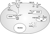
Ca2+ is an essential and versatile intracellular messenger. Normally low (submicromolar) levels of Ca2+ within the cytoplasm are interrupted by Ca2+ pulses that trigger Ca2+-dependent responses. Indeed, it can be argued that virtually all reactions in excitable cells are regulated either directly or indirectly by Ca2+ (Augustine, Santamaria & Tanaka, 2003). The time-scale over which Ca2+-dependent regulation operates ranges from microseconds to hours, and Ca2+ signals are often highly localized (reviewed by Bootman, Lipp & Berridge, 2001; Augustine et al., 2003). Cells have developed exquisitely tuned components for temporal and spatial regulation of free Ca2+ levels in the cytoplasm (reviewed by Berridge, Bootman & Roderick, 2003). These mechanisms comprise a ‘Ca2+-signalling toolkit’ consisting of a large and diverse collection of signalling units that, alone or in combination with one another, distribute Ca2+ signals with varying temporal and spatial properties. These signalling units include receptors, channels, pumps and exchangers, Ca2+ buffers, Ca2+-binding proteins, and Ca2+-sensitive enzymes and processes (see Fig.1).

Fig. 1. A subset of the components that regulate intracellular Ca2+ levels. Various receptors (R) act through second messengers to effect or modulate release of Ca2+ from the endoplasmic/sarcoplasmic reticulum (ER/SR) via IP3 receptors (IP3R) and ryanodine receptors (RyR). Voltage-gated Ca2+ channels (VGCCs) and other channels which are permeable to Ca2+ (CCs; e.g., ligand-gated channels, second-messenger-operated channels, TRP channels) also increase Ca2+ levels, both directly from entry of Ca2+ from the external medium and indirectly, since release of Ca2+ from internal stores is controlled by Ca2+ itself. Ca2+ is removed from the cytoplasm by various exchangers and pumps, including the plasma-membrane Ca2+-ATPase (PMCA) and the Na+/Ca2+ exchanger (NCX), which extrude Ca2+ to the outside. The sarco(endo)plasmic reticulum Ca2+-ATPase (SERCA) pumps Ca2+ back into intracellular stores. Ca2+ buffers (Buffers) bind a large proportion of cytoplasmic Ca2+, effectively removing it from the activating pool. Mitochondria also play an important role in regulating levels of Ca2+ in the cytoplasm. They rapidly sequester Ca2+ through a uniporter, as well as releasing Ca2+ through the NCX.
The primary pathways for transient increases in intracellular Ca2+ are via entry of extracellular Ca2+ or by release of intracellular stores of Ca2+. Major intracellular stores of Ca2+ are found in the endoplasmic reticulum (ER) and, in muscle cells, the sarcoplasmic reticulum (SR). Release of Ca2+ from these stores is mediated by inositol-1,4,5-triphosphate (IP3) receptors and by ryanodine receptors, calcium release channels encoded by two distantly-related gene families. Several ligands and secondary messengers, including Ca2+ itself, also influence Ca2+ release. Entry of Ca2+ from the external medium can be mediated by several components, including voltage- and receptor-gated channels, transient receptor potential (TRP) channels, and second messenger-gated channels (e.g., cyclic nucleotide-gated channels). One of the major gateways for entry of extracellular Ca2+ is through voltage-gated Ca2+ channels. This review will concentrate primarily on these channels, which, in addition to contributing to impulse propagation, couple membrane depolarization to rapid influxes of Ca2+ that can regulate fast Ca2+-dependent cellular responses.
CALCIUM SIGNALLING IN FLATWORMS
Several components that are essential for maintenance of Ca2+ homeostasis have been described in schistosomes and other platyhelminths (reviewed by Nöel et al. 2001), representing a subset of the cellular factors likely responsible for modulating intracellular Ca2+ levels in worms (see Fig. 1 in Redman et al. 1996). For example, sarco(endo)plasmic reticulum Ca2+-ATPases (SERCAs), intracellular ATP-powered pumps that sequester Ca2+ into the SR, have been identified in schistosomes. Two S. mansoni SERCAs can be distingushed physiologically (Cunha, Reis & Noël, 1996) and two different S. mansoni SERCA-like cDNAs have been reported (de Mendonça et al. 1995). Expression of one of these SERCA cDNAs, SMA2, rescues SERCA-deficient yeast, and is associated with an internal-membrane-associated, Ca2+-dependent ATPase activity with kinetic and pharmacological properties similar to mammalian SERCA isoforms (Talla et al. 1998).
Ryanodine receptors are gated by a ligand (Ca2+) and/or by mechanical coupling with voltage-gated Ca2+ channels, and are found in the ER of noncontractile tissues as well as in the SR of muscle cells (reviewed by Shoshan-Barmatz & Ashley, 1998; Rossi & Sorrentino, 2002). [3H]ryanodine binding (Silva et al. 1998) and pharmacological experiments on dissociated muscle fibres (Day et al. 2000) provide evidence for ryanodine receptors in schistosomes and other flatworms, and ESTs coding for homologues of this channel are found in the transcriptomes of both S. mansoni and S. japonicum.
Several receptors and channels on the plasma membranes of schistosomes likely also modulate levels of intracellular Ca2+. For example, both FMRFamide-related peptides (Day et al. 1994) and serotonin (Day, Bennett & Pax, 1994) alter Ca2+-dependent contraction in dissociated muscle fibres from S. mansoni. Recently, Agboh et al. (2004) have cloned and expressed an ATP-gated P2X receptor from S. mansoni that has high permeability to Ca2+ and may play an important role in Ca2+-dependent processes such as neurotransmission and muscle contraction. In addition, several Ca2+-binding proteins from schistosomes that have been described may serve to buffer or sequester Ca2+ as well as acting as targets of Ca2+ regulation. Information gleaned from the schistosome genome and transcriptome projects should over the next several years provide material for post-genomic studies of the cellular components that participate in regulation of Ca2+ homeostasis.
PRAZIQUANTEL
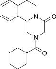
Praziquantel (Fig. 2), discovered in the 1970s, was subsequently introduced for the treatment of schistosomiasis (reviewed by Andrews et al. 1983). It is a pyrazinoisoquinoline with an asymmetric centre, and standard preparations are composed of equal proportions of the active, laevo (−) and the inactive, dextro (+) optical isomers. The activity of the (−) enantiomer has been established in experiments performed both in vivo (Andrews et al. 1983; Liu et al. 1986; Tanaka et al. 1989; Wu et al. 1991; Xiao et al. 1999) and in vitro (Staudt et al. 1992; Xiao & Catto, 1989).

Fig. 2. Chemical structure of praziquantel.
Praziquantel has activity against all species of schistosomes and shows minimal side effects. As a consequence, it has become the drug of choice against schistosomiasis. Indeed, with the added benefit of dramatic reductions in price, praziquantel has in essence become the sole antischistosomal agent that is available commercially (Fenwick et al. 2003; Hagan et al. 2004). Praziquantel is also active against other trematode and cestode infections, though generally not against nematodes (reviewed by Andrews, 1985), and schistosomes show stage- and sex-dependent differences in praziquantel sensitivity (Xiao, Catto & Webster, 1985; Sabah et al. 1986; Pica-Mattoccia & Cioli, 2004).
Praziquantel effectiveness has been proven repeatedly in large-scale schistosomiasis control efforts in different regions. However, the drug has been severely under-utilized in sub-Saharan Africa (reviewed by Fenwick et al. 2003; Hagan et al. 2004). The recently inaugurated Schistosomiasis Control Initiative (www.schisto.org) was launched as a response to this problem and has as its goal the establishment of sustainable schistosomiasis control programs in this neglected region.
Since praziquantel serves in effect as the only antischistosomal treatment in widespread use, the possibility of emerging drug resistance is troubling. Others (see e.g. Cioli, 2000; Doenhoff et al. 2002) have discussed this problem in detail. It is clear, however, that the potential for emerging resistance to praziquantel by schistosomes is of special concern considering that the molecular target and mode of action of the drug remain uncertain. Thus, despite the widespread use of praziquantel and nearly three decades of research, the exact mechanism of praziquantel action is still unresolved (reviewed by Day, Bennett & Pax, 1992; Redman et al. 1996; Harder, 2002; Cioli & Pica-Mattoccia, 2003). This review will focus on recent advances in identifying the molecular target of praziquantel, highlighting those experiments that point to a critical role for voltage-gated calcium (Ca2+) channel proteins.
Praziquantel acts selectively against members of the phylum Platyhelminthes. Accordingly, the molecular target (or targets) for praziquantel might be encoded by a novel gene (or genes) found exclusively in the flatworms. Schistosome genomes and transcriptomes contain several sequences that show no clear-cut homology with genes found in other phyla (reviewed by Hu et al. 2004; LoVerde et al. 2004; McManus et al. 2004; Verjovski-Almeida et al. 2004). On the other hand, the target for praziquantel might be a member of a gene family found in other phyla as well as in the platyhelminths, but with platyhelminth-specific structural signatures required for interaction with the drug. Even minor differences in critical domains of a protein, including single amino acid alterations, can have major consequences for the functional and pharmacological properties of typical receptors and channels (see e.g. Heinemann, Terlau & Imoto, 1992; Satin et al. 1992).
Though elucidating the mode of action of praziquantel has proved a daunting task, the effects of the drug on adult schistosomes do provide clues to potential targets for the drug. Praziquantel produces a well-documented effect on intracellular Ca2+ levels in adult schistosomes (reviewed by Andrews, 1985; Day et al. 1992; Redman et al. 1996). Within seconds of exposure to the drug, adult schistosomes exhibit a rapid, sustained contraction of the musculature (Fetterer, Pax & Bennett, 1980) and vacuolization and disruption of the parasite tegument (Becker et al. 1980; Mehlhorn et al. 1981), an effect associated with the subsequent exposure of parasite antigens on the surface of the worm (Harnett & Kusel, 1986). Both of these responses are thought to be linked to a praziquantel-dependent disruption of Ca2+ homeostasis (reviewed by Day et al. 1992; Redman et al. 1996).
Praziquantel elicits a rapid uptake of 45Ca2+ in adult male schistosomes (as well as a much slower influx of Na+; Pax, Bennett & Fetterer, 1978). The effects of praziquantel on both contraction of the worm musculature and disruption of the parasite tegument are Ca2+-dependent processes. Removal of Ca2+ from the medium blocks both responses (Pax et al. 1978; Wolde Mussie et al. 1982; Xiao et al. 1984). However, neither of these inhibitory effects appear immediately. For example, inhibition of the praziquantel-dependent contraction of the musculature requires at least 10 minutes to occur, a delay thought to correspond to the time required for depletion of sequestered intracellular Ca2+ stores. These results indicate that though extracellular Ca2+ is not required for the initiation of praziquantel-dependent action, it is required for maintenance of the response.
Based on comparisons between the praziquantel response in intact and detegumented parasites, it appears that both the tegument and the sarcolemma contain praziquantel-sensitive sites (Blair, Bennett & Pax, 1992). Thus, intact worms that are bathed in a medium with a high magnesium (Mg2+)[ratio ]Ca2+ ratio exhibit a praziquantel-dependent biphasic muscle contraction instead of the tonic contraction that occurs in standard media. Detegumented worms continue to respond to praziquantel, but they show only a single, pronounced phasic contraction in high Mg2+, indicating that a tegumental site is necessary for the full response. Furthermore, unlike intact worms, which show a transient response to praziquantel in Ca2+-free medium, application of praziquantel to detegumented worms in Ca2+-free medium produces no muscular contraction. Interestingly, praziquantel (1–2 μM) has been reported to interact with both sarcolemmal and intracellular sites to produce a sustained Ca2+-dependent contraction in the penile retractor muscle from the mollusc Lymnaea stagnalis (Gardner & Brezden, 1984).
The effects of praziquantel on Ca2+ homeostasis could point to a direct action of the drug on membrane permeability to Ca2+. However, early experiments indicated that praziquantel is not acting as a Ca2+ ionophore (Pax et al. 1978). On the other hand, it has been reported that praziquantel alters the structure of membrane bilayer phospholipids or membrane fluidity (Harder, Goossens & Andrews, 1988; Lima et al. 1994), which could result in changes in membrane permeability to Ca2+ or to indirect effects on membrane receptors and channels.
The target of praziquantel might also be one of the several cellular factors involved in regulating intracellular levels of Ca2+ discussed above and reviewed by Redman et al. (1996) To date, there is little direct evidence implicating or eliminating these factors as possible targets in praziquantel action. However, Cunha & Noël (1997) have reported that high concentrations of praziquantel (100 μM) have no effect on schistosome (Na+-K+)-ATPase or (Ca2+-Mg2+)-ATPase activities.
Recently, voltage-gated Ca2+ channels have been identified as candidate targets of praziquantel action (Kohn et al. 2001a, 2003a, b). As important entry sites for extracellular Ca2+, voltage-gated Ca2+ channels play a critical role in regulating levels of intracellular Ca2+. However, until recently, the role of voltage-gated Ca2+ channels in praziquantel action had not been tested directly, as Ca2+ currents had never been recorded from schistosome cells (see below). Nevertheless, pharmacological studies by Blair et al. (1992) on praziquantel-induced contraction in both intact and detegumented worms led them to suggest that Ca2+ channels might be involved in the action of the drug. Interestingly, high concentrations (50 μM) of praziquantel prolong the Ca2+-dependent plateau phase of the cardiac action potential in rats, which is carried by voltage-gated Ca2+ channels (Chubb et al. 1978). On the other hand, methoxyverapamil (D-600), an inhibitor of one class of mammalian Ca2+ channels (L-type), does not block the praziquantel-dependent Ca2+ influx in schistosomes, though it does block the tonic contraction of these cells resulting from elevated extracellular K+ concentrations (Fetterer et al. 1980). However, recent results from expression of cloned Ca2+ channel proteins indicates a significant role for voltage-gated Ca2+ channels in praziquantel action.
VOLTAGE-GATED CA2+CHANNELS
Voltage-gated ion channels, which are part of the ion channel superfamily, underlie electrical excitability in cells. They form an ion-selective pore through the membrane which, when activated by a change in membrane potential, allows ions to flow down the electrochemical gradient across the cell membrane. Voltage-gated channels include those selective for potassium, sodium, and Ca2+. Voltage-gated Ca2+ channels are membrane protein complexes that form Ca2+-selective pores gated by depolarization. Like other voltage-gated channels, Ca2+ channels contribute to impulse propagation, but they are also essential regulators of intracellular Ca2+ levels. By providing a pathway for rapid Ca2+ influxes, Ca2+ channels couple depolarization of the cell to a wide array of Ca2+-dependent responses including muscle contraction and secretion in muscles, nerves, and other excitable cells (Hofmann, Lacinova & Klugbauer, 1999; reviewed by Catterall, 2000). Thus, voltage-gated Ca2+ channels are essential to the behaviour and survival of the animal. This point is further underscored by the fact that organisms as diverse as fish-hunting cone snails and spiders produce toxins targeted against specific Ca2+ channel subtypes to immobilize prey.
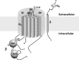
The pore-forming subunit of voltage-gated Ca2+ channels is the α1 subunit. In addition to the α1 subunit, voltage-gated Ca2+ channels typically contain associated auxiliary subunits that modulate the properties of the channel (see Fig. 3). The α1 subunit is made up of four linked homologous domains, each of which contains six transmembrane regions (S1–S6). The predicted structure of the α1 subunit fits within the ion channel superfamily (Doyle et al. 1998). The most basic ion channels are tetrameric structures comprised of the fifth and sixth transmembrane regions (S5 and S6, respectively) and the P loop, a region between S5 and S6 that forms the selectivity filter of the pore. The residues that define a channel's ionic selectivity and many of its pharmacological properties reside in this region. In the voltage-gated channels, the fourth transmembrane segment within each domain (S4) contains a positively charged amino acid (lysine or arginine) at every third residue and is thought to serve as the voltage sensor of the channel.

Fig. 3. Structure of voltage-gated Ca2+ channels. This simplified representation shows the pore-forming α1 subunit, which consists of four homologous domains surrounding a central pore. Domains II–IV are shown, and Domain I is cut away to show the structure. Each of the four domains is composed of six transmembrane regions (S1–S6). The P loop dips into the membrane between S5 and S6, forming the selectivity filter of the channel. The S4 transmembrane region contains a series of regularly spaced, positively charged residues (+) and is thought to form the voltage sensor of the channel. Also shown are associated β and α2δ subunits, which modulate α1 function. The γ subunit has been omitted. Domains of the β subunit defined by recent homology modeling and high resolution structural analysis (see text) are shown. The β subunit interacts via its guanylate kinase (GK) domain with the Alpha Interaction Domain (AID) on the I–II loop of the α1 subunit. The size of subunits and domains are not to scale in this cartoon.
Two major classes of Ca2+ currents that have been characterized in both vertebrate and invertebrate cells are Low Voltage Activated (LVA; t-type) and High Voltage-Activated (HVA). HVA currents can be further subdivided into L-type, which, in vertebrates, are sensitive to the dihydropyridine class of Ca2+ channel antagonists (nifedipine, nimodipine, etc.) and a variety of dihydropine-insensitive currents which are collectively known as non-L-type. Heterologous expression of cloned Ca2+ channel subunits has been used to demonstrate a correspondence between these different Ca2+ currents and particular subtypes of Ca2+ channel α1 subunits. Thus, LVA currents are gated by the Cav3 class of α1 subunits (reviewed by Perez-Reyes, 2003), while HVA L-type currents and non-L-type currents are gated by Cav1 α1 subunits and Cav2 α1 subunits, respectively. Similar studies have indicated that the pharmacological differences found between vertebrate L-type and non-L-type channnels (e.g. in dihydropyridine sensitivity) are not as clear-cut in invertebrates (reviewed by Jeziorski, Greenberg & Anderson, 2000a). Thus, expression of invertebrate α1 subunits that, based on structure, are clearly members of the L-type family, produces currents that are relatively insensitive to dihydropyridines and other potent modulators of vertebrate L-type channels.
In the HVA Ca2+ channels, the α1 subunit is associated with and modulated by auxiliary subunits (reviewed by Hofmann et al. 1999; Catterall, 2000; Arikkath & Campbell, 2003) that include α2/δ and β subunits (Fig. 3), as well as γ subunits (not shown). Ca2+ channel β subunits (Cavβs) are cytoplasmic proteins that have been studied extensively and are critical components of Ca2+ channel complexes. When coexpressed with β subunits, α1 subunits show increases in current density and ligand binding. Cavβs appear to play a role in membrane trafficking of the α1 subunit, likely in part by masking an ER retention site on the α1 subunit (Bichet et al. 2000). β subunits also affect a variety of the biophysical properties of Ca2+ channels, including the voltage-dependence of channel activation and steady state inactivation, rates of inactivation (Birnbaumer et al. 1998; reviewed by Walker & DeWaard, 1998; Hanlon & Wallace, 2002; Dolphin, 2003), and the rate of recovery from inactivation (Jeziorski et al. 2000b).
The primary site on the α1 subunit for binding of Cavβs is the Alpha Interaction Domain (AID; Pragnell et al. 1994), an 18 amino acid region in the intracellular loop between Domains I and II of the α1 subunit. Recent X-ray crystallographic studies (Chen et al. 2004; Opatowski et al. 2004; Petegem et al. 2004) indicate that the AID indeed does complex with Cavβs, forming an amphipathic helix with the most highly conserved residues on one side of the helix in contact with a complementary groove on the β subunit.
Using homology modelling, Hanlon et al. (1999) proposed that Cavβs are members of the membrane-associated guanylate kinase (MAGUK) family of proteins. MAGUKs are scaffolding proteins, often concentrated at synapses where they play important roles in clustering of ion channels and neurotransmitter receptors (reviewed by Dimitratos et al. 1999). Typically, MAGUKs contain one or more PDZ domains located N-terminal to a Src-homology 3 (SH3) domain, a bridging region (the HOOK domain), and a guanylate kinase (GK)-like domain. Resolution of the crystal structure of the conserved core of β subunits, both alone and in complex with the AID of the α1 subunit, are consistent with Cavβs indeed being members of the MAGUK family, though with distinct characteristics (Chen et al. 2004; Opatowski et al. 2004; Petegem et al. 2004; commentary by Yue, 2004). For example, the PDZ domains typically found in other MAGUKs appear to be absent in Cavβs, and the orientation of the SH3 and GK domains has been modified. In addition, the GK domain of Cavβs does not contain a nucleotide-binding site. Instead, the modified GK domain forms a deep hydrophobic groove (the AID-binding pocket, or ABP) with which the AID of the α1 subunit interacts.
Several lines of evidence indicated that a highly conserved ~35 amino acid region in Cavβs dubbed the Beta Interaction Domain (BID) serves as the primary site for β subunit interaction with the α1 subunit (DeWaard, Pragnell & Campbell, 1994). However, the recent crystal structures have cast doubt on this hypothesis. Instead, the BID region is found buried within the β subunit protein, and is therefore unlikely to be involved directly in protein-protein interactions such as binding to the AID. However, as Chen et al. (2004) have noted, the BID nonetheless appears to play an essential structural role in β subunits, spanning the SH3 and GK domains and their connecting HOOK region and containing two β-strands that are integral parts of the SH3 and GK domains. Indeed, the BID and surrounding areas are particularly highly conserved regions of β subunit proteins.
SCHISTOSOME AND OTHER FLATWORM Ca2+CURRENTS
Do voltage-gated Ca2+ channels in schistosomes play a role in praziquantel action? Are they the molecular targets of praziquantel? These questions may be answered by characterizing the structure, function and pharmacological sensitivities of Ca2+ channels from schistosomes and other flatworms. These studies may additionally provide insights into the physiology of excitable cells in platyhelminths, about which relatively little is currently known, and should also provide clues about the evolution of ion channels. Furthermore, structural and functional characterization of flatworm Ca2+ channels might provide highly specific targets for new antiparasitic agents. For example, a recent patent (Walter & Kuris, 2003; see also Bonn, 2004) claimed that high concentrations of verapamil and nifedipine, two compounds that block L-type voltage-gated Ca2+ channels in vertebrates, suppress egg production in S. mansoni and another trematode, Echinostoma caproni. Clearly, a more thorough understanding of the pharmacological properties of platyhelminth Ca2+ channels will be required to determine whether these compounds are acting on worm voltage-gated Ca2+ channels or other targets.
Isolated muscle cells from S. mansoni exhibit Ca2+-dependent contractility (Day, Bennett & Pax, 1994). To date, however, there have been no published reports of native Ca2+ currents from schistosome cells. Indeed, Day et al. (1993) found no inward currents of any type in their voltage-clamp analysis of muscle cells isolated from adult S. mansoni, most likely because of technical limitations with these cells.
In contrast, Ca2+ currents have been recorded from other flatworms. For example, neurons from the polyclad flatworm Notoplana acticola contain a normal complement of ionic currents that are implicated in the generation of action potentials. These include cadmium-sensitive Ca2+ currents (Keenan & Koopowitz, 1984). Similarly, using voltage-clamp analysis, voltage-gated Ca2+ currents have been recorded in nerve and muscle cells of Bdelloura candida, a triclad ectoparasitic flatworm that resides on the legs and gills of horseshoe crabs (Limulus polyphemus; Blair & Anderson, 1993, 1994). Both cell types possess Ca2+ currents that activate at −30 mV, reach peak amplitude in approximately 5 ms, and inactivate slowly. The neuronal Ca2+ current shows relatively little sensitivity to organic Ca2+ channel blockers such as nifedipine and verapamil, and is not blocked by the cone snail toxin ω-conotoxin GVIA. This neuronal Ca2+ current also exhibits no sensitivity to 10 μM praziquantel (Blair & Anderson, 1996). Unfortunately, the muscle Ca2+ current was too unstable for determination of pharmacological sensitivities. More recently, Cobbett & Day (2003) recorded Ca2+ currents from muscle cells of the triclad turbellarian Dugesia tigrina. However, these currents were also too unstable for systematic pharmacological analysis.
Clearly, determining the properties of native Ca2+ currents in schistosomes and other flatworms presents major challenges. These difficulties may be bypassed by using a molecular approach. Expression of cloned Ca2+ channel subunits from schistosomes and other flatworms in heterologous systems may present an alternative means of elucidating the physiological properties and pharmacological sensitivities of these channels. However, analysis of native currents within schistosome cells will eventually be necessary for a thorough understanding of the properties of these channels and their physiological roles within the parasite.
SCHISTOSOME Ca2+CHANNEL SUBUNITS
Adult schistosomes express at least three subtypes of HVA Ca2+ channel α1 subunits (Kohn et al. 2001b). Phylogenetic analysis shows that two of these subtypes cluster with the non-L-type class of HVA α1 subunits, while the third is most closely related to the L-type class of α1 subunit. Although mammals contain multiple subtypes of L-type and non-L-type α1 subunits, other invertebrates examined to date contain only a single representative of each of these classes (Littleton & Ganetzky, 2000; Jeziorski et al. 2000a). Schistosomes (and presumably other platyhelminths) therefore appear to be unique among the invertebrates in that they have two subtypes of non-L-type α1 subunits rather than one.
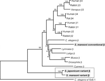
Schistosomes also express at least two subtypes of Cavβs (Kohn et al. 2001a, 2003b). This finding is also unprecedented among the invertebrates; to date, only a single β subunit gene has been identified in the genomes of other invertebrate species. Even more noteworthy, one of these schistosome β subunit subtypes has particularly unusual structural features and functional properties. To date, no representatives of this variant subtype have been identified in any vertebrates or invertebrates other than the platyhelminths (see Fig. 4).

Fig. 4. Phylogenetic tree of Ca2+ channel β subunits. Amino acid sequences were aligned using Clustal X (Thompson et al. 1997), and a tree constructed using the neighbour-joining method, as implemented in MEGA 2.1 (Kumar et al. 2001). The schistosome conventional and variant β subunits are shaded, and the variant β subunits are boxed with a dashed line. Numbers represent bootstrap values (1000 replications). Sequences and NCBI accession numbers are: Human β1, NP_954856; Rabbit β1, AAA31180; Human β2, NP_000715; Rabbit β2, CAA45576; Human β3, NP_000716; Rabbit β3, CAA45578; Xenopus laevis (toad) β3, AAA75519; Human β4, NP_000717; Rat β4, A45982; Caenorhabditis elegans (nematode) β, AAB53056; Schistosoma mansoni conventional β, AY033599; Lymnaea stagnalis (snail) β, AAO83844; Loligo bleekeri (squid) β, BAB88219; Musca domestica (housefly) β, A54844; Drosophila melanogaster (fruitfly) β, AAF21096; Cyanea capillata (jellyfish) β, AAB87751; S. japonicum variant β, AAK51116; S. mansoni variant β, AAK51117; C. elegans β-like sequence (w10c8.1), AAK21500.
The variant Cavβ subtype proteins are clearly part of the broader β subunit family, although they have very distinct structural features. For example, they are from 25%–50% larger than other β subunits, including the conventional S. mansoni Cavβ. However, the most striking feature of these variant Cavβs can be found in the BID, where two serine residues that are conserved in other Cavβs and that correspond to consensus protein kinase C (PKC) phosphorylation sites are replaced by cysteine and alanine (Fig. 5).

Fig. 5. Comparison of the amino acid sequence of the BID from the variant schistosome β subunits with a consensus β subunit BID sequence. The cysteine and alanine residues which substitute in the variant BIDs for the conserved serines are shaded. The two consensus PKC sites conserved in the consensus BID sequence are underlined, with the serines in bold.
The variant schistosome β subunits also exhibit distinctive functional properties (Kohn et al. 2001a). Conventional β subunits enhance currents when coexpressed with α1 subunits. In contrast, coexpression of a variant β subunit in Xenopus oocytes with a jellyfish (CyCav1) or human (Cav2.3) α1 subunit results in a dramatic reduction in current compared to the levels found when the α1 subunit is expressed alone. Yet, other than this novel effect on current levels, the modulatory effects of the variant schistosome Cavβs are similar to those of conventional Cavβs. For instance, like other Cavβs, the variant β subunits shift the current/voltage relationship of α1 subunits in a hyperpolarizing direction. Thus, although the variant Cavβs have some highly unusual properties, they appear to behave in most respects as genuine, functional β subunits.
A particularly exciting property of the variant schistosome Cavβs is that they can confer praziquantel sensitivity to an otherwise praziquantel-insensitive mammalian α1 subunit (Kohn et al. 2001a). When the mammalian Cav2.3 α1 subunit is expressed alone in Xenopus oocytes, it does not respond to 100 nM praziquantel. On the other hand, when Cav2.3 is coexpressed in Xenopus oocytes with one of the variant schistosome Cavβs, peak currents are increased 1·5- to 2-fold in the presence of 100 nM praziquantel. Other Cavβs, including the conventional schistosome β subunit, do not confer praziquantel sensitivity (Kohn et al. 2003b). This praziquantel-dependent increase in Ca2+ influx found with Ca2+ channels containing the variant β subunit is consistent with the effects of the drug on Ca2+ homeostasis in schistosomes and implicates the variant schistosome Cavβs in the action of praziquantel.
Using site-directed mutagenesis, the specific amino acid residues involved in determining the unusual functional properties and pharmacological sensitivities of the variant β subunits have been localized. Both the reduction in current levels and the responsiveness to praziquantel map to the two conserved consensus PKC sites in the BID that are not present in these variant β subunits (Kohn et al. 2003a,b). If serine residues are substituted at either or both of these sites in the variant Cavβ from S. mansoni the consensus PKC sites are restored (see Fig. 5). This mutated β subunit, like conventional Cavβs, now enhances currents through α1 subunits and does not confer sensitivity to praziquantel. A subunit with a double mutation containing a serine at one of these sites, but also containing a second mutation that eliminates the consensus PKC sequence, behaves like the wildtype schistosome subunit. Thus, the absence of PKC sites in the BID region, rather than simply the presence of residues other than serines, appears to be responsible for the novel effects of the variant subunit. Similarly, elimination of both PKC sites in the BID of a conventional β subunit (mammalian β2a) results in a β subunit that can now confer praziquantel sensitivity to the mammalian α1 subunit (Kohn et al. 2003b).
Thus, the absence of consensus PKC sites in the β subunit BID is associated with the capability of a β subunit to confer susceptibility to praziquantel, while the presence of a single consensus PKC site in the BID is sufficient to transform a variant Cavβ into a β subunit with characteristics typically found for conventional β subunits. Based on these results, we have hypothesized that the unusual modulatory properties and pharmacological sensitivities of the variant Cavβs from schistosomes are dependent on the absence of the consensus PKC phosphorylation sites found in the BIDs of other β subunits.
Phosphorylation of voltage-gated Ca2+ channel subunits by PKC and other protein kinases plays a central role in regulating channel properties (reviewed by Rossie, 1999; Kamp & Hell, 2000; Keef, Hume & Zhong, 2001). A recombinant mammalian β2a subunit has been shown to be phosphorylated by PKC, with a stoichiometry estimated to be 1–2 moles of phosphate per mole of β2a protein (Puri et al. 1997). There are several consensus PKC sites in β subunits, but the exact sites that are phosphorylated by PKC have yet to be defined either in vitro or in vivo.
CONCLUSIONS AND FUTURE QUESTIONS
The variant Cavβs found in schistosomes appear to be involved in praziquantel action, while the addition of a PKC site in the BID of these subunits can abolish susceptibility to the drug. It therefore follows that one mechanism for schistosomes to acquire praziquantel resistance might be by acquiring one or both of these PKC sites by mutating the cysteine or alanine residues to serine. Schistosome isolates with reported reduction in praziquantel sensitivity have been tested for the presence of these (and other) mutations (Valle et al. 2003). These isolates did not exhibit changes in primary structure of schistosome β subunits, nor did they show changes in expression levels of those subunits. Thus, a reduction in praziquantel susceptiblity in these strains apparently does not depend on altered Cavβ structures or expression levels. However, since various Egyptian isolates with reduced praziquantel susceptibility show differential stability (William et al. 2001), there may be alternative pathways for acquiring praziquantel resistance, with different strains of parasites acquiring reduced susceptibility to praziquantel via a variety of mechanisms.
There are several unresolved issues regarding schistosome Ca2+ channels and their role in praziquantel action. Of primary importance is the need to characterize schistosome Ca2+ channel α1 subunits, either alone or in combination with the two schistosome Cavβ subtypes. These experiments will answer several questions, including whether all possible schistosome α1/β combinations are capable of forming functional channels. More specifically, are only certain α1/β combinations capable of forming praziquantel-sensitive channels, or can any schistosome channel that contains a variant β subunit respond to the drug? Furthermore, which schistosome cells express these combinations? Once specific praziquantel-sensitive channels are identified, the question of precisely how praziquantel acts on these channels will still remain. For example, praziquantel might be interacting directly with the variant β subunit, or it might instead interfere with α1/β subunit interactions, or it could be acting indirectly, via effects on interacting proteins or on the membrane itself to affect Ca2+ channels that contain these variant Cavβs. Finally, what are the downstream players in the cascade of events that ultimately leads to paralysis and tegumental disruption? As described above, the components that regulate Ca2+ homeostasis in cells are connected with one another through a network of interaction and regulation. The components comprising these interdependent networks might also be affected by praziquantel. In schistosomes, these interactions are further complicated by the complexity of the adult tegument and sub-tegumental compartments (see Fig. 1 of Redman et al. 1996). The tegument of the adult is formed by a membrane that has a double-bilayer structure (McClaren & Hockley, 1977) and is electrically coupled to underlying muscle cells (Thompson, Pax & Bennett, 1982).
Independent of their role in praziquantel action, the biological function of these variant schistosome Cavβs is particularly intriguing. All other known β subunits enhance Ca2+ currents, while these variant β subunits inhibit currents. Are there special characteristics of schistosome α1 subunits or other components of the Ca2+ signalling system that might favour the emergence of this unusual type of Ca2+ channel modulation by β subunits? Are there any other organisms that have developed a similar strategy? Answers to some of these questions may result from expression of channel subunits in heterologous systems, while other insights may come from the schistosome genome and transcriptome. Ultimately, however, a full understanding of the role played by Ca2+ channels in schistosome physiology, Ca2+ signalling and praziquantel action will require a thorough understanding of the properties of native schistosome Ca2+ (and other) currents.
ACKNOWLEDGEMENTS
I thank Lynn Milstead for help with preparation of figures. RMG is supported by NIH grant #AI 40522, by the Woods Hole Center for Oceans and Human Health, and by the Neal Cornell Research Fund at the Marine Biological Laboratory.