Introduction
Prosorhynchoides gracilescens (Rudolphi, 1819) (syn. Bucephaloides gracilescens) belongs to the family Bucephalidae within the subclass Digenea. Recently the species was divided into P. borealis Bartoli, Gibson & Bray, 2006 and P. gracilescens (sensu stricto) for the North-East Atlantic and the Mediterranean forms of the parasite, respectively (Bartoli et al., Reference Bartoli, Gibson and Brey2006). The only hitherto known first intermediate host of P. gracilescens (sensu lato) is the white furrow shell Abra alba (Wood, 1802) (Bivalvia: Semelidae), in which infective cercariae develop within sporocyst tubules (Matthews, Reference Matthews1974). Definitive hosts of P. gracilescens (s. l.) are angler fish (Lophius piscatorius Linnaeus and L. budegassa Spinola) in which adult digeneans inhabit the digestive tract (Afonso-Dias & MacKenzie, Reference Afonso-Dias and MacKenzie2004). Prosorhynchoides gracilescens (s. l.) is found in the North-East Atlantic Ocean and adjacent seas, including the Mediterranean, the Barents Sea and Black Sea. Fish species within the families Gadidae, e.g. cod (Gadus morhua) and haddock (Melanogrammus aeglefinus), Merlucciidae and Lotidae (all in order Gadiformes) and bullrout (Myoxocephalus scorpius, Scorpaeniformes) act as natural second intermediate hosts (Karlsbakk, Reference Karlsbakk1995; Køie, Reference Køie2000; Ruus et al., Reference Ruus, Skaare and Ingebrigtsen2001). The occurrence of the species in the second intermediate hosts coincides with the geographical distribution of the final hosts (Køie, Reference Køie1984; Bartoli et al., Reference Bartoli, Gibson and Brey2006).
Matthews (Reference Matthews1968, Reference Matthews1974) described the larval stages, sporocysts and cercariae, of P. gracilescens (s. l.) from the first intermediate host A. alba from Scottish waters and elucidated the life cycle of the species. Since then P. gracilescens (s. l.) sporocysts and cercariae have only been reported by Johnston & Halton (Reference Johnston and Halton1982), also from A. alba off Scotland. The distribution of A. alba extends from the Norwegian Sea and the Baltic, south to the Iberian Peninsula, into the Mediterranean and Black Seas, and south along the coast of Africa to Senegal (Tebble, Reference Tebble1966). It is not found in Icelandic waters.
In 1994 P. gracilescens (s. l.) was discovered for the first time in Icelandic waters when metacercariae of the species were found in cod and haddock (Eydal et al., Reference Eydal, Bambir and Helgason1995). In the years that followed we conducted further studies to elucidate which host species play a part in the life cycle of this parasite in Icelandic waters. This included studies on the final host, which showed 97% infection prevalence in angler fish (Eydal & Ólafsdóttir, 2002–Reference Eydal and Ólafsdóttir2003) and screening for metacercariae in gadoid fish around Iceland (Eydal et al., Reference Eydal, Helgason, Kristmundsson and Bambir1999).
The present paper reports the adult stage of P. borealis from angler fish in Icelandic waters and infections with larval stages of the parasite (sporocyst tubules containing cercariae) and histopathology in the bivalve species Abra prismatica (Montagu, 1808) (Mollusca: Semelidae). Abra alba, the only previously described first intermediate host, is not found around Iceland, but two alternative Abra species are present in Icelandic waters, A. prismatica found at depths of 15–164 m and A. nitida (Müller, 1776) (Mollusca: Semelidae) at depths of 3–326 m. They are found on mud and sandy seabeds. Abra prismatica is common south and west of Iceland and A. nitida is confined to the south and south-western coast of the country (Madsen, Reference Madsen1949).
To confirm the identity of the digenean parasite from Icelandic waters the small subunit ribosomal DNA (SSU rDNA) was amplified and sequenced. To confirm that all three life cycle stages (cercaria, metacercaria and adults) found in the various hosts in Icelandic waters represent the same species, the internal transcribed spacer 1 (ITS1), 5.8S and internal transcribed spacer 2 (ITS2) parts of the rDNA were sequenced and compared.
Materials and methods
Prosorhynchoides specimens from Lophius piscatorius
We re-examined mounted Prosorhynchoides specimens from the intestine of L. piscatorius, sampled at depths of 152–170 m south of Iceland in 1995, location 63°54′N–15°07′W (Eydal & Ólafsdóttir, 2002–Reference Eydal and Ólafsdóttir2003). The digeneans were serially dehydrated in 70%, 90%, 99% and 100% ethanol, stained in aceto-carmine, cleared in xylol and mounted in Canada balsam. A total of 20 ovigerous specimens were examined. Morphometrical diagnostic features used to differentiate the two species, P. gracilescens (s. str) and P. borealis (see Bartoli et al., Reference Bartoli, Gibson and Brey2006) were determined. Eggs (n= 50) were measured only if they were uncollapsed and clearly in a horizontal position. The digeneans were examined using differential interference contrast (DIC) microscopy (Leica DMLB, Leica Microsystems, Wetzlar, Germany).
Bivalve collection
Bivalves were collected from a sandy seabed close to the island of Heimaey, off the south coast of Iceland (within the area 63°30′N–20°12′W, 63°30′N–20°35′W, 63°20′N–20°12′W, 63°20′N–20°35′W) using either a van Veen grabber or a bottom sledge. These tools were used to collect bottom samples at about 100 stations. A total of 107 A. prismatica specimens were sampled at depths between 34 and 93 m: 24 in May–June 1996, 41 in late September 1999 and 42 in early October 2000. A limited number of A. nitida bivalves (n= 18) were found in proximal sampling sites to the south-east of this area (Háfadýpi) in late July 1997 and 1998 at depths of about 70 m.
Examination of fresh bivalve material
Live bivalves were dissected and examined for the presence of bucephalid digenean sporocysts and cercariae under a stereoscopic microscope. Live cercariae were put on a slide glass with a coverslip greased under the corners to hamper flattening of the cercariae. The cercariae were examined under a compound microscope. This included measurements as described by Matthews (Reference Matthews1968, Reference Matthews1974), including the body, penetration organ, pharynx and furcae and, in addition to the original description, the tail stem and the intestinal caecum were also measured. Length of furcae was measured by drawing a straight line from the tail stem to the tip of furcae. In order to further examine the distribution of the parasite within the host, A. prismatica specimens were fixed in Davidson's seawater fixative. Subsequently, 4 μm paraffin sections were prepared, using conventional histological methods, and stained with Giemsa for microscopic examination.
Molecular analyses of the ribosomal RNA gene
Samples of all three life cycle stages (cercariae, metacercariae and adults) of the Prosorhynchoides digenean found in hosts in Icelandic waters were examined and fixed in 95% ethanol for the DNA study. Cercariae specimens were isolated from the A. prismatica specimens described above. Metacercariae were dissected from cysts sampled from the cauda of two Atlantic cod (Gadus morhua L.) caught in Faxafloi, West Iceland in May 2010. Adult digeneans were isolated from the intestines of four L. piscatorius sampled close to the island of Heimaey, off the south coast of Iceland, in June 2008.
Total DNA was extracted using a GeneMATRIX kit (EURx, Gdansk, Poland) following the tissue protocol and used as template DNA for polymerase chain reactions (PCR). The universal SSU rDNA primers 18e, 390f, 870f and 18gM (Freeman et al., Reference Freeman, Yokoyama and Ogawa2004; Freeman & Ogawa, Reference Freeman and Ogawa2010) were used to amplify the SSU rDNA from metacercariae and adult samples. The primers BD1 and BD2 (Chen et al., Reference Chen, Wang, Yao and Nie2007) were used to amplify the ITS1, 5.8S and ITS2 region of the rDNA and the nested primers BD1-IF 5'TTGGTGCGGCTGTCGGCAACG3' and BD2-IR 5'TCGCACACATGCACCCTGGTC3' were designed to amplify an internal region of the BD1/BD2 amplicon, to confirm sequence identity for samples with low template concentrations (cercariae). PCR conditions were as described previously (Freeman et al., Reference Freeman, Yokoyama and Ogawa2004; Chen et al., Reference Chen, Wang, Yao and Nie2007; Freeman & Ogawa, Reference Freeman and Ogawa2010); for the nested PCR (BD1-IF/BD2-IR) an initial denaturation at 95°C for 4 min preceded 35 cycles of amplification (denaturation at 94°C for 30 s, primer annealing at 58°C for 45 s, and extension at 72°C for 1 min), followed by a 7 min terminal extension at 72°C. PCR bands of the correct size were recovered from the PCR products using a GeneMATRIX PCR products extraction kit (EURx Poland). PCR reactions were performed in triplicate (three separate DNA extractions) and sequencing reactions were performed using BigDye™ terminator cycle sequencing chemistry, utilizing the same oligonucleotide primers that were used for the original PCRs. DNA sequencing was performed in both forward and reverse directions for all PCR products and contiguous sequences obtained manually using CLUSTAL_X (Thompson et al., Reference Thompson, Gibson, Plewniak, Jeanmougin and Higgins1997) and BioEdit (Hall, Reference Hall1999). All DNA sequences obtained were compared to sequences available in the GenBank databases using nucleotide–nucleotide BLAST searches (Altschul et al., Reference Altschul, Gish, Miller, Myers and Lipman1990) to verify a digenean origin.
Voucher specimens
The following P. borealis specimens were deposited in The Icelandic Institute of Natural History, Reykjavik, Iceland: adults (catalogue number: NI-2203), cercariae (catalogue number: NI-26 260) and metacercariae (catalogue number: NI-26 261).
Results
Adult digeneans from Lophius piscatorius
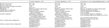
The Prosorhynchoides specimens from Icelandic waters were identified as P. borealis on the grounds of morphometric features (table 1), which are in agreement with the description of the species made by Bartoli et al. (Reference Bartoli, Gibson and Brey2006). Additional description of the specimens: mean body length is 1.81 mm, range 1.1–2.8 mm; mean body width is 0.61 mm, range 0.35–1.00 mm; mean size of rhynchus is 209 × 230 μm, range 138–290 × 162–300 μm; mean pharynx size is 126 × 145 μm, range 90–140 × 90–195 μm; mean egg size is 29.0 × 18.3 μm, range 25–33 × 15–21 μm. In 5 out of 20 specimens the size of the pharynx could not be determined and therefore the rhynchus/pharynx length ratio was only calculated for 15 individuals. In seven individuals ovary–anterior testis and anterior testis–posterior testis configuration could not be verified.
Morphometric diagnostic features used to differentiate between the two species of Prosorhynchoides from Lophius piscatorius. Results of the present study (first column) are compared with those of Bartoli et al. (Reference Bartoli, Gibson and Brey2006) (second and third columns) (all measurements in micrometres).

% value denotes the percentage of specimens with that feature.
Cercariae from bivalve molluscs
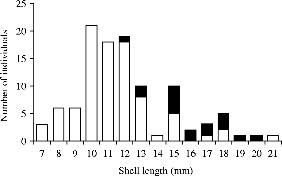
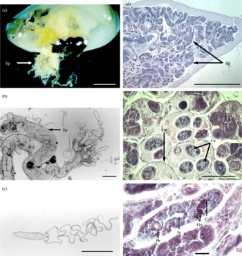
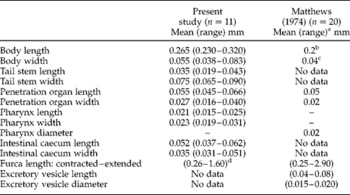
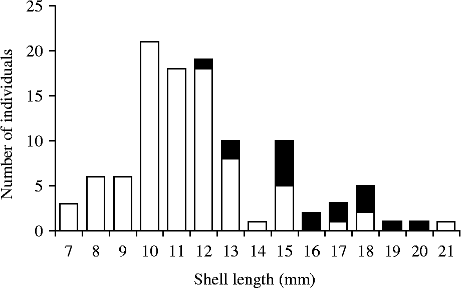
The shell lengths of A. prismatica and A. nitida ranged from 7 to 21 mm (mean 12.1 mm) and from 8 to 11 mm (mean 9.5 mm), respectively. The largest A. prismatica specimens attained the maximum size reported for the species from Iceland, but A. nitida individuals did not reach the maximum size (15 mm) reported from Iceland (Madsen, Reference Madsen1949). No bucephalid larvae were found in A. nitida, but 17 of 107 A. prismatica individuals (prevalence 16%) were found to be infected with Prosorhynchoides larval stages, sporocysts and cercariae, which were identified as P. borealis. Infected individulas were sampled from any depth within the range 34–93 m. Only larger bivalves (12–20 mm long, median = 16 mm) were infected, with an infection prevalence of 44% among the 39 specimens in this size category (fig. 1). No A. prismatica individuals collected in the year 1996 (n = 24) were parasitized, but three infected bivalves were obtained in 1999 (n = 41) and 14 in the year 2000 (n = 42). None of the bivalves sampled in May and June were infected, but the prevalence of infection was 7% and 33% in specimens sampled in September and October, respectively. The size distribution and average length of bivalves collected was similar each year; 13, 19 and 22 bivalves were 12 mm or longer, in May/June, September and October respectively. After separation of the shell valves, sporocyst tubules were often seen macroscopically protruding from internal organs (fig. 2a). Microscopy showed sporocysts filled with cercariae at different developmental stages, i.e. germinal balls, early cercariae without furcae, early cercariae with developing furcae and fully developed cercariae with very long contractile furcae (fig. 2b,c and fig. 3). Measurements of fully developed infective cercariae and comparison with original descriptions of P. gracilescens (s. l.) (i.e. P. borealis) are given in table 2.
Frequency distribution of non-infected (white bars) (n= 90) and infected (black bars) (n= 17) A. prismatica with P. borealis sporocysts and cercariae, in relation to bivalve size.
Prosorhynchoides borealis larval stages from the new host species A. prismatica; (a–c) fresh material, (d–f) histological transverse sections stained with Giemsa. (a) Sporocysts (arrow), a white mass, composed of many tubules, protruding from the internal organs of A. prismatica (scale bar 3 mm). (b) Sporocyst tubules (arrow) filled with cercariae at different developmental stages, some released from the ruptured tubule (scale bar 200 μm). (c) Fully developed infective cercaria (scale bar 200 μm). (d) Numerous sporocysts filled with cercariae occupy the haemocoel of the gonads in the entire bivalve foot (scale bar 1 mm). (e) Higher magnification of the foot (scale bar 100 μm). (f) Section of a cercaria in the bivalve foot (scale bar 25 μm). C, Cirrus; Ce, cercariae; Go, gonads; In, intestine; Ph, pharynx; Sp, sporocysts; Sw, sporocyst wall. (A colour version of this figure can be found online at http://www.journals.cambridge.org/jhl)

Dimensions of P. borealis cercariae and internal organs compared to original data of Matthews (Reference Matthews1974).

a Range was only given for furcae and excretory vesicle by Matthews (Reference Matthews1974).
b When contracted 0.08 mm, when extended 0.5 mm.
c When contracted 0.08 mm, when extended 0.02 mm.
d Extended furcae were observed to reach a length of even more than 1.60 mm, but were not measured in that state.
In parasitized A. prismatica many organs were infected; fig. 2d and e show histological sections from the foot. Extensive sporocyst infection in the haemocoel of the foot has caused mechanical muscle damage with subsequent degeneration and necrosis. Other tissues, such as the digestive gland, nephridia, gills and intestine, were less heavily infected. Only focal necrosis was observed in the digestive gland, nephridia and gills, and local atrophy in the intestine. Cercariae were also observed in the lumen of both the stomach and intestine. The arrangement of internal organs of a cercaria in histological section is shown in fig. 2f. The gonads and cirrus are well developed at this stage. The unicellular layers of the walls of the pharynx and intestine are distinct.
Molecular analyses of the ribosomal RNA gene
The SSU rDNA sequences amplified from metacercariae (from G. morhua) and adult (from L. piscatorius) samples of Prosorhynchoides were found to be identical to each other over the entire length of 1927 base pairs. BLAST searches of this SSU sequence showed a >99.9% similarity (1914/1915 of comparable sequence data) to P. gracilescens (AJ228789) which was derived from L. piscatorius taken in the North Sea, UK (Littlewood et al., Reference Littlewood, Rohde and Clough1998). The ITS1, 5.8S and ITS2 sequences amplified from metacercariae and adult samples of Prosorhynchoides were also found to be identical to each other over a length of 1014 base pairs. The nested PCR successfully amplified DNA from cercariae, metacercariae and adults of Prosorhynchoides, which all showed 100% identity over 734 bases of comparable sequence data. BLAST searches of this gene region showed the highest similarity to Bucephalus spp., ranging from 88 to 91% identity.
The novel sequences generated in this study have been submitted to GenBank with the following accession numbers: GenBank BankIt submission ID: 1462548 (accession numbers JN182208–JN182212).
Discussion
Comparing the Prosorhynchoides specimens from our study in Iceland to the description of P. borealis from the North Sea (Bartoli et al., Reference Bartoli, Gibson and Brey2006) shows clear similarities (table 1). Arrangement of organs and gonad configurations are consistent. The only noticeable difference between parasite dimensions is that the body of the Iceland specimens is on average somewhat shorter, 1.81 mm (range 1.1–2.8 mm), than that of the specimens from the North Sea, 2.50 mm (range 1.96–3.37 mm), and mean lengths of rhyncus and pharynx in Iceland specimens are also slightly shorter (table 1). Mean body width is similar in specimens from both studies (0.610 mm versus 0.594 mm) and so is the mean width of rhynchus (table 1). The mean width of the pharynx in Icelandic specimens is slightly shorter than in specimens from the North Sea (table 1). The most significant morphological features used by Bartoli et al. (Reference Bartoli, Gibson and Brey2006) to differentiate between the two species P. borealis and P. gracilescens (s. str.), listed in table 1, distinctly places our specimens as P. borealis. Our assignment of the Prosorhynchoides specimens from Icelandic waters to P. borealis confirms a northerly distribution of this recently described species, and supports the assumption (Bartoli et al., Reference Bartoli, Gibson and Brey2006) that it should be considered as a North-East Atlantic form of the P. gracilescens (s.l.) complex.
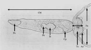
A photograph of P. gracilescens cercaria, with some of the morphological characteristics sharpened (drawn in pencil). Cbl, Cercaria body length (200 μm); Po, penetration organ; Ph, pharynx; In, intestinal caecum; Cp, cirrus pouch; Ts, tail stem; Tsl, tail stem length; Az, adhesive zone of the tail stem; Fu, furcae.

The Prosorhynchoides cercariae from A. prismatica in the present study are assigned to P. borealis based on the dimensions of the body, and dimensions, shapes and relative positions of organs which match the original description of cercariae (Matthews, Reference Matthews1974), which most likely belonged to the North-East Atlantic form P. borealis. Further support is that adult P. borealis, as verified in the present study, are very common in angler fish, L. piscatorius, in Icelandic waters (Eydal & Ólafsdóttir, 2002–Reference Eydal and Ólafsdóttir2003). Furthermore, Prosorhynchoides metacercariae, typically found in the nerves and brain cavity of gadids, are abundant in four gadoid species in Icelandic waters, with high intensities, in particular, found in fish off southern Iceland, from where infected bivalves were sampled (Eydal et al., Reference Eydal, Bambir, Helgason and Ólafsdóttir1998, Reference Eydal, Helgason, Kristmundsson and Bambir1999). Accordingly, no gadoid fish were found infected off the northern and north-eastern coasts of Iceland, in waters where A. prismatica is not found (Eydal et al., Reference Eydal, Helgason, Kristmundsson and Bambir1999).
Prosorhynchoides gracilescens (s. l.) cercariae have until now solely been found in the bivalve A. alba and only from Scottish waters (Matthews, Reference Matthews1974; Johnston & Halton, Reference Johnston and Halton1982). Since these cercariae originated from the North-East Atlantic, they should be considered as belonging to P. borealis. To date, there are no reports of other Abra species being examined specifically for the presence of bucephalid infections. Matthews (Reference Matthews1968, Reference Matthews1974) examined 300 specimens of A. alba sampled in April and November in the Firth of Clyde, western Scotland at depths of 10–40 fathoms (equivalent to 18–73 m). Of these, 15% were infected; differences between sampling dates were not given. Prevalence of infection increased with age: 0, 10, 26 and 50% being recorded in groups of shells claimed to be 1, 2, 3 and 4 years old, respectively (shell lengths were not given). This is in agreement with our results for A. prismatica; 44% of bivalves 12 mm long or larger were infected but none which were 7–11 mm long. This indicates that the infection accumulates with time and age of the bivalves. Johnston & Halton (Reference Johnston and Halton1982) reported approximately 30% infection prevalence in A. alba (sampled in October–February, size of bivalves not given) and also noted castration of infected hosts. In our study no bivalves were found to be infected in May and June but the prevalence of infection was 7% in September and 33% in October. This might indicate a seasonal variation in infection rates. However, it is not practical to draw decisive conclusions from the differences observed in prevalence between dates because of the relatively small sample size and the fact that sampling stations within the study area were not exactly the same between years/months.
Matthews (Reference Matthews1974) described infection sites and cercarial release in A. alba, accordingly: the sporocysts occupy the haemocoel of the gonad, which occupies the foot, and extend into the nephridia. The sporocysts break through the body wall at points of weakness, and protrude into the mantle cavity. The sporocysts rupture and release cercariae into the exhalant chamber, which are then discharged by the host through the exhalant canal. Johnston & Halton (Reference Johnston and Halton1982) reported sporocysts occurring in the digestive gland and dorsal foot haemocoel. Our observations indicate that a similar pattern applies to infections in A. prismatica as reported for A. alba. Dauvin & Gentil (Reference Dauvin and Gentil1989) describe parasitic castration in females of both A. alba and A. prismatica from the western English Channel, but without giving any further description of the parasite or remarks on its identity. They reported much higher prevalence of parasitic castration in females of A. prismatica (39%) than in A. alba (4%). In our view it is quite likely that P. borealis larvae were the causative agents in both bivalve species, indicating that A. prismatica may function as intermediate host in their study area. Køie (Reference Køie1984) examined hundreds of A. alba from northern Øresund and western Kattegat in Danish waters and did not find any infected with P. gracilescens (s. l.). Køie (Reference Køie1984, Reference Køie1985, Reference Køie2000) claims, based on depth preferences of individual Abra species, that one or more of the other three known subtidal species (A. longicallus, A. nitida, A. prismatica) in the North-East Atlantic Ocean must act as the alternative first intermediate hosts for P. gracilescens (s. l.). It is commonly asserted that digeneans are very specific concerning their first intermediate molluscan host. However, many publications show that it is not uncommon for two or more molluscan species to function as a first intermediate host for a given digenean species. For example, Galaktionov & Bustnes (Reference Galaktionov and Bustnes1999) found that 11 out of 14 digenean species from their studies in the Barents Sea had 2–6 different molluscan species acting as first intermediate hosts, and Galaktionov & Skirnisson (Reference Galaktionov and Skirnisson2000) found that 11 out of 19 digenean species from intertidal molluscs of south-western Iceland had 2–5 different species as first intermediate hosts.
The DNA sequencing of the SSU rDNA shows that Prosorhynchoides metacercariae from G. morhua and adults from L. piscatorius have the same SSU rDNA sequence (>99.9% identity) to that previously reported for P. gracilescens, sampled from L. piscatorius in the North Sea, UK (Littlewood et al., Reference Littlewood, Rohde and Clough1998). Since that time, P. gracilescens found in such northern locations have been reassigned to the species P. borealis (Bartoli et al., Reference Bartoli, Gibson and Brey2006) and we can assume that the SSU rDNA sequences available in GenBank (AJ228789 and from the present study) are for P. borealis. Currently, no SSU rDNA sequence data are available for the more southern species, P. gracilescens (s. str.), and it remains to be seen what variability exists in the SSU rDNA sequences between the two species. Chen et al. (Reference Chen, Wang, Yao and Nie2007) used ITS1, 5.8S and ITS2 regions of the ribosomal DNA to successfully distinguish between closely related bucephalid digeneans from different localities, hosts and microhabitats. They found that these molecular markers could discriminate between closely related species and identify single nucleotide polymorphisms within the same species. In the present study, the 100% homology seen across this gene region, in the Prosorhynchoides found in Icelandic waters from three separate hosts, strongly supports the morphological data presented herein, that they represent the same species, P. borealis.
Based on our species assignment, we report a new intermediate host record for P. borealis. Consequently, two bivalve species, A. alba and A. prismatica, are now identified as natural first intermediate hosts for this digenean. The distribution of A. prismatica extends from the Mediterranean Sea and the Atlantic coast of Morocco, along the European coast of the North-East Atlantic Ocean to northern Norway, and reaches the British Isles, Faeroe Islands and Iceland (Tebble, Reference Tebble1966). Our assumption is that A. prismatica is an important first intermediate host in European waters where P. borealis is present. The first intermediate bivalve host species for P. gracilescens (s. str.) still has to be confirmed.
Acknowledgements
We acknowledge a grant awarded by the Research Fund of the University of Iceland. Molecular studies were conducted in Malaysia and supported by a University of Malaya HIR grant no:UMC/625/1/HIR/027.