


Introduction
Goto and Ozaki (Reference Goto and Ozaki1929) proposed the microscaphidiid genus Hexangium Goto & Ozaki, 1929 for H. sigani Goto & Ozaki, 1929, collected from Siganus fuscescens (Houttuyn) (Siganidae) from two localities (Misaki and Takamatsu) off Japan. A further eight species have been described in the genus. Just two of these, Hexangium brayi Hassanine & Gibson, 2005 and Hexangium saudii Al-Jahdali, 2013 are presently recognised (WoRMS 2026). Three nominal species, Hexangium affinum Tubangui & Masiluñgan, Reference Tubangui and Masiluñgan1944, Hexangium loossi (Nagaty, 1954) Yamaguti, 1958 and Hexangium secundum Annereaux, 1947 are presently accepted as junior synonyms of H. sigani. Two species have been convincingly transferred to other genera; Hexangium elongatum Manter, 1963 is now accepted as Parawardula elongata (Manter, 1963) Jones & Blair, 2005 (Mesometridae) and Hexangium leptosomum Machida & Uchida, 1990 is now accepted as Pseudohexangium leptosomum (Machida & Uchida, 1990) Blair, 2005 (Microscaphidiidae). Finally, Hexangium ecsomi Al-Zanbagi & Hassan, 2013 is considered a taxon inquirendum having been proposed in an unpublished thesis.
The type-species, H. sigani, together with its presently recognised synonyms, has been reported from Japan (Yamaguti Reference Yamaguti1934, Machida et al. Reference Machida, Ichihara and Kamegai1970), the Philippines (Tubangui and Masiluñgan Reference Tubangui and Masiluñgan1944, Annereaux Reference Annereaux1947, Velasquez Reference Velasquez1961, Fischthal and Kuntz Reference Fischthal and Kuntz1964, Villahermosa et al. Reference Villahermosa, Estaño, Calagui and Paylangco2022), the Red Sea (Nagaty Reference Nagaty1954, El-Labadi et al. Reference El-Labadi, Ismail and Khalaf2006, Al-Zanbagi and Hassan Reference Al-Zanbagi and Hassan2013, Hassanine et al. Reference Hassanine, Al-Zahrani, Touliabah and Youssef2016, Khalifa et al. Reference Khalifa, Hassan, Mohamadain and Karar2018, Abd-Elmonsef Mahmoud et al. Reference Abd-Elmonsef Mahmoud, Osman and Abdel-Hakeem2020, Abo Elhussien et al. Reference Abo Elhussien, Hussein, Rabie and Abuelwafa2025), Andaman and Nicobar Islands (Gupta and Miglani Reference Gupta and Miglani1976), the Bay of Bengal (Gupta and Tandon Reference Gupta and Tandon1985), Sri Lanka (Parukhin Reference Parukhin1976), Arabian Gulf (Al-Yamani and Nahhas Reference Al-Yamani and Nahhas1981, Kardousha Reference Kardousha2003, Sey et al. Reference Sey, Nahhas, Uch and Vang2003), Kenya (Martens and Moens Reference Martens and Moens1995, Geets and Ollevier Reference Geets and Ollevier1996), Madagascar (Razarihelisoa Reference Razarihelisoa1959), China (Ku and Shen Reference Ku and Shen1965), Malaysia (Fischthal and Kuntz Reference Fischthal and Kuntz1965), New Caledonia (Durio and Manter Reference Durio and Manter1968), Indonesia (Yamaguti Reference Yamaguti1953), the Great Barrier Reef (Durio and Manter Reference Durio and Manter1968, Motson et al. Reference Motson, Hutson and Hoey2023) and the Seychelles (Toman Reference Toman1977). Possible Lessepsian migration can be inferred as H. sigani has also been reported (ambiguously from either Lake Manzala and the Mediterranean coast in Port Said or Lake Timsah in the Suez Canal of Egypt) (El-Ella et al. Reference El-Ella, Youssef, Sallam, Hassanin and Abouelhassan2023). The host list arising from these records comprises single species of Acanthuridae, Caesionidae, Engraulidae, Holocentridae, scarine Labridae, Lutjanidae and Sciaenidae together with 12 species of Siganus: S. argenteus (Quoy & Gaimard), S. canaliculatus (Houttuyn), S. doliatus Guérin-Méneville, S. fuscescens, S. guttatus (Bloch), S. javus (Linnaeus), S. luridus (Rüppell), S. rivulatus Forsskål & Niebuhr, S. spinus (Linnaeus), S. stellatus Forsskål, S. sutor (Valenciennes) and S. vermiculatus (Valenciennes). Durio and Manter (Reference Durio and Manter1968) stated that they identified six Siganus species as hosts for H. sigani from the Great Barrier Reef but did not list them. The other two presently recognised species, H. brayi from Siganus luridus (see Hassanine and Gibson Reference Hassanine and Gibson2005) and H. saudii from Siganus rivulatus (see Al-Jahdali Reference Al-Jahdali2013), were both described from the Red Sea.
Here we use an integrated approach, incorporating molecular, morphological, host and geographical data, to assess richness in this genus.
Materials and methods
Collecting
Fishes were collected from a range of sites in the tropical Indo-West Pacific over the past three decades (Table 1). Fishes were caught by line, spear or seine nets. Some fishes from Japan were purchased from fish markets. Those from the Tomari fish market in Naha (Okinawa) were caught from around Okinawa Island, and those from the Minabe fish market (Wakayama Prefecture) were caught from nearby waters. Digeneans were collected from freshly killed fishes as described by Cutmore et al. (Reference Cutmore, Bray, Huston, Martin, Miller, Wee, Yong and Cribb2025), fixed by pipetting into near-boiling saline, and immediately preserved in either formalin (early collections) or 80% ethanol (recent collections). Some individual worms preserved in 80% ethanol were processed for parallel morphological and molecular analyses (hologenophores and paragenophores sensu Pleijel et al. Reference Pleijel, Jondelius, Norlinder, Nygren, Oxelman, Schander, Sundberg and Thollesson2008).
Siganids examined for Hexangium species from Indo-Pacific localities. Samples pooled for Moreton Bay and from off Noosa as southeast (SE) Queensland, from Okinawa and Shirahama as Japan, and Lizard Island and Yonge Reef as northern Great Barrier Reef. FP – French Polynesia; Qld – Queensland; WA – Western Australia

Table 1. Long description
The table consists of 13 columns and 14 rows.
Column headers from left to right: Species name, Qld Heron Island, Qld Northern G B R, Qld S E Queensland, W A Ningaloo Reef, F P Gambiers, F P Marquesas, F P Moorea, F P Tuamotus, Japan, New Caledonia, Palau, and Total.
Data rows for species:
* S dot argenteus: 15 in Northern G B R, 9 in Ningaloo, 4 in Gambiers, 3 in Marquesas, 4 in Moorea, 6 in Tuamotus, 1 in New Caledonia, 2 in Palau. Total 44.
* S dot corallinus: 33 in Heron Island, 19 in Northern G B R, 1 in New Caledonia, 2 in Palau. Total 55.
* S dot doliatus: 27 in Heron Island, 24 in Northern G B R, 6 in New Caledonia, 3 in Palau. Total 60.
* S dot fuscescens: 46 in Heron Island, 4 in Northern G B R, 119 in S E Queensland, 15 in Ningaloo, 6 in Japan, 3 in New Caledonia. Total 192.
* S dot lineatus: 74 in Heron Island, 51 in Northern G B R. Total 125.
* S dot puellus: 17 in Heron Island, 22 in Northern G B R, 1 in New Caledonia, 3 in Palau. Total 43.
* S dot punctatissimus: 13 in Northern G B R, 2 in Palau. Total 15.
* S dot punctatus: 31 in Heron Island, 13 in Northern G B R, 2 in Palau. Total 46.
* S dot spinus: 3 in Heron Island, 14 in Northern G B R, 18 in Moorea, 3 in Japan, 3 in New Caledonia, 3 in Palau. Total 45.
* S dot trispilos: 11 in Ningaloo. Total 11.
* S dot virgatus: 12 in Ningaloo. Total 12.
* S dot vulpinus: 23 in Heron Island, 9 in Northern G B R, 1 in Palau. Total 33.
The final row provides totals for each locality:
* Heron Island: 254
* Northern G B R: 184
* S E Queensland: 119
* Ningaloo Reef: 47
* Gambiers: 4
* Marquesas: 3
* Moorea: 22
* Tuamotus: 6
* Japan: 9
* New Caledonia: 15
* Palau: 18
* Grand Total: 681
The key host family for species of Hexangium is the Siganidae. All siganid species belong to just one genus, Siganus. Siganus species are recognised as forming four clades as demonstrated by Kuriiwa et al. (Reference Kuriiwa, Hanzawa, Yoshino, Kimura and Nishida2007). All four clades are represented in the present samples: Clade 1 – S. argenteus; Clade 2 – S. fuscescens and S. spinus; Clade 3.1 – S. puellus (Schlegel), S. punctatissimus Fowler & Bean, S. punctatus (Schneider & Forster) and S. vulpinus (Schlegel & Müller); Clade 3.2 – S. corallinus (Valenciennes), S. doliatus, S. lineatus (Valenciennes), S. trispilos Woodland & Allen and S. virgatus (Valenciennes). Notably, S. trispilos has not yet been sequenced, but was originally described as closely related to S. corallinus (see Woodland and Allen Reference Woodland and Allen1977) and on that basis we have recognised it as belonging to Clade 3.2. These clades are referred to in consideration of patterns of host-specificity.
Morphology
Trematodes for morphological examination were washed in distilled water, stained in Mayer’s haematoxylin, destained in 1% hydrochloric acid, and neutralised in 1% ammonium hydroxide. Worms were then dehydrated in a graded ethanol series, cleared in methyl salicylate, and mounted on glass slides in Canada balsam. Measurements were taken using an Olympus SC50 digital camera attached to an Olympus BX-53 compound microscope with cellSens v1.13 software. Our approach to measurement was to measure as many specimens as possible for the characters that appeared to have some utility but to ignore some frequently measured features. Testes and the ovary were not measured on the basis that their size was clearly highly variable within species and showed no prospect of distinguishing between species.
Drawings were made using a drawing tube connected to the same compound microscope and subsequently digitised using a drawing pad and Adobe Illustrator 2026. Measurements are given in micrometres as the range and mean. Because the posterior end of many specimens was removed for sequencing, the length from the anterior end to the anterior testis was used as a proxy of total body length in morphometric analyses. Measurements of specimens of H. sigani from Japan were calculated from photographs of specimens collected by Yamaguti (Reference Yamaguti1934) and Machida et al. (Reference Machida, Ichihara and Kamegai1970). Type-specimens and voucher material are deposited in the Queensland Museum (QM), Brisbane, Australia, the Western Australian Museum (WAM), Perth, Australia, and the Meguro Parasitological Museum (MPM), Tokyo, Japan.
Molecular analyses
Specimens for molecular analyses were processed according to the protocols used by Cribb et al. (Reference Cribb, Bray, Justine, Reimer, Sasal, Shirakashi and Cutmore2022). Three genetic markers were sequenced: the second internal transcribed spacer region (ITS2 rDNA), the large (28S rDNA) ribosomal subunit RNA coding region, and the cytochrome c oxidase subunit 1 (cox1) mitochondrial region. The complete ITS2 region, with flanking 5.8S and 28S, was amplified and sequenced using the primers 3S (Morgan and Blair Reference Morgan and Blair1995) and ITS2.2 (Cribb et al. Reference Cribb, Anderson, Adlard and Bray1998), the partial D1–D3 region 28S using LSU5 (Littlewood Reference Littlewood1994), 300F (Littlewood et al. Reference Littlewood, Curini-Galletti and Herniou2000), ECD2 (Littlewood et al. Reference Littlewood, Rohde and Clough1997) and 1500R (Snyder and Tkach Reference Snyder and Tkach2001), and the partial cox1 region using Dig_cox1Fa (Wee et al. Reference Wee, Cribb, Bray and Cutmore2017) and Dig_cox1R (Wee et al. Reference Wee, Cribb, Bray and Cutmore2017). Geneious® version 10.2.6 (Kearse et al. Reference Kearse, Moir, Wilson, Stones-Havas, Cheung, Sturrock, Buxton, Cooper, Markowitz, Duran, Thierer, Ashton, Meintjes and Drummond2012) was used to assemble and edit contiguous sequences.
The three sequence datasets generated during this study were each aligned with MUSCLE in MEGA 7 (Kumar et al. Reference Kumar, Stecher and Tamura2016) using UPGMA clustering for iterations 1 and 2. No comparative ITS2 or cox1 sequences were available on GenBank, and just a single congeneric 28S sequence was available (AY222215 for Hexangium sp. from the Great Barrier Reef). The ends of the ITS2 and 28S alignments were trimmed, without filtering ambiguously aligned regions, for final datasets of 460 and 1,338 base positions (bp), respectively. The cox1 alignment was transferred to Mesquite v.3.31 (Maddison and Maddison Reference Maddison and Maddison2025), translated (echinoderm/flatworm mitochondrial code) and inspected for internal stop codons. After the correct reading frame was determined, the first base position was then removed so that the reading frame began at position one, simplifying position-coding in downstream analyses. The final cox1 dataset was 474 bp. All codon positions in the cox1 dataset were evaluated for substitution saturation using the ‘Test of substitution saturation by Xia et al.’ function (Xia et al. Reference Xia, Xie, Salemi, Chen and Wang2003, Xia and Lemey Reference Xia, Lemey, Lemey, Salemi and Vandamme2009) as implemented in DAMBE v. 7.2 (Xia Reference Xia2018); no significant substitution saturation was detected. Unrooted neighbour joining analyses were conducted using MEGA 7 for each dataset to explore species boundaries, with the following parameters: ‘Model/Method = No. of differences’, ‘Substitutions to Include = d: Transitions + Transversions’, ‘Rates among Sites = Uniform rates’ and ‘Gaps/Missing Data Treatment = Pairwise deletion’. Nodal support was estimated by performing 10,000 bootstrap replicates.
Pairwise differences were estimated for each dataset to demonstrate how different each of the combinations of species are in terms of bases and p-distance using the following conditions: ‘Variance Estimation Method = None’, ‘Model/Method = No. of differences’ and ‘Substitutions to Include = d: Transitions + Transversions’ and ‘Gaps/Missing Data Treatment = Pairwise deletion’.
Species recognition
All specimens considered here were identified as consistent with Hexangium using the classification of Blair (Reference Blair, Jones, Bray and Gibson2005). We used an integrative approach to develop our taxonomic hypothesis, considering molecular, morphological, host and distributional data. The final species recognition hypothesis is proposed using, as a starting point, the criteria proposed by Bray et al. (Reference Bray, Cutmore and Cribb2022).
Results
Samples
Specimens consistent with Hexangium were collected from 695 individuals of 12 Siganus species from 11 localities. Hexangium species were found in all 12 species of Siganus and in all collecting localities except for French Polynesia (35 individuals of 2 species) and Palau (18 individuals of 8 species). From our broader sampling effort of marine fishes in the region (see Cutmore et al., Reference Cutmore, Bray, Huston, Martin, Miller, Wee, Yong and Cribb2025), four Hexangium specimens were detected in fishes other than siganids: one each from two acanthurids (Acanthurus triostegus, n = 57; Prionurus maculatus, n = 3), one zanclid (Zanclus cornutus, n = 69) and one scarine labrid (Chlorurus microrhinos, n = 2).
Following consideration of morphological, genetic, host and geographical distribution, the material is interpreted to comprise six species. Our case to support this hypothesis and the names applied to the species is made in the Discussion. Descriptions or new data for the six species, including four new, are given below. Descriptions are followed by summaries of molecular data and morphological and biological distinctions between the putative species. Given the difficulty of distinction between some of the combinations of species, type-series for newly described species are comprised principally of hologenophores and paragenophores; only small numbers of unsequenced voucher specimens are designated as types for two of the species.
Species accounts
Hexangium sigani Goto & Ozaki, 1929 (Figures 1A, B)
Gravid adult Hexangium sigani from Siganus fuscescens. A, Presumed holotype, Japan, redrawn from Goto and Ozaki (Reference Goto and Ozaki1929); B, Hologenophore, Okinawa, Japan. Scale bars = 1 mm.

Figure 1. Long description
Panel A shows a full-body line drawing of the presumed holotype. At the anterior tip is a circular oral sucker. A narrow esophagus leads to a long, winding central intestine that extends nearly the length of the body. Flanking the intestine are two longitudinal bands of dark, solid-black vitelline follicles. Near the posterior end are two large, stippled circular testes arranged diagonally, followed by a smaller, more densely stippled ovary. A small Y-shaped excretory vesicle is at the posterior tip. A vertical scale bar at the bottom left represents 1 mm.
Panel B shows a hologenophore specimen, focusing on the anterior and mid-body regions. It features a similar oral sucker and esophagus. The central intestine is narrower and more convoluted than in Panel A. The dark vitelline follicles are clustered more densely around the mid-section. The posterior portion is cut off, showing only the upper edges of the stippled testes. A vertical scale bar to the left of the body represents 1 mm.
Synonyms: Hexangium loossi (Nagaty, 1954) Yamaguti, 1958; Arthurloossia loossi Nagaty, 1954.
Type-host: Siganus fuscescens (Houttuyn), Mottled spinefoot (Siganidae).
Type-locality: Misaki or Takamatsu, Japan.
Records: 1. Goto and Ozaki (Reference Goto and Ozaki1929); 2. Yamaguti (Reference Yamaguti1934); 3. Machida et al. (Reference Machida, Ichihara and Kamegai1970). Further putative records discussed below in Remarks.
Hosts: S. fuscescens (1, 2, 3).
Localities: Japan (1, 2, 3).
New collections
Known host: Siganus fuscescens.
New localities: Tomari fish market (26°13′N, 127°40′E), Naha, Okinawa Prefecture, Japan.
Site in host: Intestine.
Deposition of specimens: 5 voucher specimens, MPM 25,532.
Molecular sequence data: cox1 mtDNA, PZ466217–18; ITS2 rDNA, PZ469408; 28S rDNA, PZ469389.
ZooBank LSID: urn:lsid:zoobank.org:act:3F2AEF6D-1B1B-49FA-8F52-6B74CFFD3759.
Remarks
We did not recover any new material of a suitable standard to allow useful morphological description; the worms were already dead and macerating when collected but did allow reliable sequencing. Egg measurements are given in Supplementary Table 1.
In our interpretation of this species, we accept as presently reliable only the original description by Goto and Ozaki (Reference Goto and Ozaki1929) from S. fuscescens from Japanese waters and the subsequent reports by Yamaguti (Reference Yamaguti1934) and Machida et al. (Reference Machida, Ichihara and Kamegai1970) from the same area and host. The many other reports from other localities and hosts (including several non-siganids) fall into three broad categories: (i) plausible but requiring molecular testing; (ii) inconsistent with what is understood for H. sigani; or (iii) too incompletely characterised to allow interpretation relative to what is known for morphology and host distribution of H. sigani. Multiple non-Japanese reports (Nagaty Reference Nagaty1954, Velasquez Reference Velasquez1961, Al-Yamani and Nahhas Reference Al-Yamani and Nahhas1981, Kardousha Reference Kardousha2003, Al-Zanbagi and Hassan Reference Al-Zanbagi and Hassan2013, Hassanine et al. Reference Hassanine, Al-Zahrani, Touliabah and Youssef2016, Khalifa et al. Reference Khalifa, Hassan, Mohamadain and Karar2018, Villahermosa et al. Reference Villahermosa, Estaño, Calagui and Paylangco2022, Abo Elhussien et al. Reference Abo Elhussien, Hussein, Rabie and Abuelwafa2025) are broadly plausible. The report of two specimens of H. sigani from an Acanthurus sp. from the Andaman and Nicobar Islands by Gupta and Miglani (Reference Gupta and Miglani1976) shows the vitelline follicles as exceptionally distant from the oesophageal bulb and an exceptionally small pharynx; it seems highly unlikely that these specimens represent H. sigani. The report of H. loossi, which was interpreted as a synonym of H. sigani by Velasquez (Reference Velasquez1961), is confused by the fact that the species was reported from both a siganid and a scarine labrid and that the figured samples appear significantly different without attribution to the two hosts. Numerous studies (Yamaguti Reference Yamaguti1953, Fischthal and Kuntz Reference Fischthal and Kuntz1964, Fischthal and Kuntz Reference Fischthal and Kuntz1965, Ku and Shen Reference Ku and Shen1965, Durio and Manter Reference Durio and Manter1968, Gupta and Miglani Reference Gupta and Miglani1976, Parukhin Reference Parukhin1976, Toman Reference Toman1977, Gupta and Tandon Reference Gupta and Tandon1983, Martens and Moens Reference Martens and Moens1995, Geets and Ollevier Reference Geets and Ollevier1996, Dzikowski et al. Reference Dzikowski, Paperna and Diamant2003, Sey et al. Reference Sey, Nahhas, Uch and Vang2003, El-Labadi et al. Reference El-Labadi, Ismail and Khalaf2006, Abd-Elmonsef Mahmoud et al. Reference Abd-Elmonsef Mahmoud, Osman and Abdel-Hakeem2020, El-Ella et al. Reference El-Ella, Youssef, Sallam, Hassanin and Abouelhassan2023, Motson et al. Reference Motson, Hutson and Hoey2023) are variously lacking in details, figures or report improbable (especially non-siganid) hosts and cannot presently be considered convincing. In our view, these results all require reconsideration, especially in the light of molecular data. At present, H. sigani is best considered an exceptionally large species of Hexangium seemingly restricted to S. fuscescens and perhaps other closely related species.
Hexangium affinum Tubangui & Masiluñgan, 1944 (Figures 2A–L)
Gravid Hexangium affinum Tubangui & Masiluñgan, 1944. A–C, Heron Island: A, from S. corallinus; B, from S. punctatus; C, from S. doliatus (hologenophore). D–J, Lizard Island: D, from S. argenteus (hologenophore); E, from S. corallinus (hologenophore); F–H, from S. doliatus (H and I are hologenophores); J, from S. puellus. K–L, Ningaloo Reef: K, from S. doliatus (hologenophore); L, from S. virgatus (hologenophore). Scale bars = 1 mm.

Figure 2. Long description
A multi-panel set of 12 line drawings (A through L) illustrating the morphology of the flatworm Hexangium affinum. Each drawing shows an elongated, translucent body with a small circular oral sucker at the anterior tip.
* Panels A through F are arranged in the top row.
* Panels G through L are arranged in the bottom row.
Each specimen features a central digestive tract and a prominent uterus filled with dark, oval-shaped eggs concentrated in the mid-section. At the posterior end of each worm, there are two large, rounded testes and a smaller, stippled ovary.
* Specimen A is the longest and most complete, showing a clear distribution of eggs along the lateral margins of the midbody.
* Specimens C, D, I, K, and L are partial drawings, focusing on the anterior and mid-sections while the posterior tips are omitted.
* Specimen J is notably wider and more robust than the others.
Vertical scale bars representing 1 mm are placed to the left of each specimen to indicate relative size. The drawings highlight variations in body length, width, and egg density among specimens collected from Heron Island (A to C), Lizard Island (D to J), and Ningaloo Reef (K to L).
Synonym: Hexangium secundum Annereaux, 1947
Type-host: Siganus javus (Linnaeus), Streaked spinefoot (Siganidae).
Type-locality: Manila, the Philippines.
Records: 1. Tubangui and Masiluñgan (Reference Tubangui and Masiluñgan1944); 2. Annereaux (Reference Annereaux1947).
Hosts: Siganidae – Siganus guttatus (Bloch), Orange-spotted spinefoot (2); Siganus javus (1).
Localities: Manila, the Philippines (1); near Mercedes, Samar, the Philippines (2).
ZooBank LSID: urn:lsid:zoobank.org:act:2E3A8F6C-533A-47B7-9E65-6FC7F1FE78FE.
New collections
New hosts: Siganidae – Siganus argenteus (Quoy & Gaimard), Streamlined spinefoot; Siganus corallinus (Valenciennes), Blue-spotted spinefoot; Siganus doliatus Guérin-Méneville, Barred spinefoot; Siganus puellus (Schlegel), Masked spinefoot; S. punctatissimus Fowler & Bean, Peppered spinefoot; Siganus punctatus (Schneider & Forster), Goldspotted spinefoot; Siganus trispilos Woodland & Allen, Threeblotched spinefoot; Siganus virgatus (Valenciennes), Barhead spinefoot. Zanclidae – Zanclus cornutus (Linnaeus), Moorish Idol.
New localities: Off Heron Island (23°27′S, 151°55′E), southern Great Barrier Reef; off Lizard Island (14°40′S, 145°27′E), northern Great Barrier Reef, Australia; Ningaloo Reef (22°42′S, 113°40′E), Western Australia; off Noumea (21°30′S, 165°30′E), New Caledonia.
Host/locality combinations: Heron Island – S. corallinus, S. doliatus and S. punctatus. Lizard Island – S. argenteus, S. corallinus, S. doliatus, S. punctatissimus, S. punctatus, S. puellus and Z. cornutus. Ningaloo Reef – S. argenteus, S. trispilos and S. virgatus. New Caledonia – S. argenteus, S. corallinus and S. doliatus.
Site in host: Intestine.
Deposition of specimens: 96 voucher specimens QM G242,643–738; 14 voucher specimens, WAM 13,794–807.
Molecular sequence data: cox1 mtDNA, PZ466219–39; ITS2 rDNA, PZ469409–16; 28S rDNA, PZ469384–88.
Description
[Based on all specimens identified as H. affinum. Measurements are of eight hologenophore and three paragenophore specimens. 98 voucher specimens (no molecular data) examined and identified but not incorporated in main measurements. Measurements for each host/locality combination in Supplementary Table 2. Egg measurements given in Supplementary Table 1.]
Body elongate, frequently conspicuously narrow, with margins ranging from almost parallel to clearly widest in middle or posterior third of body, 2,871–3,193 (3,065) × 405–670 (518). Length to anterior testis 1,867–2,910 (2220). Length to anterior testis: body width ratio 3.76–5.12 (4.33):1. Largest gravid specimens approximately twice length of smallest. Tegument armed with fine spines; spines most clearly visible at anterior extremity. Eye-spot pigment not detected. Pharynx typically slightly wider than long, 106–157 (130) × 115–168 (148). Oesophagus conspicuously elongate, 447–650 (531) in length. Oesophagus length: pharynx length ratio 3.60–4.64 (4.10):1. Oesophageal bulb distinct. Intestinal caeca terminate close to anterior margin of anterior testis, highly variable in width.
Testes two, entire, contiguous, occasionally partly flanked by intestinal caeca, usually somewhat oblique but sometimes tandem or opposite. Seminal vesicle winding from well posterior to intestinal bifurcation to genital pore, typically largely obscured by uterus. Poorly developed ‘cirrus-sac’ present. Genital pore median, midway between pharynx and intestinal bifurcation.
Ovary entire, usually ventral to and partially overlapping posterior testis. Uterus normally well-developed from about anterior margin of anterior testis to about intestinal bifurcation, in smaller specimens appears to follow helical path but broadly irregular in fully mature specimens, typically ventrally overlapping intestinal caeca when full of eggs. Ventral vitelline follicles in two lateral fields, ventral to intestinal caeca, typically extend anteriorly from just anterior to ends of intestinal caeca (occasionally reaching ends) to 256–555 (380) from intestinal bifurcation, rarely almost reaching it; dorsal vitelline follicles medially confluent, dorsal to intestinal caeca and uterus but usually largely obscured by uterus, sometimes include overall most anterior follicles. Eggs 62–89 (77) × 38–56 (49) (n = 137, all samples).
Excretory pore dorsally subterminal, close to posterior extremity of body. ‘Rosette organ’ represented only by a pair of distinct diverticula that unite and open at excretory pore. Excretory vesicle thin-walled, voluminous, usually extends to posterior margin of ovary.
Remarks
These specimens are considered identifiable as the known species H. affinum (see Discussion). Specimens interpreted as this species are moderately consistent in their morphology, most importantly in their possession of a characteristically long oesophagus. Some of the specimens, including hologenophores, are amongst the narrowest seen among all six species in this study (e.g. Figures 2H, I), but this character is not unique to the species or entirely reliable within it. The largest specimen interpreted as this species (Figure 2J) from S. puellus at Lizard Island was anomalous in having no eggs in the uterus. We suspect that it was senescent, and as such perhaps a good indicator of the maximum size reached by this species.
Hexangium urabeae n. sp. (Figures 3A–I)
Gravid Hexangium urabeae n. sp. A–G, Lizard Island: A–D, from S. lineatus (C is a hologenophore); E, from S. corallinus (hologenophore); F, from S. doliatus (hologenophore); G, from S. punctatus (hologenophore). H, Heron Island, from S. lineatus. I, New Caledonia, from S. doliatus. Scale bars = 1 mm.

Figure 3. Long description
A multi-panel line drawing containing nine specimens labeled A through I. Each drawing depicts an elongated, worm-like body with a distinct oral sucker at the anterior end.
* Anatomical structure from anterior to posterior: A circular oral sucker leads to a narrow esophagus, which bifurcates into two long intestinal caeca extending down the sides of the body. The central region contains a convoluted tubular uterus. Dark, solid-black circular spots representing vitelline follicles are distributed along the lateral fields. Near the posterior end, two large oval testes are situated side-by-side, followed by a smaller ovary and a terminal excretory pore.
* Panel A: A full specimen from S. lineatus at Lizard Island.
* Panel B: A smaller, narrower specimen from S. lineatus at Lizard Island.
* Panel C: A close-up of the anterior and mid-section of a hologenophore specimen.
* Panel D: A complete specimen from S. lineatus at Lizard Island.
* Panel E: A specimen from S. corallinus at Lizard Island.
* Panel F: A specimen from S. doliatus at Lizard Island.
* Panel G: A specimen from S. punctatus at Lizard Island.
* Panel H: A specimen from S. lineatus at Heron Island.
* Panel I: The largest specimen shown, from S. doliatus at New Caledonia, featuring a more robust body and denser vitelline follicles.
Vertical scale bars representing 1 mm are placed to the left of each specimen.
Type-host: Siganus lineatus (Valenciennes), Golden-lined spinefoot (Siganidae).
Type-locality: off Lizard Island (14°40′S, 145°27′E), northern Great Barrier Reef, Australia.
Other hosts: Siganidae – Siganus argenteus (Quoy & Gaimard), Streamlined spinefoot; Siganus corallinus (Valenciennes), Blue-spotted spinefoot; Siganus doliatus Guérin-Méneville, Barred spinefoot; Siganus puellus (Schlegel), Masked spinefoot; Siganus punctatus (Schneider & Forster), Goldspotted spinefoot.
Other localities: off Heron Island (23°27′S, 151°55′E), southern Great Barrier Reef; off Noumea (21°30′S, 165°30′E), New Caledonia.
Host/locality combinations: Lizard Island – S. argenteus, S. corallinus, S. doliatus, S. lineatus, S. puellus and S. punctatus. Heron Island – S. lineatus. New Caledonia – S. doliatus.
Site in host: Intestine.
Deposition of specimens: Holotype, QM G242,739; 17 paratypes, QM G242,740–56; 72 voucher specimens, QM G242,757–828.
Molecular sequence data: cox1 mtDNA, PZ466203–16; ITS2 rDNA, PZ469405–07; 28S rDNA, PZ469393–94.
ZooBank LSID: urn:lsid:zoobank.org:act:93F818E4-46D7-43A7-92B8-8EB7DF12BAA3.
Etymology: This species is named for Dr Misako Urabe in recognition of her major contribution to the field.
Description
[Based on 10 hologenophore and four paragenophore specimens. 64 voucher specimens (no molecular data) examined and identified but not incorporated in main measurements. Measurements for each host/locality combination in Supplementary Table 2. Egg measurements given in Supplementary Table 1.]
Body relatively elongate, with margins ranging from rarely almost parallel to typically clearly widest in middle or posterior third of body, 2,895–3,788 (3,338) × 595–1,025 (770). Length to anterior testis 2,040–3,443 (2,667). Length to anterior testis: body width ratio 3.05–3.94 (3.48):1. Largest gravid specimens over twice length of smallest. Tegument armed with fine spines; spines most clearly visible at anterior extremity. Eye-spot pigment not detected in anterior body. Pharynx typically slightly wider than long, 175–289 (235) × 209–314 (265). Oesophagus well-developed but conspicuously short, 376–541 (450) in length. Oesophagus length: pharynx length ratio 1.55–2.19 (1.93):1. Oesophageal bulb distinct. Intestinal caeca terminate close to anterior margin of anterior testis, highly variable in width.
Testes two, entire, usually contiguous, occasionally partly flanked by intestinal caeca, always oblique with sinistral testis more anterior. Seminal vesicle winding from well posterior to intestinal bifurcation to genital pore, typically largely obscured by uterus. Poorly developed ‘cirrus-sac’ present. Genital pore median, midway between pharynx and intestinal bifurcation.
Ovary entire, usually ventral to and partially overlapping posterior testis. Uterus normally well-developed from about anterior margin of anterior testis to about intestinal bifurcation, in smaller specimens appears to follow helical path but broadly irregular in fully mature specimens, sometimes ventrally overlapping intestinal caeca when full of eggs. Ventral vitelline follicles in two lateral fields, ventral to intestinal caeca, typically extend anteriorly from just anterior to ends of intestinal caeca (occasionally reaching ends) to 215–764 (494) from intestinal bifurcation, rarely almost reaching it; dorsal vitelline follicles medially confluent, dorsal to intestinal caeca and uterus but usually largely obscured by uterus, only rarely appears to include most anterior vitelline follicles. Eggs 69–95 (81) × 43–63 (51) (n = 97, all samples).
Excretory pore dorsally subterminal close to posterior extremity of body. ‘Rosette organ’ represented only by a pair of distinct diverticula that unite and open at excretory pore. Excretory vesicle thin-walled, voluminous, usually occupies space between rosette organ and ovary.
Remarks
This species has been found at the three Pacific sites where suitable hosts, principally belonging to Siganus Clades 3.1 and 3.2, have been examined. Notably, it has not yet been detected at Ningaloo Reef in the total of 23 S. trispilos and S. virgatus examined there. It is partly recognisable morphologically by its vitelline follicles being well-separated from the intestinal bifurcation and by its relatively short oesophagus.
Hexangium kuramochii n. sp. (Figures 4A–H)
Gravid Hexangium kuramochii n. sp. A–B, Moreton Bay from S. fuscescens. C–F, Lizard Island: C, from S. argenteus; D, from S. fuscescens; E, from S. lineatus; F, from S. spinus. G–H, Ningaloo Reef: G, from S. fuscescens; H, from S. trispilos. Scale bars = 1 mm.

Figure 4. Long description
The multi-panel illustration consists of eight line drawings (A-H) of the trematode Hexangium kuramochii n. sp. Each drawing features a vertical scale bar representing 1 mm.
* Panel A: A small, slender specimen from S. fuscescens in Moreton Bay.
* Panel B: A larger specimen from the same host and location as A, showing a more developed internal structure.
* Panel C: An anterior portion of a specimen from S. argenteus at Lizard Island.
* Panel D: A specimen from S. fuscescens at Lizard Island with a distinct indentation on the right side of the body wall.
* Panel E: A shorter specimen from S. lineatus at Lizard Island.
* Panel F: A specimen from S. spinus at Lizard Island.
* Panel G: A large, robust specimen from S. fuscescens at Ningaloo Reef.
* Panel H: A specimen from S. trispilos at Ningaloo Reef with a constricted mid-body.
Common anatomical features across all specimens include:
* An anterior oral sucker at the top.
* A long esophagus leading to a bifurcated intestine.
* Numerous dark, rounded vitelline follicles distributed along the lateral margins of the mid-body.
* A central, coiled uterus filled with eggs.
* Large, rounded gonads (testes and ovary) located in the posterior third of the body.
Type-host: Siganus fuscescens (Houttuyn), Mottled spinefoot (Siganidae).
Type-locality: Moreton Bay (27°22′S, 153°13′E), southeastern Queensland, Australia.
Other hosts: Acanthuridae – Acanthurus triostegus (Linnaeus), Convict surgeonfish; Prionurus maculatus Ogilby, Yellowspotted sawtail. Siganidae – Siganus argenteus (Quoy & Gaimard), Streamlined spinefoot; Siganus lineatus (Valenciennes), Golden-lined spinefoot; S. spinus (Linnaeus), Little spinefoot; S. trispilos Woodland & Allen, Threeblotched spinefoot.
Other localities: Hell’s Gates, Sunshine Coast, southern Queensland (26°22′S, 153°07′E); off Lizard Island (14°40′S, 145°27′E), northern Great Barrier Reef, Australia; Ningaloo Reef (22°42′S, 113°40′E), Western Australia.
Host/locality combinations: Moreton Bay – A. triostegus, P. maculatus and S. fuscescens. Hell’s Gates – S. fuscescens. Lizard Island – S. argenteus, S. fuscescens, S. lineatus and S. spinus. Ningaloo Reef – S. argenteus, S. fuscescens and S. trispilos.
Site in host: Intestine.
Deposition of specimens: Holotype, QM 242,829; 10 paratypes, QM G242,830–9; 50 voucher specimens, QM G242,840–89; 18 paratypes, WAM 13,808–25.
Molecular sequence data: cox1 mtDNA, PZ466240–69; ITS2 rDNA, PZ469397–404; 28S rDNA, PZ469390–92.
ZooBank LSID: urn:lsid:zoobank.org: act:1BE021AC-7B5D-4A39-BA79-ECBCFC072708.
Etymology: This species is named for Dr Toshiaki Kuramochi in recognition of his significant contribution to this field.
Description
[Based on 24 hologenophore and four paragenophore specimens. 48 voucher specimens (no molecular data) examined and identified but not incorporated in main measurements. Measurements for each host/locality combination in Supplementary Table 2. Egg measurements given in Supplementary Table 1.]
Body relatively elongate, with margins ranging from rarely almost parallel to typically clearly widest in middle or anterior third of body, 3,470–6,839 (4885) × 698–1,288 (928). Length to anterior testis 2,463–5,144 (3,628). Length to anterior testis: body width ratio 3.16–4.45 (3.91):1. Largest gravid specimens over 2.5 times length of smallest. Tegument armed with fine spines; spines most clearly visible at anterior extremity. Eye-spot pigment not detected. Pharynx typically slightly wider than long, 202–364 (284) × 199–385 (307). Oesophagus well-developed but conspicuously short, 433–756 (588) in length. Oesophagus length: pharynx length ratio 1.79–2.65 (2.09):1. Oesophageal bulb distinct. Intestinal caeca terminate close to anterior margin of anterior testis, highly variable in width.
Testes two, entire, contiguous, occasionally partly flanked by intestinal caeca, usually oblique but ranging from almost opposite to tandem. Seminal vesicle winding from well posterior to intestinal bifurcation to genital pore, typically largely obscured by uterus. Poorly developed ‘cirrus-sac’ present. Genital pore median, midway between pharynx and intestinal bifurcation.
Ovary entire, usually ventral to and partially overlapping posterior testis. Uterus normally well-developed (full of eggs) from about anterior margin of anterior testis to about intestinal bifurcation, although sometimes not extensive as far anteriorly; in smaller specimens appears to follow helical path but broadly irregular in fully mature specimens, sometimes partially overlapping intestinal caeca ventrally when full of eggs. Ventral vitelline follicles in two lateral fields, ventral to intestinal caeca, typically extend anteriorly from just anterior to ends of intestinal caeca (occasionally reaching ends) to 269–868 (545) from intestinal bifurcation, rarely almost reaching it; dorsal vitelline follicles medially confluent, dorsal to intestinal caeca and uterus but usually largely obscured by uterus, appears to include most anterior follicles only rarely. Eggs 73–95 (84) × 43–65 (53) (n = 120, all samples).
Excretory pore dorsally subterminal close to posterior extremity of body. ‘Rosette organ’ represented only by pair of distinct diverticula that unite and open at excretory pore. Excretory vesicle thin-walled, voluminous, usually occupies space between rosette organ and ovary.
Remarks
This species has a seemingly disjunct distribution, known from southern Queensland and Ningaloo Reef, but not from suitable hosts at Heron Island on the Great Barrier Reef.
Hexangium robustum n. sp. (Figures 5A–I)
Gravid Hexangium robustum n. sp. A–C, Lizard Island: A, from S. vulpinus (hologenophore); B–C, from S. punctatus (Hologenophores). D–I, Heron Island: D, from S. corallinus; E–F, from S. doliatus; G, from S. lineatus; H, from S. puellus; I, from S. vulpinus. Scale bars = 1 mm.

Figure 5. Long description
A multi-panel set of nine line drawings labeled A through I. Each drawing depicts the elongated, spindle-shaped body of a gravid fluke.
Anatomical features common to all specimens from anterior to posterior include:
* A small oral sucker at the very top tip.
* A long, thin esophagus leading to a bifurcated intestine that runs down the lateral sides of the body.
* Numerous dark, solid black circular vitelline follicles arranged in two longitudinal rows along the intestinal caeca.
* A central uterus containing coiled structures.
* Two large, stippled oval testes located in the posterior third of the body, usually arranged in a tandem or slightly diagonal orientation.
* A small, circular ovary with distinct internal spotting located just posterior to the testes.
* A small excretory vesicle at the posterior-most tip.
Variations across panels:
* Panels A, B, and C show specimens from Lizard Island. B and C are noticeably wider than A.
* Panels D through I show specimens from Heron Island.
* Panel H is the largest specimen, showing a much higher density and larger size of black vitelline follicles and more prominent posterior organs.
* Panel I is the smallest and most slender specimen.
* Vertical scale bars representing 1 mm are placed to the left of each specimen to indicate relative size differences.
Type-host: Siganus vulpinus (Schlegel & Müller), Foxface (Siganidae).
Type-locality: off Lizard Island (14°40′S, 145°27′E), northern Great Barrier Reef, Australia.
Other hosts: Labridae – Chlorurus microrhinos (Bleeker), Steephead parrotfish. Siganidae – Siganus doliatus Guérin-Méneville, Barred spinefoot; Siganus lineatus (Valenciennes), Golden-lined spinefoot; Siganus puellus (Schlegel), Masked spinefoot; Siganus punctatus (Bloch & Schneider), Goldspotted spinefoot; Siganus punctatissimus Fowler & Bean, Peppered spinefoot.
Other localities: Yonge Reef (14°35′S, 145°37′E), northern Great Barrier Reef; off Heron Island (23°27′S, 151°55′E); southern Great Barrier Reef, Australia.
Host/locality combinations: Heron Island – C. microrhinos, S. doliatus, S. lineatus, S. puellus, S. punctatus and S. vulpinus. Lizard Island – S. puellus, S. punctatissimus and S. vulpinus. Yonge Reef – S. punctatus.
Site in host: Intestine.
Deposition of specimens: Holotype, QM G242,890; 12 paratypes, QM G242,891–902; QM G 242,903–62.
Molecular sequence data: cox1 mtDNA, PZ466270–74; ITS2 rDNA, PZ469419–21; 28S rDNA, PZ469395.
ZooBank LSID: urn:lsid:zoobank.org:act:8EF3D6AA-F56D-40E7-A2B2-22530B475330.
Etymology: This species is named for its distinctly robust form.
Description
[Based on six hologenophore specimens and five whole specimens (all from the type-host/type-locality combination). 68 voucher specimens (no molecular data) examined and identified but not incorporated in main measurements. Measurements for each host/locality combination in Supplementary Table 2. Egg measurements given in Supplementary Table 1.]
Body relatively plump, rarely with almost parallel margins, in larger specimens frequently conspicuously widest at level of testes, 2,400–2,976 (2,658) × 612–1,267 (822). Length to anterior testis 1,716–2,700 (2,093). Length to anterior testis: body width ratio 2.04–3.18 (2.60):1. Largest gravid specimens over 3 times length of smallest. Tegument armed with fine spines; spines most clearly visible at anterior extremity. Eye-spot pigment not detected. Pharynx typically slightly wider than long, 138–228 (181) × 156–273 (210). Oesophagus well-developed, relatively long, 457–718 (603) in length. Oesophagus length: pharynx length ratio 3.05–4.04 (3.37):1. Oesophageal bulb distinct. Intestinal caeca terminate close to anterior margin of anterior testis, highly variable in width.
Testes two, entire, typically contiguous, occasionally partly flanked by intestinal caeca, usually oblique but also often nearly opposite. Seminal vesicle winding from well posterior to intestinal bifurcation to genital pore, typically largely obscured by uterus. Poorly developed ‘cirrus-sac’ present. Genital pore median, midway between pharynx and intestinal bifurcation.
Ovary entire, usually ventral to and partially overlapping posterior testis. Uterus normally well-developed from about anterior margin of anterior testis to about intestinal bifurcation, in smaller specimens appears to follow helical path but broadly irregular in fully mature specimens, sometimes partially overlapping intestinal caeca ventrally when full of eggs. Ventral vitelline follicles in two lateral fields, ventral to intestinal caeca, typically extend anteriorly from just anterior to ends of intestinal caeca (occasionally reaching ends) to as far as 39 anterior to or 247 posterior to intestinal bifurcation it (mean 76 posterior); dorsal vitelline follicles medially confluent, dorsal to intestinal caeca and uterus but usually largely obscured by uterus, never detected to include most anterior vitelline follicles. Eggs 65–97 (82) × 41–59 (51) (n = 63, all samples).
Excretory pore dorsally subterminal close to posterior extremity of body. ‘Rosette organ’ represented only by a pair of distinct diverticula that unite and open at excretory pore. Excretory vesicle thin-walled, voluminous, usually occupies space between rosette organ and ovary.
Remarks
This species has a distinctive host-specificity, being concentrated in Clade 3.1 siganids; no other species is found as commonly in these fishes. It is also relatively morphologically distinct in that the vitelline follicles normally extend to close to the intestinal bifurcation.
Hexangium reimeri n. sp. (Figures 6C–G)
Hexangium reimeri n. sp. from Siganus fuscescens. A–D, Heron Island. E, Ningaloo Reef. Scale bars = 1 mm.

Figure 6. Long description
A multi-panel line drawing displays five specimens of the parasite Hexangium reimeri n. sp. Panels A, B, C, and D represent specimens from Heron Island, while panel E represents a specimen from Ningaloo Reef. Each specimen is elongated and cylindrical with a distinct oral sucker at the anterior tip.
* Specimen A is a large, mature individual. Below the oral sucker, a long esophagus leads to a bifurcated intestine. Large, black, circular vitelline follicles are densely packed along the lateral margins of the mid-body. Two large, stippled testes are located in the posterior third, followed by a smaller, spotted ovary and a terminal excretory pore.
* Specimen B is a significantly smaller, narrower juvenile form. It contains fewer vitelline follicles and less developed reproductive organs. A vertical scale bar is positioned to its left.
* Specimen C is an elongated mature form similar to A but with a more slender body. The vitelline follicles are more spaced out along the sides. A vertical scale bar is at the bottom left.
* Specimen D shows the anterior and mid-section of a specimen, focusing on the oral sucker, esophagus, and the beginning of the vitelline follicle rows. A vertical scale bar is at the bottom left.
* Specimen E is a large mature individual nearly identical in structure to A, showing the full complement of internal organs including the oral sucker, esophagus, lateral vitelline follicles, posterior testes, and ovary. A vertical scale bar is at the bottom left.
All scale bars represent 1 mm.
Type-host: Siganus fuscescens (Houttuyn), Mottled spinefoot (Siganidae).
Type-locality: off Heron Island (23°27′S, 151°55′E), southern Great Barrier Reef, Australia.
Other localities: Ningaloo Reef (22°42′S, 113°40′E), Western Australia.
Site in host: Intestine.
Deposition of specimens: Holotype, QM G242,963; 12 paratypes QM G242,964–75; 60 voucher specimens, QM G242,976–3,020; 1 paratype, WAM 13,826.
Molecular sequence data: cox1 mtDNA, PZ466275–79; ITS2 rDNA, PZ469417–18; 28S rDNA, PZ469396.
ZooBank LSID: urn:lsid:zoobank.org:act:FC893E09-947D-4D0F-8016-176D9BF4171F.
Etymology: This species is named for Dr James Reimer in recognition of his help in this study and his significant contribution to the understanding of Indo-Pacific biodiversity.
Description
[Based on two hologenophore specimens and 39 whole specimens (no molecular data) from the type-host/type-locality combination. 30 voucher specimens (no molecular data) examined and identified but not incorporated in main measurements. Measurements for each host/locality combination in Supplementary Table 2. Egg measurements given in Supplementary Table 1.]
Body relatively elongate, typically with almost parallel margins, widest point highly variable, 2,762–6,472 (3856) × 441–1,464 (696). Length to anterior testis 2,036–4,747 (2,842). Length to anterior testis; body width ratio 3.24–4.93 (4.16):1. Largest gravid specimen approximately 2.5 times length of smallest. Tegument armed with fine spines; spines most clearly visible at anterior extremity. Eye-spot pigment not detected in anterior body. Pharynx typically slightly wider than long, 154–365 (212) × 172–377 (236). Oesophagus well-developed, relatively elongate, 495–943 (660) in length. Oesophagus length: pharynx length ratio 2.37–3.85 (3.13):1. Oesophageal bulb distinct. Intestinal caeca terminate close to anterior margin of anterior testis, highly variable in width.
Testes entire, contiguous, occasionally partly flanked by intestinal caeca, usually oblique but ranging from almost opposite to directly tandem. Seminal vesicle winding from well posterior to intestinal bifurcation to genital pore, typically largely obscured by uterus. Poorly developed ‘cirrus-sac’ present. Genital pore median, midway between pharynx and intestinal bifurcation.
Ovary entire, usually ventral to and partially overlapping posterior testis. Uterus normally well-developed from about anterior margin of anterior testis to about intestinal bifurcation, in smaller specimens appears to follow helical path but broadly irregular in fully mature specimens, sometimes partially overlapping intestinal caeca ventrally when full of eggs. Ventral vitelline follicles in two lateral fields, ventral to intestinal caeca, typically extend anteriorly from just anterior to ends of intestinal caeca (occasionally reaching ends) to 111–476 (249) from intestinal bifurcation, rarely almost reaching it; dorsal vitelline follicles medially confluent, dorsal to intestinal caeca and uterus but usually largely obscured by uterus, never detected to include most anterior vitelline follicles. Eggs 71–89 (81) × 38–61 (49) (n = 70, all samples).
Excretory pore dorsally subterminal close to posterior extremity of body. ‘Rosette organ’ represented only by a pair of distinct diverticula that unite and open at excretory pore. Excretory vesicle thin-walled, voluminous, usually occupies space between ‘rosette organ’ and ovary.
Remarks
This species is morphologically relatively unspecialised and is difficult to distinguish from H. affinum with which it shares a relatively long oesophagus. It is, however, seemingly distinct from H. affinum in its host distribution.
Molecular analyses
Sequences for cox1 (n = 113) distinguish eight strongly supported clades that differ from each other at p-distances of 3.79–16.00 (18–76 bp) (Figure 7, Table 2). Each of the species recognised above relates to one clade except for H. affinum, which has three subclades. Sequence differences within each of the eight clades range from 0 to 9 bp with no or negligible host or locality correlation; these differences are interpreted as (almost entirely) uninformative intraspecific variation. The exception with minor structuring is H. urabeae, for which 18 of 19 sequences from Lizard Island differ from each other at just 0–4 bp and at 5–6 bp from the five sequences from Heron Island; this distinction is interpreted as representing weak intraspecific geographic variation. The level of distinction in p-distance between the eight clades ranges from 3.79–16.00 (18–76 bp) (Table 2). The three most similar clades involve p-distance differences of 3.79–5.47 (18–26 bp) and ultimately form the highly supported clade interpreted as relating to H. affinum.
Phylogram from the unrooted neighbour-joining analysis of the cox1 mtDNA dataset. Bootstrap support values are shown at the nodes, with values of <85 not shown. The scale bar indicates the number of base differences.

Figure 7. Long description
The phylogram branches from a single root on the left into six distinct major clades. Each clade is labeled on the far right with the species name.
* The top clade is Hexangium urabeae, containing numerous specimens from Siganus lineatus and other hosts at Lizard Island and Heron Island.
* Below it is a smaller clade for H. sigani, primarily from Siganus fuscescens in Okinawa, with a bootstrap support value of 100.
* The third clade is Hexangium affinum, showing specimens from various Siganus species across Lizard Island, Heron Island, and Ningaloo Reef, with internal bootstrap values of 98 and 99.
* The fourth and largest clade is Hexangium kuramochii, containing a high density of specimens from Ningaloo Reef, Lizard Island, and Moreton Bay.
* The fifth clade is H. robustum, with a bootstrap value of 100, including specimens from Yonge Reef and Lizard Island.
* The bottom-most clade is H. reimeri, also with a bootstrap value of 100, featuring specimens from Heron Island and Ningaloo Reef.
A scale bar at the bottom left represents 10 base differences. Bootstrap support values are indicated at major nodes, including 98 at the primary divergence point for the top three clades.
Genetic differences between eight Hexangium cox1 clades. p-distances are shown above the diagonal, base pair differences below the diagonal, and intra-clade differences on the diagonal

Table 2. Long description
The table compares eight clades of Hexangium. The diagonal shows intra-clade differences. Values above the diagonal represent p-distances (percentages), and values below the diagonal represent base pair differences.
* Clade 1: H. urabeae (n=24). Intra-clade: 0–7. p-distances to clades 2-8 range from 4.84 to 14.32.
* Clade 2: H. sigani (n=7). Intra-clade: 0–1. Base pair diff to clade 1: 23–27. p-distances to clades 3-8 range from 5.68 to 14.95.
* Clade 3: H. affinum subclade 1 (n=12). Intra-clade: 0–9. Base pair diff to clades 1-2: 32–40. p-distances to clades 4-8 range from 3.79 to 14.74.
* Clade 4: H. affinum subclade 2 (n=7). Intra-clade: 0–5. Base pair diff to clades 1-3: 21–34. p-distances to clades 5-8 range from 3.79 to 15.16.
* Clade 5: H. affinum subclade 3 (n=7). Intra-clade: 0–5. Base pair diff to clades 1-4: 18–38. p-distances to clades 6-8 range from 11.16 to 16.00.
* Clade 6: H. kuramochii (n=43). Intra-clade: 0–8. Base pair diff to clades 1-5: 45–61. p-distances to clades 7-8 range from 12.63 to 15.16.
* Clade 7: H. robustum (n=7). Intra-clade: 0. Base pair diff to clades 1-6: 60–76. p-distance to clade 8: 15.16–15.37.
* Clade 8: H. reimeri (n=7). Intra-clade: 0–4. Base pair diff to clades 1-7: 61–73.
Sequences for ITS2 rDNA (n = 48) show a low overall level of distinction between samples; the maximum p-distance was 2.89 (11 bp) (Figure 8, Table 3). The distinctions suggested by these data are generally compatible with those from cox1 sequences with two exceptions. First, sequences corresponding to H. urabeae again form two clades (relating to Lizard and Heron Islands and differing at 4 bp) which do not, however, themselves form a clade. Second, ITS2 sequences corresponding to cox1 sequences for H. kuramochii show a consistent 1 bp difference between samples from Moreton Bay and those from Lizard Island and Ningaloo. Differences between corresponding cox1 sequences from samples from the two areas are negligible. Hexangium sigani differs consistently from all other species in ITS2, but by as few as 2 bp relative to H. urabeae and H. kuramochii. ITS2 sequences relating to the three cox1 variants of H. affinum form a clade differing from all other species at 3–11 bp and differ from each other at only 1–2 bp. Hexangium robustum differs at 7–11 bp from all other species. Hexangium reimeri differs at 3–9 bp from all other species. For H. sigani and H. robustum, there was no intraspecific variation. Hexangium affinum, H. kuramochii, H. reimeri and H. urabeae each showed low-level geographic variation.
Phylogram from the unrooted neighbour-joining analysis of the ITS2 rDNA dataset. Bootstrap support values are shown at the nodes, with values of <85 not shown. The scale bar indicates the number of base differences.

Figure 8. Long description
The phylogram originates from a single root on the left, branching into several distinct clades.
* The uppermost clade contains Hexangium kuramochii specimens from Moreton Bay, Lizard Island, and Ningaloo Reef, primarily hosted by Siganus fuscescens and Siganus argenteus.
* Below this is a smaller group of Hexangium urabeae from Lizard Island.
* The middle section features a large clade of Hexangium sigani from Okinawa, followed by a diverse clade of Hexangium affinum from Ningaloo Reef, Lizard Island, and Heron Island.
* The lower section includes a separate cluster of Hexangium urabeae from Heron Island (bootstrap value 98), followed by Hexangium reimeri from Ningaloo Reef and Heron Island (bootstrap value 90 and 91).
* The bottom-most clade consists of Hexangium robustum from Yonge Reef, Lizard Island, and Heron Island (bootstrap value 99).
Terminal labels include the species name, host species, location, and accession number. A scale bar at the bottom left represents 1 base difference.
Genetic differences between six Hexangium species for ITS2 rDNA. p-distances are shown above the diagonal, base pair differences below the diagonal, and intra-clade differences on the diagonal. Two species (H. kuramochii and H. urabeae) are subdivided by locality. HI – Heron Island; LI – Lizard Island; MB – Moreton Bay; NR – Ningaloo Reef

Table 3. Long description
The table consists of 8 rows and 8 numbered columns representing species groups.
Row 1: H. urabeae L I. n equals 5. Diagonal: 0. P-distances to others: 0.87, 0.27 to 0.43, 0.43 to 0.65, 0.22 to 0.43, 0.22 to 0.43, 1.52 to 1.74, 0.65 to 1.30.
Row 2: H. urabeae H I. n equals 3. Base pair difference to group 1: 4. Diagonal: 0. P-distances to others: 1.09, 1.30 to 1.52, 1.09, 0.87 to 1.09, 2.39, 1.52 to 1.96.
Row 3: H. sigani. n equals 7. Base pair differences to groups 1 to 2: 1 to 2, 5. Diagonal: 0. P-distances to others: 0.65 to 1.09, 0.87, 0.43 to 0.65, 1.96, 1.09 to 1.52.
Row 4: H. affinum. n equals 9. Base pair differences to groups 1 to 3: 2 to 3, 6 to 7, 3 to 5. Diagonal: 0 to 2. P-distances to others: 0.87 to 1.09, 0.65 to 1.09, 2.17 to 2.39, 1.30 to 1.96.
Row 5: H. kuramochii M B. n equals 7. Base pair differences to groups 1 to 4: 1 to 2, 5, 4, 4 to 5. Diagonal: 0. P-distances to others: 0.22 to 0.43, 2.17, 1.30 to 1.74.
Row 6: H. kuramochii L I or N R. n equals 9. Base pair differences to groups 1 to 5: 1 to 2, 4 to 5, 2 to 3, 3 to 5, 1 to 2. Diagonal: 0 to 1. P-distances to others: 1.74 to 1.96, 0.87 to 1.52.
Row 7: H. robustum. n equals 5. Base pair differences to groups 1 to 6: 7 to 8, 11, 9, 10 to 11, 10, 8 to 9. Diagonal: 0. P-distance to group 8: 1.74 to 2.17.
Row 8: H. reimeri. n equals 3. Base pair differences to groups 1 to 7: 3 to 6, 7 to 9, 5 to 7, 6 to 9, 6 to 10, 4 to 7, 8 to 10. Diagonal: 0 to 2.
Sequences for 28S rDNA (n = 13) were generated for the eight clades recognised from cox1 sequences (Figure 9, Table 4). All combinations of clades recognised from cox1 data were distinguished by this marker except for two of those ultimately identified as H. affinum, which were identical for this marker. Notably, samples of H. urabeae from Heron and Lizard Islands form a clade (as for cox1), whereas they do not in the ITS2 analysis. Of the four species that show inferred intraspecific ITS2 variation (H. affinum, H. kuramochii, H. reimeri and H. urabeae), all but H. reimeri (sequenced only from Heron Island) varied at a single bp for 28S. All other combinations differed at p-distances of 0.30–1.20 (3–16 bp). Hexangium robustum and H. reimeri are by far the most distantly related species, differing from all other species at p-distances of 0.90–1.20 (12–16 bp).
Phylogram from the unrooted neighbour-joining analysis of the 28S rDNA dataset. Bootstrap support values are shown at the nodes, with values of <85 not shown. The scale bar indicates the number of base differences.

Figure 9. Long description
A neighbor-joining phylogram with a scale bar at the bottom left representing 1 base difference. The tree originates from a single root on the left, splitting into two primary lineages.
Top Lineage:
- A major node with a bootstrap value of 99 leads to several sub-clades.
- The uppermost sub-clade contains five entries of Hexangium affinum from Siganus argenteus, Siganus virgatus, Siganus corallinus, and Siganus doliatus across Ningaloo Reef, Lizard Island, and Heron Island.
- Below this is a single branch for Hexangium sigani from Siganus fuscescens in Okinawa.
- A node with a bootstrap value of 85 leads to a clade of three Hexangium kuramochii samples from Prionurus maculatus and Siganus fuscescens in Moreton Bay and Ningaloo Reef.
- A node with a bootstrap value of 96 leads to two Hexangium urabeae samples from Siganus lineatus at Heron Island and Lizard Island.
Bottom Lineage:
- This lineage splits into two distinct branches.
- The upper branch leads to Hexangium robustum from Siganus punctatus at Yonge Reef.
- The lower branch, supported by a bootstrap value of 99, leads to two Hexangium reimeri samples from Siganus fuscescens at Heron Island.
Genetic differences between six Hexangium species for 28S rDNA. p-distances above the diagonal, base pair differences below the diagonal, and intra-clade differences on the diagonal. Two species (H. kuramochii and H. urabeae) are subdivided by locality. HI – Heron Island; LI – Lizard Island; MB – Moreton Bay; NR – Ningaloo Reef

Table 4. Long description
A matrix table comparing eight groups across six Hexangium species. The columns are numbered 1 through 8, corresponding to the rows: 1. H. urabeae (L I), 2. H. urabeae (H I), 3. H. sigani, 4. H. affinum, 5. H. kuramochii (M B), 6. H. kuramochii (L I, N R), 7. H. robustum, and 8. H. reimeri.
Values above the diagonal represent p-distances. Notable values include 0.07 between H. urabeae L I and H I, and higher distances reaching 1.20 between H. robustum and several other species.
Values below the diagonal represent base pair differences. For example, there is 1 base pair difference between the two H. urabeae localities, while H. robustum (row 7) shows 14 to 16 differences compared to species 1 through 6.
The diagonal represents intra-clade differences, with H. affinum and H. kuramochii (M B) showing a value of 0.
Sample sizes (n) for each row are: 1, 1, 1, 5, 2, 1, 1, 1 respectively. Locality abbreviations are L I for Lizard Island, H I for Heron Island, M B for Moreton Bay, and N R for Ningaloo Reef.
Morphology
The six species recognised above are morphologically broadly similar. Potential distinctions were sought by the examination, drawing and measurement of hologenophores, paragenophores and corresponding (mainly same host and same locality) intact specimens. Some specimens were identified despite representing host/locality combinations for which no sequences were obtained. Allocations from host/locality combinations for which molecular data indicate the presence of more than one species were challenging. We found two or three Hexangium species in nine of the 12 Siganus species relating to 12 host/locality combinations. Our case-by-case approach to delineation is detailed in Table 5. For none of the species was a single morphological character completely reliably diagnostic. However, combinations of several characters, in the light of molecular distinction and host distribution, support variably clear distinctions between the putative species. We recognise the following progressive but imperfect morphological and biological distinction between what is interpreted as six species.
Basis of distinction of multiple species of Hexangium from individual host/locality combinations. V – vouchers

Table 5. Long description
The table consists of four columns: Host, Locality, Species and material, and Voucher identification.
* S. argenteus at Lizard Island: Includes H. urabeae (1 c o x 1), H. affinum (4 c o x 1, 9 V), and H. kuramochii (5 c o x 1). Identified by vitelline follicle distribution and oesophagus to pharynx length ratio.
* S. corallinus at Lizard Island: Includes H. urabeae (1 c o x 1, 12 V) and H. affinum (3 c o x 1, 4 V). Identified by oesophagus to pharynx length ratio.
* S. doliatus at Lizard Island: Includes H. urabeae (4 c o x 1, 16 V) and H. affinum (9 c o x 1, 28 V). Identified by oesophagus to pharynx length ratio.
* S. doliatus at New Caledonia: Includes H. urabeae (2 V) and H. affinum (1 V). Identified by oesophagus to pharynx length ratio.
* S. fuscescens at Lizard Island: Includes H. sigani (1 c o x 1) and H. kuramochii (1 c o x 1, 8 V). Identified by rarity of H. sigani versus abundance of H. kuramochii.
* S. fuscescens at Ningaloo Reef: Includes H. kuramochii (18 c o x 1) and H. reimeri (1 c o x 1). No unsequenced vouchers.
* S lineatus at Lizard Island: Includes H. urabeae (12 c o x 1, 26 V) and H. kuramochii (1 c o x 1). Identified by probability from c o x 1 sequencing.
* S. puellus at Lizard Island: Includes H. urabeae (3 V), H. affinum (9 V), and H. robustum (1 c o x 1). Identified by vitelline follicle distribution and oesophagus to pharynx length ratio.
* S. punctatissimus at Lizard Island: Includes H. affinum (1 c o x 1, 7 V) and H. robustum (2 c o x 1). Identified by vitelline follicle distribution.
* S. punctatus at Heron Island: Includes H. affinum (7 V) and H. robustum (16 V). Identified by vitelline follicle distribution.
* S. punctatus at Lizard Island: Includes H. urabeae (1 c o x 1), H. affinum (15 V), and H. robustum (2 c o x 1). Identified by vitelline follicle distribution and oesophagus to pharynx length ratio.
* S. trispilos at Ningaloo Reef: Includes H. affinum (1 c o x 1, 2 V) and H. kuramochii (3 c o x 1). Identified by oesophagus to pharynx length ratio.
Hexangium sigani is recognised based on multiple sequences from S. fuscescens from Japan (from where we have one macerated hologenophore together with several intact but poor specimens). These sequences thus relate to the type-host and are close to the type-locality of H. sigani, and the specimens are broadly morphologically consistent with the original description. Measurements of specimens of H. sigani from the original descriptions of Goto and Ozaki (Reference Goto and Ozaki1929) and others from S. fuscescens from Japanese waters reported by Yamaguti (Reference Yamaguti1934) and Machida et al. (Reference Machida, Ichihara and Kamegai1970) are large; many are substantially longer than any of those of the other five species recognised here (Figure 10). The 25–75% quartiles of the length distribution of available H. sigani specimens do not overlap with those of any other species. Figure 11 shows a large (often the largest available) specimen of each of the six species, all at the same scale; the size distinction between H. sigani and the other species is striking. Although size is not informative for the identification of smaller specimens of H. sigani, this size skewing appears to support the recognition of H. sigani as both morphologically and genetically distinct from the other species in our collection. As originally described, H. sigani differs from H. robustum in having the vitelline follicles well-separated from the oesophageal bulb, and from H. affinum and H. reimeri by its relatively short oesophagus. These differences are also seen in the single available (but macerated) hologenophore specimen from Japan. Except by maximum size, H. sigani is not presently morphologically distinguishable from H. kuramochii and H. urabeae.
Box and whisker plots of body lengths (in μm) of gravid specimens of six Hexangium species: 1, H. urabeae; 2, H. sigani; 3, H. affinum; 4, H. kuramochii; 5, H. robustum; 6, H. reimeri.

Figure 10. Long description
The Y axis represents body length in mu m, ranging from 1,000 to 11,000 in increments of 1,000. The X axis lists six species numbered 1 to 6. Each entry consists of a box plot showing the median, quartiles, and whiskers, with individual data points plotted vertically along the center line.
* Species 1 H. urabeae: Median is approximately 2,800. The box spans from 2,200 to 3,400. Whiskers reach from 1,200 to 4,500.
* Species 2 H. sigani: This species is the largest. Median is approximately 7,200. The box spans from 6,000 to 8,100. Whiskers reach from 3,900 to 10,300.
* Species 3 H. affinum: Median is approximately 2,600. The box spans from 2,300 to 3,500. Whiskers reach from 1,100 to 5,000.
* Species 4 H. kuramochii: Median is approximately 3,900. The box spans from 2,600 to 5,000. Whiskers reach from 1,500 to 6,900.
* Species 5 H. robustum: Median is approximately 2,700. The box spans from 2,100 to 3,200. Whiskers reach from 1,000 to 4,700.
* Species 6 H. reimeri: Median is approximately 3,200. The box spans from 2,700 to 4,000. Whiskers reach from 1,300 to 6,000, with one outlier near 6,500.
Examples of largest or near-largest representatives of six putative Hexangium species. A, H. urabeae from S. corallinus, Lizard Island. B, Presumed holotype H. sigani from S. fuscescens, Japan. C, H. affinum from S. puellus, Lizard Island. D, H. kuramochii from S. fuscescens, Ningaloo Reef. E, H. robustum from S. puellus, Heron Island; F, H. reimeri from S. fuscescens, Ningaloo Reef. Scale bars = 1 mm.

Figure 11. Long description
A series of six anatomical line drawings labeled A through F, each representing a different Hexangium species. Each specimen is elongated and leaf-shaped with a small circular oral sucker at the anterior tip.
* Panel A shows H. urabeae, the largest specimen, with dense black vitelline follicles lining the lateral margins and two large stippled testes in the posterior third.
* Panel B shows H. sigani, a smaller specimen with fewer, more scattered follicles.
* Panel C shows H. affinum, characterized by a slender body and small, concentrated follicles.
* Panel D shows H. kuramochii, featuring a medium-sized body with follicles extending from the mid-body to the posterior.
* Panel E shows H. robustum, with a wider mid-section and large, prominent testes.
* Panel F shows H. reimeri, displaying large, dark follicles that are more circular in shape compared to the other specimens.
Common internal structures across all panels include a long, winding esophagus leading to a central intestinal cecum, and a small, stippled ovary located just posterior to the testes. At the very bottom of each specimen is a small Y-shaped excretory pore. Vertical scale bars representing 1 mm are positioned to the bottom-left of each drawing.
Hexangium robustum is distinguishable from the other five species in having the vitelline follicles reach close to the posterior margin of the oesophageal bulb, rarely slightly exceeding it anteriorly (Figure 12A); only one specimen of H. robustum (a hologenophore from S. punctatus, Lizard Island) is inconsistent with this measure. One specimen from S. corallinus (Heron Island), seven specimens from S. doliatus (Lizard Island, two individual fish), and two specimens from S. lineatus (one Heron Island, one Lizard Island), are morphologically identified as this species based largely on this character. These fishes belong to Siganus Clade 3.2 and are thus somewhat distant from the other identified hosts (S. puellus, S. punctatus, S. punctatissimus and S. vulpinus), which all belong to Clade 3.1 and harboured most of the specimens identified as this species. Unfortunately, no relevant material is available for the molecular testing of the identity of the S. corallinus, S. doliatus and S. lineatus samples. Specimens of H. affinum and H. kuramochii have small numbers of specimens in which the vitelline follicles approach relatively close to the oesophageal bulb. One specimen identified as H. affinum is exceptional in this respect, but the length of the oesophagus and the body shape suggest identity as H. affinum. Similarly, two specimens from S. fuscescens from off Noosa (southern Queensland) have vitelline follicles reaching close to the oesophageal bulb. In this case, the host (S. fuscescens) and the shortness of the oesophagus both suggest identity as H. kuramochii. The anterior distribution of the vitellarium for specimens of H. reimeri is adjacent to that for H. robustum. In large specimens, the body shape of H. robustum is typically distinctly more rounded than for all the other Hexangium species (Figure 12B), but this feature is not completely reliable and is not pronounced in smaller specimens. Hexangium robustum differs marginally from H. affinum by its relatively larger pharynx, and from H. kuramochii, H, reimeri and H. urabeae, by its proportionally more elongate oesophagus. None of these latter distinctions are completely reliable. Overall, although there are some issues with character overlap, H. robustum is the most distinctive of the six Hexangium species considered here.
Morphometric separation of five putative Hexangium species. A, Vitelline follicles separation from oesophageal bulb vs body length to anterior testis. B, Body width vs body length to anterior testis. C, Oesophagus length: pharynx length ratio vs body length to anterior testis (H. urabeae, H. affinum, H. kuramochii, and H. reimeri). D, Vitelline follicles separation from oesophageal bulb vs body length to anterior testis (H. affinum and H. reimeri). E, Oesophagus length: pharynx length ratio vs body length (H. affinum and H. reimeri). F, Vitelline follicles separation from oesophageal bulb vs body length to anterior testis (H. urabeae and H. kuramochii). H. urabeae +; H. affinum Δ; H. kuramochii □; H. robustum ♦; H. reimeri.

Figure 12. Long description
A six-panel scatter plot series labeled A through F.
Panel A: X-axis ranges from 1000 to 5500. Y-axis ranges from 0 to 1000. It shows vitelline follicles separation from oesophageal bulb versus body length to anterior testis. H. robustum (solid diamonds) clusters at the bottom left. H. kuramochii (open squares) is widely dispersed at the top right. H. urabeae (plus signs), H. affinum (open triangles), and H. reimeri (open circles) overlap in the center.
Panel B: X-axis 1000 to 5500. Y-axis 250 to 1450. Body width versus body length to anterior testis. H. robustum shows a steep linear increase. Other species show a more gradual linear trend with H. kuramochii at the highest body length values.
Panel C: X-axis 1000 to 5500. Y-axis 2.00 to 6.00. Oesophagus length to pharynx length ratio versus body length to anterior testis. H. affinum (triangles) occupies the highest ratio values (3.0 to 5.5), while H. urabeae (plus signs) and H. kuramochii (squares) cluster at the bottom (2.0 to 3.0).
Panel D: X-axis 1000 to 5500. Y-axis 0 to 1000. Focuses on H. affinum (triangles) and H. reimeri (circles). H. affinum generally shows higher vitelline follicle separation values than H. reimeri across similar body lengths.
Panel E: X-axis 1000 to 5000. Y-axis 2.00 to 6.00. Oesophagus length to pharynx length ratio versus body length for H. affinum and H. reimeri. H. affinum maintains a higher ratio (3.0 to 5.5) compared to H. reimeri (2.5 to 3.5).
Panel F: X-axis 1000 to 5500. Y-axis 0 to 1000. Vitelline follicles separation versus body length for H. urabeae (plus signs) and H. kuramochii (circles). H. urabeae shows higher separation values (200 to 800) compared to H. kuramochii (100 to 450).
Comparison of the ratio of oesophagus length to pharynx length for specimens of H. affinum, H. kuramochii, H. reimeri and H. urabeae (Figure 12C) generates a compelling case for distinguishing two groups of species. In H. urabeae and H. kuramochii the ratios are relatively low at 1.55–2.90:1 and 1.74–2.76:1, respectively, whereas in H. affinum and H. reimeri they are substantially higher, at 2.87–5.27:1 and 2.37–3.85:1, respectively; there is only marginal overlap between the two pairs of species. This distinction is driven mainly by the length of the oesophagus so that H. urabeae and H. kuramochii are ‘short oesophagus’ forms and H. affinum and H. reimeri are ‘long oesophagus’ forms. Hexangium robustum also has a relatively high ratio range (2.53–4.04:1) but is distinguished from H. affinum and H. reimeri by the distribution of its vitelline follicles.
Hexangium affinum and H. reimeri (‘long oesophagus’ forms) have completely non-overlapping host distributions. Specimens of H. affinum have been sequenced from S. argenteus (n = 6 cox1 sequences), S. corallinus (n = 4), S. doliatus (n = 10), S. punctatissimus (n = 1), S. trispilos (n = 1) and S. virgatus (n = 4). Morphology-only voucher specimens have also been identified from S. puellus, S. punctatus and Zanclus cornutus. In contrast, specimens of H. reimeri have been sequenced only from S. fuscescens (n = 7), and no other hosts are suspected from voucher specimens. Hexangium affinum has been found at Heron Island, Lizard Island and Ningaloo Reef, whereas H. reimeri has been predominantly found at Heron Island and is represented by a single hologenophore from Ningaloo Reef. Morphologically, these two forms are partly distinguished by how closely the vitelline follicles approach the posterior margin of the oesophageal bulb (Figure 12D), the oesophagus to pharynx length ratio (Figure 12E), and the pharynx length itself. None of these distinctions is absolute; multiple specimens overlap in their ranges for all three characters. As such, these distinctions serve to suggest a biological distinction between the populations of these two species, but individuals of the two may be indistinguishable by morphology.
Samples identified as H. affinum form three clearly distinct cox1 clades and show some division in ITS2 and 28S sequences. Samples were found from 12 host/locality combinations, more than for any other species. Overall, specimens of this putative species are moderately distinctive, especially in their long oesophagus and strong separation of the vitellarium from the oesophageal bulb, and partly by their relatively narrow bodies. They are surprisingly variable in the ratio of oesophagus length to pharynx length (2.87–5.27:1), including between specimens of similar overall size from the same host and locality. The large range in this ratio arises from variability in the length of both the pharynx and the oesophagus. Variation of this character does not align with the three cox1 clades; we found no morphological variation that correlated with the three clades. All three subtypes are concentrated in siganids of Clade 3.1.
Hexangium urabeae and H. kuramochii (‘short oesophagus’ species) co-occur at Lizard Island; both are also found elsewhere but so far not together. They have been identified in largely non-overlapping ranges of hosts. Specimens of H. urabeae have been sequenced from S. argenteus (n = 1 cox1 sequence), S. corallinus (n = 1), S. doliatus (n = 4), S. lineatus (n = 13) and S. punctatus (n = 1); unsequenced vouchers have been identified from S. corallinus (n = 12), S. doliatus (n = 19) and S. lineatus (n = 31). In contrast, specimens of H. kuramochii have been sequenced from S. argenteus (n = 6 cox1 sequences), S. fuscescens (n = 26), S. lineatus (n = 1), S. spinus (n = 9), S. trispilos (n = 3) and the acanthurid Prionurus maculatus (n = 1); unsequenced identified vouchers are from Acanthurus triostegus (n = 1), S. fuscescens (n = 50) and S. spinus (n = 2). Morphologically, these two forms are highly similar. Notably, based on cox1 sequence data, S. argenteus and S. lineatus may be infected by both of these species. All specimens from S. argenteus were sequenced and are thus not ambiguous. Unsequenced specimens from S. lineatus were interpreted as representing H. urabeae because 13 of 14 cox1 sequences from that host related to H. urabeae and only one related to H. kuramochii. We found no reliable morphological distinction between these two species. Hexangium kuramochii has a noticeably larger size range [1,665–5,144 (3,392)] than H. urabeae [1,493–3,443 (2,377)], but although perhaps biologically significant, this distinction is of no value for the identification of smaller specimens. The only morphological distinction that we found is an imperfect tendency for the vitelline follicles to approach closer to the posterior margin of the oesophageal bulb in H. kuramochii than in H. urabeae (Figure 12F). Although the overall pattern of this character again suggests a distinction between these two species, many specimens overlap with regards to it.
Specimens of H. urabeae are sharply divided into distinct locality-based clades (Heron and Lizard Islands) based on ITS2 data (4 bp distinction) but show only slight distinction based on the cox1 (2–7 bp) and 28S (1 bp) datasets. Both clades overwhelmingly infect S. lineatus. We detected no morphological distinctions between them. Specimens of H. kuramochii from Moreton Bay and Lizard Islands differ consistently at a single bp in both the ITS2 and 28S datasets. Both clades have been found predominantly in S. fuscescens. We detected no morphological differences between samples from the two localities.
Eggs
Egg size was considered as a possible independent marker of distinction between species after initial distinction on the basis of molecular, host and other morphological data. We measured the length and width of 547 eggs selected on the basis of the extent to which they were orientated to allow accurate length measurement and still in a realistic oval shape. Means of length and width for the six species are charted in Figure 13A. The greatest separation of means was between those for H. sigani (87.3 × 51.9) and H. affinum (76.8 × 49.0); the most similar means, those for H. urabeae and H. robustum, differed by <1 μm in each dimension. When all the data points were plotted for pairwise combinations of Hexangium species, only those for H. sigani and H. affinum were convincingly separated, although still with a few overlapping data points (Figure 13B). Three other pairs of species (H. affinum + H. kuramochii; H. sigani + H. reimeri; and H. kuramochii + H. reimeri) show moderate separation but with considerable overlap. For all other pairs, the overlap between ranges largely or completely obscures any signal of distinction. These data fail to resolve three pairs of species that are otherwise the most difficult to distinguish by morphology: H. urabeae + H. sigani, H. affinum + H. reimeri, and H. urabeae + H. kuramochii. Thus, egg size provides support for some of the distinctions recognised here, but in no case facilitates completely unambiguous species delineation.
Egg size of six Hexangium species. A, Mean length and width of eggs of six Hexangium OTUs. B, Lengths and widths of individual eggs. H. urabeae ▲; H. sigani □; H. affinum ●; H. kuramochii ♦; H. robustum +; H. reimeri ■.

Figure 13. Long description
A two-panel scatter plot.
Panel A is titled with a capital A in the top left. The Y axis is labeled Width and ranges from 48 to 53. The X axis is labeled Length and ranges from 76 to 88. Six distinct symbols represent mean values. A solid circle is at approximately length 77 width 49. A solid square is at length 81 width 49. A solid triangle is at length 81.5 width 51. A plus sign is slightly above and to the right of the triangle. A solid diamond is at length 84 width 52.8. An open square is at length 87.5 width 52.
Panel B is titled with a capital B in the top left. The Y axis ranges from 35 to 65 and the X axis ranges from 60 to 100. This plot shows a dense cluster of individual data points. Solid circles are concentrated between length 65 to 85 and width 40 to 55. Open squares are concentrated further to the right between length 80 to 95 and width 42 to 60. There is a significant overlap between the two groups in the center of the plot around length 82 width 52.
Host-specificity
Table 6 summarises the host distribution of the six Hexangium species relative to 12 examined Siganus species. This table distinguishes between identifications based on sequences and those made in the light of interpretation of morphological, host and geographical distributional data. Overall, the host-specificity of Hexangium species appears to be both variable and variably informative in this system. We observe the following key patterns. Two Hexangium species are restricted to just S. fuscescens, whereas the other four species have been found in at least five species of Siganus. No species has been found in species of all four siganid clades. No Hexangium species is restricted to S. argenteus, the sister to all other siganid species sampled; it shares its three species with the other Siganus clades. No species is found in and restricted to both the two Clade 2 siganids (S. fuscescens and S. spinus). Hexangium urabeae occurs predominantly in Clade 3.2 but also rarely in Clades 1 and 3.1. Hexangium robustum is primarily in Clade 3.1, but specimens from one S. corallinus, two S. doliatus and two S. lineatus (Clade 3.2) are identified as this species. Hexangium affinum and H. kuramochii (both in three Siganus clades) appear to have the lowest specificity, each infecting species of three siganid clades.
Host distribution of six Hexangium species in Indo-Pacific siganids. Filled circles represent species/host combinations confirmed from molecular data; empty circles represent combinations inferred without molecular data

Table 6. Long description
The table columns list Clade, Host Species, six Hexangium species (H. urabeae, H. sigani, H. affinum, H. kuramochii, H. robustum, H. reimeri), and a row-wise count. Filled circles indicate molecularly confirmed combinations; empty circles indicate inferred combinations.
* Clade 1: S. argenteus hosts H. urabeae (confirmed), H. affinum (confirmed), and H. kuramochii (confirmed). Count 3.
* Clade 2: S. fuscescens hosts H. sigani (confirmed), H. kuramochii (confirmed), and H. reimeri (confirmed). Count 3. S. spinus hosts H. kuramochii (confirmed). Count 1.
* Clade 3.1: S. puellus hosts H. urabeae (inferred), H. affinum (inferred), and H. robustum (confirmed). Count 3. S. punctatissimus hosts H. affinum (confirmed) and H. robustum (confirmed). Count 2. S. punctatus hosts H. urabeae (confirmed), H. affinum (inferred), and H. robustum (confirmed). Count 31. S. vulpinus hosts H. robustum (confirmed). Count 1.
* Clade 3.2: S. corallinus hosts H. urabeae (confirmed) and H. affinum (confirmed). Count 2. S. doliatus hosts H. urabeae (confirmed), H. affinum (confirmed), and H. robustum (inferred). Count 3. S. lineatus hosts H. urabeae (confirmed), H. kuramochii (confirmed), and H. robustum (inferred). Count 3. S. trispilos hosts H. affinum (confirmed) and H. kuramochii (confirmed). Count 2. S. virgatus hosts H. affinum (confirmed). Count 1.
Bottom row totals for parasite species: H. urabeae (6), H. sigani (1), H. affinum (8), H. kuramochii (5), H. robustum (6), H. reimeri (1).
Geographical distribution
Table 7 summarises the geographical distribution of the six putative Hexangium species relative to six collection localities, distinguishing between identifications based on cox1 sequences and those inferred only from morphological, host and geographical distributional data. French Polynesia, where moderate numbers of two siganids have been examined (S. argenteus, n = 17; S. spinus, n = 18), has not produced any infections. Eight Siganus species were examined at Palau without finding any infections, but numbers of individuals were low. Moreton Bay, where just one siganid species (S. fuscescens) has been sampled, has only a single species based on seven cox1 sequences. Japanese waters have a rich fauna of siganids (13 according to FishBase), but we have sampled only S. fuscescens, which, based on 6 sequences, has a single Hexangium species. Heron Island, Lizard Island and Ningaloo Reef are by far the best studied localities with 8, 10 and 4 siganid species sampled, respectively. All six Hexangium species have been found in at least two localities.
Geographical distribution of six Hexangium species. Filled circles indicate combinations confirmed by molecular data. Empty circles indicate combinations without molecular data but inferred from other indications. HI – Heron Island; LI – Lizard Island; NR – Ningaloo Reef

Table 7. Long description
The table consists of 8 columns and 8 rows. The columns are labeled: Species Name, L I, H I, New Cal. (New Caledonia), N R, Japan, S E Queensland, and n (total count of locations per species). Filled circles indicate molecularly confirmed data, while empty circles indicate inferred data.
* Hexangium urabeae: Filled circles at L I and H I; empty circle at New Cal.; n equals 3.
* Hexangium sigani: Filled circle at Japan; n equals 1.
* Hexangium affinum: Filled circles at L I, H I, and N R; empty circle at New Cal.; n equals 4.
* Hexangium kuramochii: Filled circles at L I, N R, and S E Queensland; n equals 3.
* Hexangium robustum: Filled circle at L I; empty circle at H I; n equals 2.
* Hexangium reimeri: Filled circles at H I and N R; n equals 2.
The bottom row provides a total species count per location: L I has 4; H I has 4; New Cal. has 2; N R has 3; Japan has 1; S E Queensland has 1.
Discussion
Species recognition
We hypothesise that integrated consideration of all available data suggests the recognition of six species of Hexangium in our collections. Four species have a recognisable genetic population structure in at least one marker, either over range or in sympatry, at a level which does not justify species level subdivision. These conclusions are driven most importantly by molecular data, especially partial mitochondrial cox1 sequences. The distinctions suggested by this marker are broadly supported by two ribosomal markers, partly supported by morphological and host distributional data, but are perhaps only confused by geographical data. Some aspects of the data sources conflict in the conclusions that they suggest. We predict that the taxonomic hypothesis will evolve significantly as further evidence accrues.
Partial mitochondrial cox1 sequences provide clear evidence for eight distinct Hexangium clades. Because the intra-clade variation (maximum of 9 bp) does not approach the level of inter-clade variation (minimum of 18 bp), the recognition of only eight clades is unambiguous. However, the level of inter-clade variation is high; p-distances range from 3.79–16.0 (18–76 bp). The lowest p-distance difference in sympatry is 3.79–5.47 (18–26 bp) and the lowest allopatric difference is 3.79–5.05 (18–24 bp). The extensive literature employing cox1 sequences to help distinguish trematodes reports a dramatic range of levels of interspecific variation. Difficulty in interpretation is associated with both high and low figures. For example, two microscaphidiids also parasitic in herbivorous Indo-Pacific fishes, Neohexangitrema crypticum Cribb, Martin & Cutmore, 2025 and N. phytophaga Cribb, Martin & Cutmore, 2025, a pair of sympatric and unambiguously morphologically distinct species (Cribb et al. Reference Cribb, Martin and Cutmore2025), differ at the unusually low p-distance of 5.7. In contrast, far higher differences have been interpreted as intraspecific: 12.48 for samples identified as the monorchiid Hurleytrematoides morandi McNamara & Cribb, 2011 and 11.60 for the lepocreadiid Preptetos laguncula Bray & Cribb, 1996, both between the Great Barrier Reef and French Polynesia (McNamara et al. Reference McNamara, Miller and Cribb2014, Bray et al. Reference Bray, Cutmore and Cribb2022). In both cases, the samples were considered morphologically indistinguishable, were highly morphologically distinctive relative to their congeners, and infected the same fishes over range. We find all three sets of interpretations convincing. Thus, low molecular variation is sometimes associated with high morphological distinction and vice versa, and intraspecific variation may exceed interspecific variation in different groups and contexts. These interpretations are thus incompatible with the use of a subjective ‘yardstick’ of a level of variation to distinguish between populations and species of trematodes. Although we are heavily influenced by the cox1 data, we consider it inadequate to enable the best hypotheses in isolation. That is, none of the levels of distinction in cox1 sequences are necessarily best interpreted as representing inter- or intraspecific distinction.
Ribosomal data (ITS2 and 28S) here provide evidence for the recognition of multiple Hexangium clades, broadly but not entirely consistent with inferences from cox1 sequences. Overall, the differences between the combinations of clades are low for these markers, with maximum p-distances of only 2.39 (11 bp) for ITS2 and 1.20 (16 bp) for 28S sequences; multiple clades are distinguished by as few as one bp for both markers. Most strikingly, these data also suggest a population distinction for H. urabeae (4 bp) and H. kuramochii (1 bp) between Heron and Lizard Islands that is only weakly evident from cox1 data. There are multiple cases in which combinations of what are considered distinct species on other criteria have identical ITS2 or 28S sequences (e.g. Cutmore et al. Reference Cutmore, Yong, Reimer, Shirakashi, Nolan and Cribb2021, Bray et al. Reference Bray, Cutmore and Cribb2023, Vermaak et al. Reference Vermaak, Smit and Kudlai2023, Tkach et al. Reference Tkach, Gasperetti, Fernandes, Carrion-Bonilla, Cook and Achatz2024), and the level of intraspecific variation that can be considered indicative of separate species is subjective.
Analysis of morphology in this system identifies a small number of distinctions that can reasonably be considered as consistent with separate species. However, none of the distinctions are entirely reliable in distinguishing all the combinations of putative species. For example, morphology suggests that H. sigani is partly distinguishable from other species by its enormous maximum size; however, this character is not diagnostic for smaller specimens of this species. Specimens of H. robustum are typically, but not unfailingly, distinguishable from the other species by the distribution of their vitelline follicles. Hexangium affinum and H. reimeri are distinguishable from the other species by the length of their oesophagus, but far less so from each other; they are only weakly distinguishable by the distribution of their vitelline follicles and overall size. Hexangium urabeae and H. kuramochii are imperfectly distinguished by the distribution of the vitelline follicles. We found no morphological basis to distinguish the clades embedded within the putative species H. urabeae, H. affinum and H. kuramochii.
Overall, we found few morphological characters informative for distinguishing Hexangium species. The position of the genital pore, the length of the intestinal caeca, and the arrangement of the gonads (significantly variable within species in the extent to which the testes are tandem or opposite) were all considered without suggesting any useful distinctions. The terminal genitalia are typically difficult to elucidate, and we detected no consistent variation from them. Finally, the excretory system is clearly highly complex in this genus but rarely reliably decipherable; we saw no evidence of interspecific variation although we do not exclude the possibility. The excretory pore, excretory vesicle and bladder were often excised in the preparation of hologenophores. However, when they were retained, the arrangement of these features appeared consistent across all the taxa. In the absence of distinctions from these characters, we found only overall size and shape, the size of the pharynx, the length of the oesophagus, and the distribution of the vitelline follicles to be discriminating. Although the distribution of the vitelline follicles is an informative and important character, it is susceptible to significant intraspecific variation of a kind that does not appear to affect the size of the pharynx or the length of the oesophagus. An important general conclusion is that the use of morphology to distinguish species in this genus is highly dependent on the capacity to assess multiple consistently prepared specimens.
Host distribution for most of the putative species is relatively distinct. Siganus fuscescens is the only or the dominant host for H. sigani, H. kuramochii, and H. reimeri. Hexangium robustum is heavily concentrated in Siganus Clade 3.1 and the only species common in those fishes. Hexangium urabeae and H. affinum are concentrated in overlapping ranges of (principally) Siganus Clade 3.2 species. These correlations between host distribution and molecular and morphological data corroborate the recognition of six species, although the distributions are only indicative, given that most siganids are infected by more than one Hexangium species and there are multiple apparent ‘stragglers’.
The imperfectly understood geographical distribution of the six putative Hexangium species provides only limited insights into identifications. Low intraspecific genetic variation over range allows the understanding that many species are geographically widespread: H. urabeae and H. affinum occur at Heron and Lizard Islands; H. kuramochii occurs at Ningaloo Reef, southern Queensland and Lizard Island; and H. reimeri occurs at Heron Island and Ningaloo Reef. These widespread distributions occur with negligible genetic divergence. By morphology alone, H. robustum occurs at Heron and Lizard Islands, and H. urabeae and H. affinum occur additionally in New Caledonia. In contrast, however, our data suggest that some species of Hexangium have patchy or localised distributions. Hexangium sigani is known convincingly only from Japan. Hexangium reimeri is known from Siganus fuscescens at Heron Island and Ningaloo. Surprisingly, it has not been detected in the significant numbers of S. fuscescens (and its relative S. spinus), examined to the south (Moreton Bay) or the north (Lizard Island) where H. kuramochii occurs. Given that some Hexangium species are widespread with low genetic diversity, these distinct clades might be interpreted as reflecting distinct species. However, no morphological differences have been identified, the hosts overlap, and two of the genotypes occur in sympatry, even in the same individual host fish. As such, they are presently mutually cryptic in every respect except for their modest molecular distinction.
In our view, the data and patterns considered above suggest that six species are clearly recognisable; evidence comes from sharp distinctions in cox1 data (notably delineating four species in sympatry), moderate support from ribosomal markers, convincing though not definitive morphological distinctions, and convincing though not definitive variation in patterns of host-specificity. Although it is sometimes difficult to identify isolated individuals of these putative species if the data are incomplete, ultimately the presence of six species is relatively clear. However, in contrast to the ease with which six species can be recognised, the best interpretation of the molecular variation within H. affinum, H. kuramochii and H. urabeae is less clear. The status applied to the subtypes is in each case entirely dependent on the interpretation of genetic data and geographic distribution; neither morphology nor host distribution suggests any basis for distinction. The three cases are contrasting in their data and interpretation.
Specimens identified as H. kuramochii from Moreton Bay differ from those from Lizard Island and Ningaloo Reef at 1–6 bp in the cox1 dataset, at 1–2 bp in the ITS2 dataset, and at 1 bp in the 28S dataset. We are comfortable to interpret these consistently low differences as intraspecific geographical variation. Such low variation is consistent with what has been interpreted as intraspecific variation over range for many species of the region.
Specimens identified as H. urabeae from Lizard Island and Heron Island differ at 2–7 bp in the cox1 dataset, at 4 bp in the ITS2 dataset, and at 1 bp in the 28S dataset. These data are partly contradictory. The cox1 distinction is trivial; specimens from Heron Island only vary as much. In contrast, the distinction in ITS2 sequences is greater than that between several combinations of species recognised here. The single bp distinction in 28S sequences adds only ambiguity. Cases where ITS2 sequence variation exceeds that in cox1 sequences are exceptional for trematodes; we are aware only of the case of Preptetos prudhoei Bray, Cutmore & Cribb, Reference Bray, Cutmore and Cribb2022 for which, between the Great Barrier Reef and French Polynesia, there were 5 bp differences in the ITS2 dataset but only 1–4 bp differences in the cox1 dataset. These conflicting data are not readily reconciled. As with P. prudhoei, we consider the conservative approach for H. urabeae to be the recognition of a single species with due attention drawn to the issue.
For specimens identified as H. affinum, three clades differ at 18–25 bp in the cox1 dataset, at 1–2 bp in the ITS2 dataset, and at 1 bp in the 28S dataset. These distinctions are broadly consistent with each other (unlike for H. urabeae) and generally higher than for H. kuramochii. The key problem arising is that the distinctions in the cox1 data are comparable to the 23–27 bp difference between H. urabeae and H. sigani. For that combination of species, the differences are 2–5 bp and 8 bp for ITS2 and 28S, respectively; these figures are, respectively, marginally and substantially higher than for the clades of H. affinum. Adding additional confusion, H. sigani and H. urabeae form an unsupported clade in the cox1 analysis but are phylogenetically distant in the two ribosomal analyses. There is seemingly a clear difference in host-specificity and some in morphology between H. urabeae and H. sigani. In contrast, for H. affinum only the moderate distinctions in cox1 sequences are susceptible to interpretation as suggesting separate species, and we consider this insufficient to justify recognition of three species.
We consider the need for combined molecular and morphological studies to understand the species-level richness of Hexangium to be clear. Following such work, in some cases specimens may not need to be sequenced for identification, especially where robust evidence suggests that only one species is present in a given host or locality. In our view, although the species of this genus are not necessarily strictly morphologically cryptic, their distinction is sufficiently problematic that it should be considered dependent on molecular analysis.
Names
Here we make the case for the names that we apply to the six species of Hexangium recognised in our collection. Four nominal species, H. sigani and three species presently considered its junior synonyms, bear significant resemblance to at least one of the present species. Hexangium sigani (type-species) was described from S. fuscescens (Siganus Clade 2) from off Japan by Goto and Ozaki (Reference Goto and Ozaki1929). This species has been reported many times subsequently, but we restrict our comparisons below to samples from the type-host and the region of the type-locality. The three species presently considered synonyms of H. sigani all certainly resemble that species, but, based on the kinds of characters that we have found informative here, it is possible that some of them are valid. Hexangium affinum was described from Siganus javus (Siganus Clade 3.1) from the Philippines. Hexangium secundum was described from Siganus guttatus (as Teuthis concatenata) (Siganus Clade 3.2) from the Philippines. Both species were synonymised with H. sigani by Velasquez (Reference Velasquez1961). Hexangium loossi was described from Siganus oramin (now S. canaliculatus and thus closely related to S. fuscescens, the type-host of H. sigani) and from a scarine labrid, Pseudoscarus harid, by Nagaty (Reference Nagaty1954), but later synonymised with H. sigani by Razarihelisoa (Reference Razarihelisoa1959). The original description is problematic because it does not designate the type-host or distinguish between specimens from the two strikingly different hosts. The two figures suggest the possibility that two species are present, but the caption does not indicate the hosts of the specimens. In the figures, the smaller of the two specimens has a distinctly larger pharynx than the larger specimen. The larger of the two specimens (Nagaty’s Figure 6) resembles H. sigani as originally described. Notably, the specimen is 8.14 mm long (calculated from the figure), which is comparable to the large size reported for H. sigani by Goto and Ozaki (Reference Goto and Ozaki1929). Our colleague Dr Yasser Karar (New Valley University, Egypt) advises us that Nagaty’s original specimens appear to have been lost. Subject to reinvestigation of the issues, we here assume that the larger of the figured specimens is most representative of H. loossi.
The identification of new samples of H. sigani is based principally on the six cox1 sequences from specimens from S. fuscescens from Japan (the type-host and from close to the type-locality of H. sigani). Our limited morphological observations of the samples are consistent with what is known for H. sigani. We note that individual species of Siganus are frequently infected by more than one species of Hexangium, sometimes over range but also in sympatry. It is therefore possible that the true H. sigani is a species that differs from those sequenced here. This possibility can only be resolved by further sampling in Japanese waters. For the present, we consider this identification conservative.
We here re-recognise H. affinum as a valid species and identify numerous new samples as this species. We conclude that this species is distinct from H. sigani (from which it is also genetically distinct) most clearly based on its relatively long oesophagus, which is evident in the original description. This feature is also seen in H. secundum, which was described after H. affinum. Both H. affinum and H. secundum were described from Clade 3 siganid species from the Philippines (original images of both species redrawn in Figure 14). Our new specimens generally agree with H. affinum, especially in the elongate oesophagus and vitelline follicles well-separated from the oesophageal bulb. However, although the reported size range of H. affinum (2.7–3.0 mm) is consistent with our samples, the pharynx width is larger (200–240) than in our specimens (93–234). Many of our specimens of H. affinum are narrower than the original description of H. affinum suggests, but the distinction might relate to flattening, which may also explain the larger pharynx of H. affinum; handling of the specimens was not reported by Tubangui and Masiluñgan (Reference Tubangui and Masiluñgan1944). Hexangium secundum was described from a single specimen reported as 4.17 mm long. This is at the upper end of the range of sizes that we have collected for H. affinum. The vitelline follicles for H. secundum are portrayed as relatively massive, but we see no other distinction between it and H. affinum, including for our new specimens. Although this hypothesis requires testing, especially with molecular data from the type-locality, we here reinstate H. affinum, and place H. secundum in synonymy with it rather than with H. sigani. A widespread distribution for H. affinum incorporating the Philippines, Ningaloo Reef, the Great Barrier Reef and New Caledonia seems plausible.
A, Hexangium affinum Tubangui & Masiluñgan, Reference Tubangui and Masiluñgan1944 redrawn from original figure by Tubangui and Masiluñgan (Reference Tubangui and Masiluñgan1944). B, Hexangium secundum Annereaux, Reference Annereaux1947 redrawn from original figure by Annereaux (Reference Annereaux1947), recognised as a synonym of H. affinum. Scale bars = 1 mm.

Figure 14. Long description
A two-panel scientific illustration of parasitic flatworms.
Panel A, on the left, depicts Hexangium affinum. At the anterior end is a circular oral sucker with radial striations. A narrow esophagus leads down to a bifurcated intestine that runs along the sides of the body. The central region contains a vertical column of small oval eggs. Flanking the intestine are dark, rounded vitelline follicles. At the posterior end, two large, stippled testes are positioned above a smaller, dark-spotted ovary. A vertical 1 mm scale bar is located at the bottom left.
Panel B, on the right, depicts Hexangium secundum. It shares a similar anatomical structure to Panel A but is elongated. The oral sucker is at the top, followed by a long esophagus. The central body is densely packed with a large cluster of oval eggs. The vitelline follicles along the lateral margins are larger and more numerous than in Panel A. The posterior region contains two large testes and a single ovary with distinct internal spotting. A vertical 1 mm scale bar is located at the bottom left of this panel.
Molecular, morphological and biological data all suggest that the other four species recognised here cannot be convincingly identified as any existing species and thus H. kuramochii, H. robustum, H. reimeri and H. urabeae are all proposed as new. Hexangium robustum resembles H. loossi. In the two figures of H. loossi provided by Nagaty (Reference Nagaty1954), the vitelline follicles approach relatively close to the oesophageal bulb. However, Nagaty’s specimens are far larger than those of H. robustum (6.3–8.2 mm vs 1.6–3.9 mm for 34 gravid specimens). In addition, H. loossi was reported from Siganus oramin (now S. canaliculatus), which is seemingly inconsistent with the strong association between H. robustum and Siganus Clade 3.1. Differences (molecular, morphological and host-specificity) for all other combinations of species have been reviewed above.
Khalifa et al. (Reference Khalifa, Hassan, Mohamadain and Karar2018) described ‘H. sigani’ from S. luridus, S. rivulatus and S. sutor from the Egyptian part of the northern Red Sea and supplied an 18S rDNA sequence (KT070706) from S. rivulatus, which, unfortunately, is not compatible with the sequences reported in this paper. The morphology of these worms is not readily comparable with those discussed in this paper, although they are small (length 3,431−3,958) and the oesophagus short (10−12% of body length). The morphometric data from the three hosts are not given separately. Villahermosa et al. (Reference Villahermosa, Estaño, Calagui and Paylangco2022) illustrated ‘H. sigani’ from S. fuscescens from the Philippines, El-Ella et al. (Reference El-Ella, Youssef, Sallam, Hassanin and Abouelhassan2023) gave a poor photomicrograph of ‘H. sigani’ from S. rivulatus in Egyptian waters, and Abo Elhussien et al. (Reference Abo Elhussien, Hussein, Rabie and Abuelwafa2025) illustrated ‘H. sigani’ from S. rivulatus in the Red Sea and gave measurements of worms from S. rivulatus and S. luridus with measurements from these hosts amalgamated; these measurements show a small worm (length 1600–3500). None of these papers allow convincing identification with the species we recognise. Gimin and Sanam (Reference Gimin and Sanam1992) recorded an unnamed Hexangium species from S. guttatus from East Nusa Tenggara, Indonesia.
Conclusions
We find the exploration of this genus challenging. The issues start with the historical background of numerous records for which none of the now key molecular data are available. Molecular data are essential for progress in delineating the species, but the widely used rDNA markers ITS2 and 28S appear to have limited utility in this group. Partial cox1 sequences generated what appears to be the most reliable data for exploring interspecific difference, but the level of distinction between clades varies dramatically, and integrated consideration of all the data suggests that not all cox1 clades are best considered separate species. We frequently found only limited morphological variation between what, based on molecular and host-specificity data, we consider to be clearly distinct species. We consider this the key barrier to interpretation of this genus. Undoubtedly the simplicity of these worms (no suckers, no cirrus-sac) is important in this regard. The levels of morphological variation cover the spectrum from combinations that are clearly distinct to some that are operationally cryptic. Host distributions differ between putative species, but a background of apparent stragglers in atypical hosts means that host identity must be considered with caution if it is to be used as part of the identification process. Finally, most of the species appear to be widespread in parts of the Indo-west Pacific, but certainly they are not all everywhere, and the underlying dataset is presently inadequate to allow a comprehensive biogeographical understanding of this genus.
Supplementary material
The supplementary material for this article can be found at http://doi.org/10.1017/S0022149X26101825.
Acknowledgements
We thank the many colleagues, students, and volunteers who have assisted in field collection, and the staff of Lizard Island, Heron Island, Moreton Bay, and Coral Bay Research Stations for enabling these collections in Australia. We thank Dr Sho Shirakashi and Dr James Reimer for facilitating collections in Japan, Dr Claude Chauvet for facilitating collections in New Caledonia, and Dr Pierre Sasal for facilitating collections in French Polynesia. We especially thank Dr Toshiaki Kuramochi for providing us with photographs of samples from the Meguro Parasitological Museum. We are grateful to Dr Yasser Karar (New Valley University, Egypt) for his insights on the status of Hexangium loossi. This project represents a contribution to Taxonomy Australia (2020), a national initiative organised under the auspices of the Australian Academy of Science that brings together the taxonomic community to develop approaches that will significantly increase the rate at which new species are discovered, resolved and named, with a view to completely documenting the Australian biota within a generation.
Financial support
This project is supported by funding from the Australian Government’s Australian Biological Resources Study National Taxonomy Research Grants Program (4-H04JDSM, awarded to SCC and THC; G046WN7, awarded to SBM and THC; and NTRGII000024, awarded to NW and THC) and the Linnean Society of New South Wales J.W. Vickery Scientific Research Fund. Collecting in Japan was supported by an Australian Society for Parasitology (ASP) Network Researcher Exchange, Training and Travel Award grant to SCC. Collection in Western Australia was supported by a Pacific Science Foundation Research Grant (APSF21048, awarded to SBM and THC), by the Nippon Foundation: Nekton Ocean Census programme, and funds provided by the Centre for Sustainable Aquatic Ecosystems, Harry Butler Institute, Murdoch University.
Competing interests
The authors declare none.
Ethical standard
The authors assert that all procedures contributing to this work comply with the ethical standards of the relevant national and institutional committees on human experimentation and with the Helsinki Declaration of 1975, as revised in 2008. The authors assert that all procedures contributing to this work comply with the ethical standards of the relevant national and institutional guides on the care and use of laboratory animals.


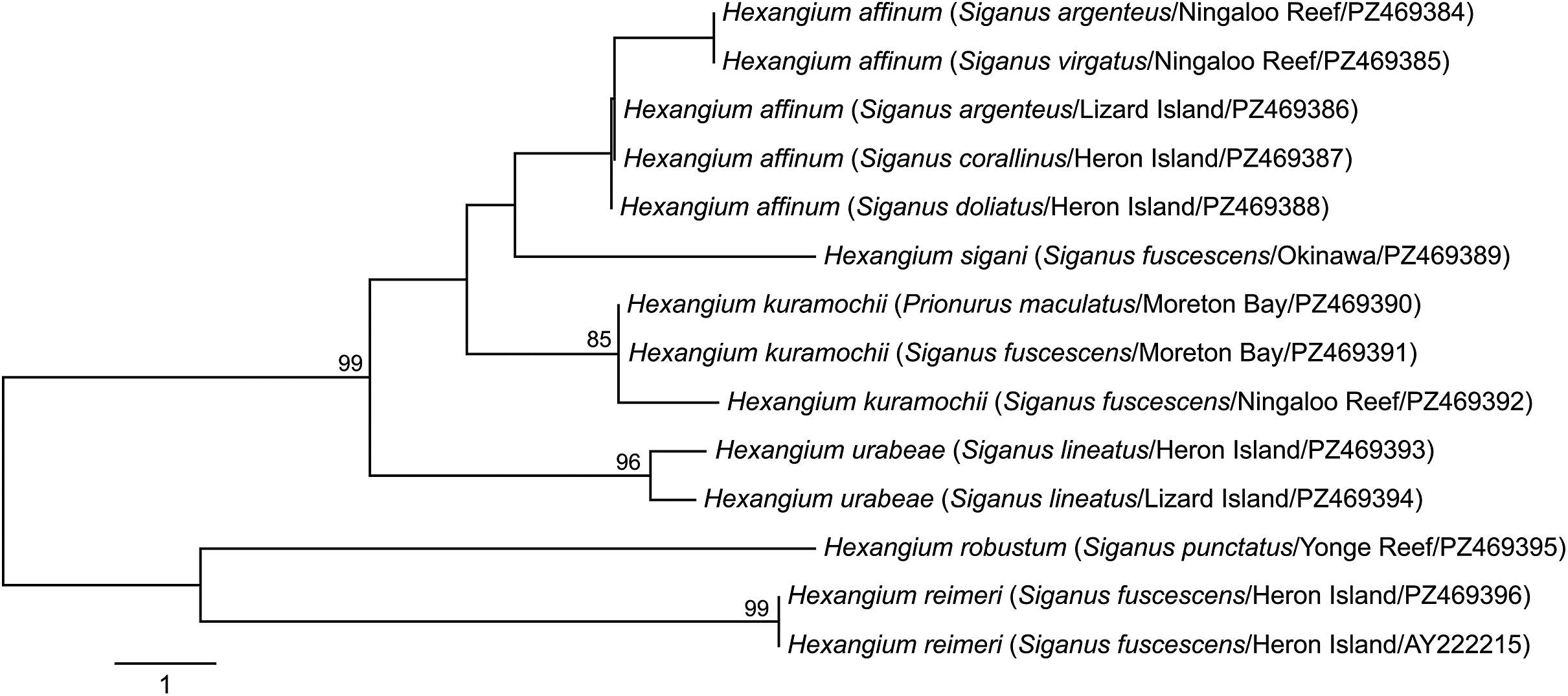


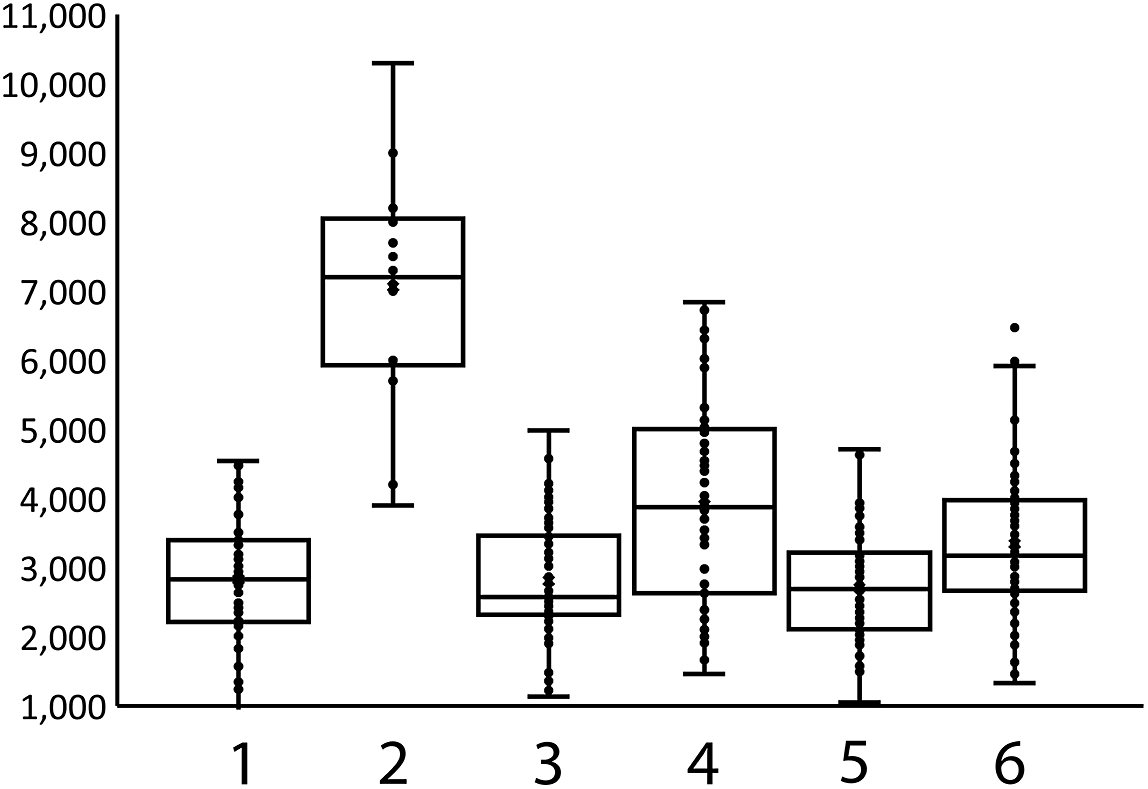


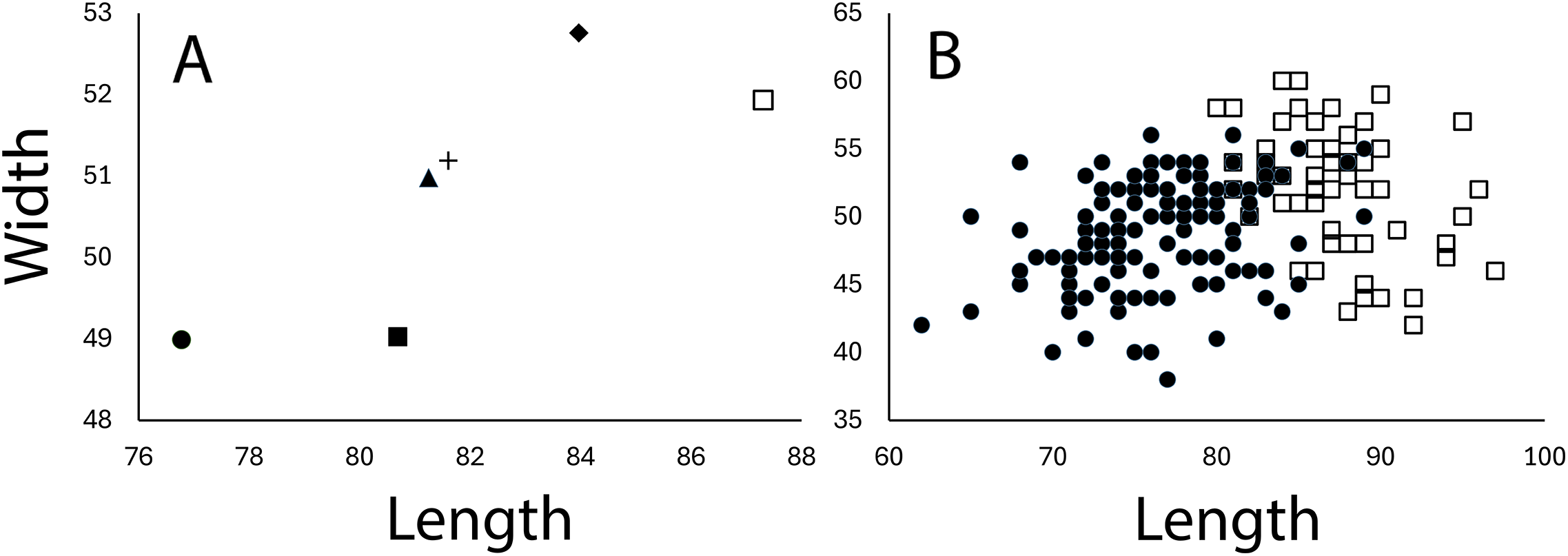

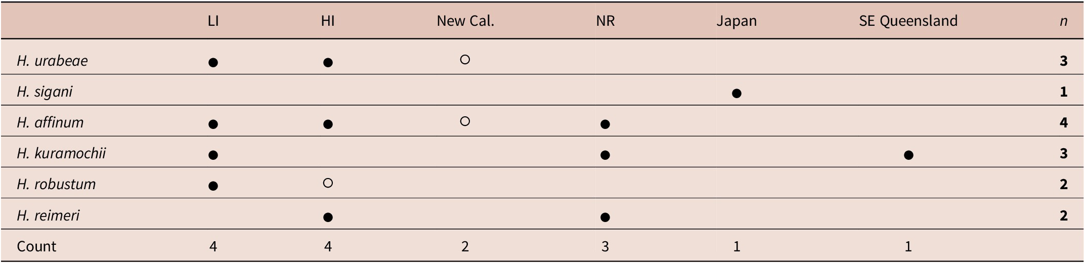


 Open access

