INTRODUCTION
Microsporidia of the order Metchnikovellida live in gregarines, inhabiting the alimentary tract of polychaetes and some other marine invertebrates. Metchnikovellids were first described in the 19th century (Caullery and Mesnil, Reference Caullery and Mesnil1897). Their affiliation with other groups of protists, Haplosporidia, Chitrids or Plasmodiophorales, was debated (Caullery and Mesnil, Reference Caullery and Mesnil1919) until Vivier proposed the microsporidian nature of metchnikovellids based on similarities in spore ultrastructure and intracellular development (Vivier, Reference Vivier1965). Since then metchnikovellids were treated as a deviated group of microsporidia. Sprague, who first claimed Microsporidia as a phylum, erected the class Rudimicrosporea, restricted exclusively to the order Metchnikovellida, in contrast to the class Microsporea embracing all other microsporidia (Sprague, Reference Sprague, Bulla and Cheng1977). The relationships of metchnikovellids with other microsporidia have been always uncertain. Metchnikovellids lack the standard characters of microsporidia, such as polaroplast and coiled polar filament; this group has unusual general spore morphology, different spore wall structure, and can produce spores endogenously inside cysts (Vivier, Reference Vivier1975) that are also called spore sacs (Larsson, Reference Larsson2000; Larsson and Køie, Reference Larsson and Køie2006). Sprague et al. (Reference Sprague, Becnel and Hazard1992) even refrained from placing metchnikovellids within their new classification system, and left the group among the taxa Incertae sedis. Recently, affiliation of Metchnikovellidae with Microsporidia, as well as their position as a basal taxon within the phylum has been provisionally validated by SSU rDNA sequence analyses (Simdianov et al. Reference Simdianov, Yudina and Aleoshin2009), although there is still no metchnikovellid sequence data in the GenBank.
Metchnikovellids have an amazingly long history of discovery, and all 3 valid genera (Metchnikovella, Amphiamblys and Amphiacantha) have been known since the first decades of the 20th century (Caullery and Mesnil, Reference Caullery and Mesnil1914, Reference Caullery and Mesnil1919), with their life cycles described ‘quite correctly’ (Vivier, Reference Vivier1975). The presence of conspicuous spore sacs (‘cysts’), produced by all metchnikovellids, explains their early (compared with most other microsporidia) discovery. Large spore sacs (20–65 μm long) were easily recognized by early microscopists and allowed the differentiation of the genera: elongated sacs with filamentous projections are characteristic for Aphiacantha, long and cylindrical – for Amphiamblus, and oval or slightly bent – for Metchnikovella (Vivier, Reference Vivier1975). Presently, 25 metchnikovellid species are known. However, progress in the study of these protists is greatly impaired by arduous sampling. Only 6 species have been investigated with electron microscopy (Vivier and Schrével, Reference Vivier and Schrével1973; Hildebrand, Reference Hildebrand1974; Desportes and Théodoridès, Reference Desportes and Théodoridès1979; Ormières et al. Reference Ormières, Loubès and Maurand1981; Larsson, Reference Larsson2000; Larsson and Køie, Reference Larsson and Køie2006). Most metchnikovellid species remain poorly described and have been documented solely with light-microscopic data.
At the same time, there are certain reasons to suggest that metchnikovellids play a key role in the evolution of microsporidia and are more closely related to the hypothetical ancestral microsporidium, than any group of the phylum Microsporidia. The presumably ancestral features of metchnikovellids include an undeveloped polar filament, lack of a polaroplast, endogenous enveloped sporogony, and absence of merogonial reproduction. The latter 2 features are shared with some chytridiopsids (Beard et al. Reference Beard, Butler and Becnel1990; Larsson Reference Larsson1993, Reference Larsson2000), the insect microsporidia that were hypothesized to be an evolutionary bridge between metchnikovellids and ‘true’ microsporidia (Weiser, 1997; Larsson, Reference Larsson2000). Another important indicator of the early origin of metchnikovellids might be the parasitizing marine aseptate gregarines, mainly archigregarines and lecudinid eugregarines. Archigregarines are among the earliest diverging lineages within Apicomplexa, and a sort of ‘polyphyletic stem’, from which all other gregarines evolved (Leander, Reference Leander2007). Noteworthy is the fact that insects which share a common ancestor with annelids are the major host group for gregarines (Perkins et al. Reference Perkins, Barta, Clopton, Peirce, Upton, Lee, Leedale and Bradbury2000) as well as for microsporidia (Becnel and Andreadis, Reference Becnel, Andreadis, Wittner and Weiss1999). Gregarines harbouring ancestral microsporidia might be a possible avenue of transferring microsporidia from marine to freshwater and terrestrial hosts. Playing a ‘Trojan horse’, gregarines might have enabled dispersion of microsporidia among marine and freshwater arthropods, and their ultimate establishment as parasites of insects.
Since 1984 metchnikovellid infections have been reported occasionally in gregarines Polyrhabdina sp. from the intestines of polychaetes Pygospio elegans inhabiting the silt littoral zone in the vicinity of the Biological Station of St. Petersburg State University at the White Sea (Rotari, Reference Rotari1988; Rotari and Paskerova, Reference Rotari and Paskerova2007). Some gregarines contained cysts, easily identified in the light microscope as Metchnikovella incurvata Caullery and Mesnil, Reference Caullery and Mesnil1914, by the characteristic boomerang-shaped (curved) sporocysts with thickenings at both ends, containing up to 16 spores (Vivier, Reference Vivier1975; Caullery and Mesnil, Reference Caullery and Mesnil1914, Reference Caullery and Mesnil1919). Only 2 species of the genus Metchnikovella, Metchnikovella hovassei (Vivier and Schrével, Reference Vivier and Schrével1973) and Metchnikovella wohlfarthi (Hildebrand and Vivier, Reference Hildebrand and Vivier1971; Hildebrand, Reference Hildebrand1974), have been studied with the electron microscope so far, the latter being quite fragmentary, without any description of the sac-bound sporogony (SBS). Here we describe the ultrastructure of the third species of the genus, M. incurvata Gaullery and Mesnil, 1914, and compare it with data on the fine structure of other metchnikovellids.
MATERIALS AND METHODS
Polychaetes, P. elegans Claparède, 1863 were collected in July–August during 2009–2011 at the silt littoral zone of the Levin reach (66°17·878′N, 33°27·774′E), Chupa Inlet, Kandalaksha Gulf of the White Sea. Polychaetes were dissected with needles and examined under a dissecting microscope. Gregarines provisionally identified as Polyrhabdina sp. (Apicomplexa: Lecudinidae) were either attached to the gut epithelium, or occurred freely in the gut lumen. Infected gregarines were observed and photographed using the Nomarski contrast optics of a Leica DM 2500 microscope equipped with Plan-Apo objective lenses. Infected gregarines were differentiated by the presence of conspicuous cysts or oval inclusions inside the cytoplasm. All gregarines from the polychaete in which at least 1 gregarine was infected, were fixed for electron-microscopic examination.
For electron microscopy small pieces of polychaete intestine with the attached gregarines, or free gregarines released from the gut lumen, were fixed in 2·5% glutaraldehyde made on 0·2 m cacodylate buffer containing 0·05% MgCl2 (pH 7·4, final osmolarity 700 mOsm) for 1–4 h, washed in filtered seawater and post-fixed in 2% osmium tetroxide solution in the same buffer for 1 h at 4 °C. Gregarines were dehydrated in an ascending ethanol series, transferred to acetone and embedded in Epon-Araldite in flat moulds. Embedded samples were examined in bright field under the 5× objective of an inverted OLYMPUS IX50 microscope, and those gregarines that contained cysts and suspicious inclusions were sectioned with an ultratome MT XL (RMC products). Thick (0·5–1 μm) sections were stained with methylene blue, examined and photographed under a Zeiss Axioplan microscope equipped with a Microfire digital camera. Thin (70–80 nm) sections were stained with uranyl acetate and lead citrate and examined in a JEOL JEM 1011 microscope with the attached HAMAMATSU ORCA-HR digital camera. Four gregarines were sectioned and examined under the electron microscope.
RESULTS
Prevalence and light microscopy
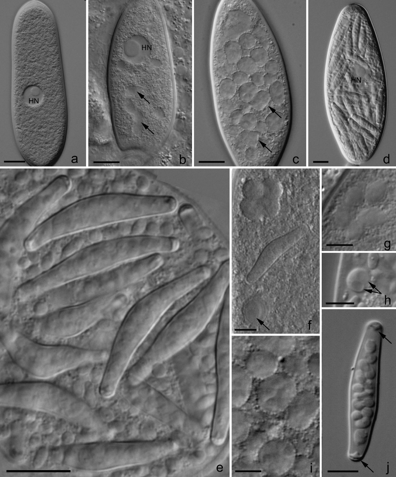
The alimentary tracts of 43·5% (145 of 333) of the dissected polychaetes were parasitized by gregarines of the genus Polyrhabdina Mingazzini, 1891 (Fig. 1a). The average number of gregarines per polychaete was 3·5, ranging from 1 to 50. Gut lumens of 7·6% (11 of 145) of the polychaetes contained at least 1 metchnikovellid-infected gregarine. Various rounded and oval inclusions inside the cytoplasm (Fig. 1b, c, f, and i) as well as conspicuous sacs with spores (Fig. 1d and e) were observed in 3·9% (20 of 509) of gregarines liberated from such polychaetes. Most gregarines with detectable inclusions contained elongated boomerang-shaped spore sacs 22–27 μm long and 4–5 μm wide (24·2 ± 2·6 × 4·6 ± 0·3 μm, n=35) with characteristic plugging structures at both ends (Fig. 1e and j). Sac-bound spores were oval and measured 3·6 ± 0·3 × 1· ± 0·1 μm (n=8) (Fig. 1j). Fresh and Epoxy resin-embedded gregarines showed up to 16 spores in each spore sac. Together with the sacs, the cytoplasm of infected gregarines contained numerous free spores (3·7 ± 0·4 × 1·8 ± 0·2 μm, n=12) with the same morphology (Fig. 1e). Infected gregarines usually were shorter and wider compared with non-infected ones; at the advanced stages of infection their nuclei were concealed by masses of spore sacs (Fig. 1d and e). Although it is hard to ascertain the maximum number of spore sacs per single gregarine, as many as 30 were visible in 1 focal plane. Spore sacs were arranged chaotically inside the host cytoplasm. Even when tightly packed with spore sacs, gregarines were able to glide.
Light microscopy, Nomarski contrast: live gregarines Polyrhabdina sp. from intestines of Pygospio elegans infected with Metchnikovella incurvata. (a) A non-infected gregarine slightly pressed with the coverslip; (b) A gregarine containing chains of interconnected cells, the earliest observed stage of the sac-bound sporogony (SBS) sequence (arrowed); (c) A gregarine filled with sporogonial plasmodia of the free sporogony (FS) sequence; 2 of many visible plasmodia are arrowed; (d) Multiple spore sacs are arranged chaotically inside the host cell. The host nucleus is concealed by a mass of spore sacs; (e) Spore sacs in the cytoplasm of the gregarine under high magnification. The space between the spore sacs is filled with free spores. Up to 16 spores are visible inside a spore sac; (f) A sac with spores, a uninucleate cell with remarkably large nucleus (arrowed), possibly ‘the sporogonial mother cell’ of FS, and a group of cells, likely corresponding to a stage of division of FS sporogonial plasmodia; (g) Another fragment of the gregarine shown on (f): a group of uninucleate cells, corresponding probably to sporonts of FS sequence; (h) A binucleate cell, one of those appearing at the onset of SBC (arrows indicate nuclei); this gregarine also contained numerous free spores and spore sacs. (i) Multinucleate sporogonial plasmodia of FS; (j) A spore sac released from the gregarine, displays an elongated boomerang shape and characteristic ‘plugging’ structures at both ends (black arrows). Scale bars, a–e, 10 μm; f–j, 5 μm. HN, nucleus of the gregarine.

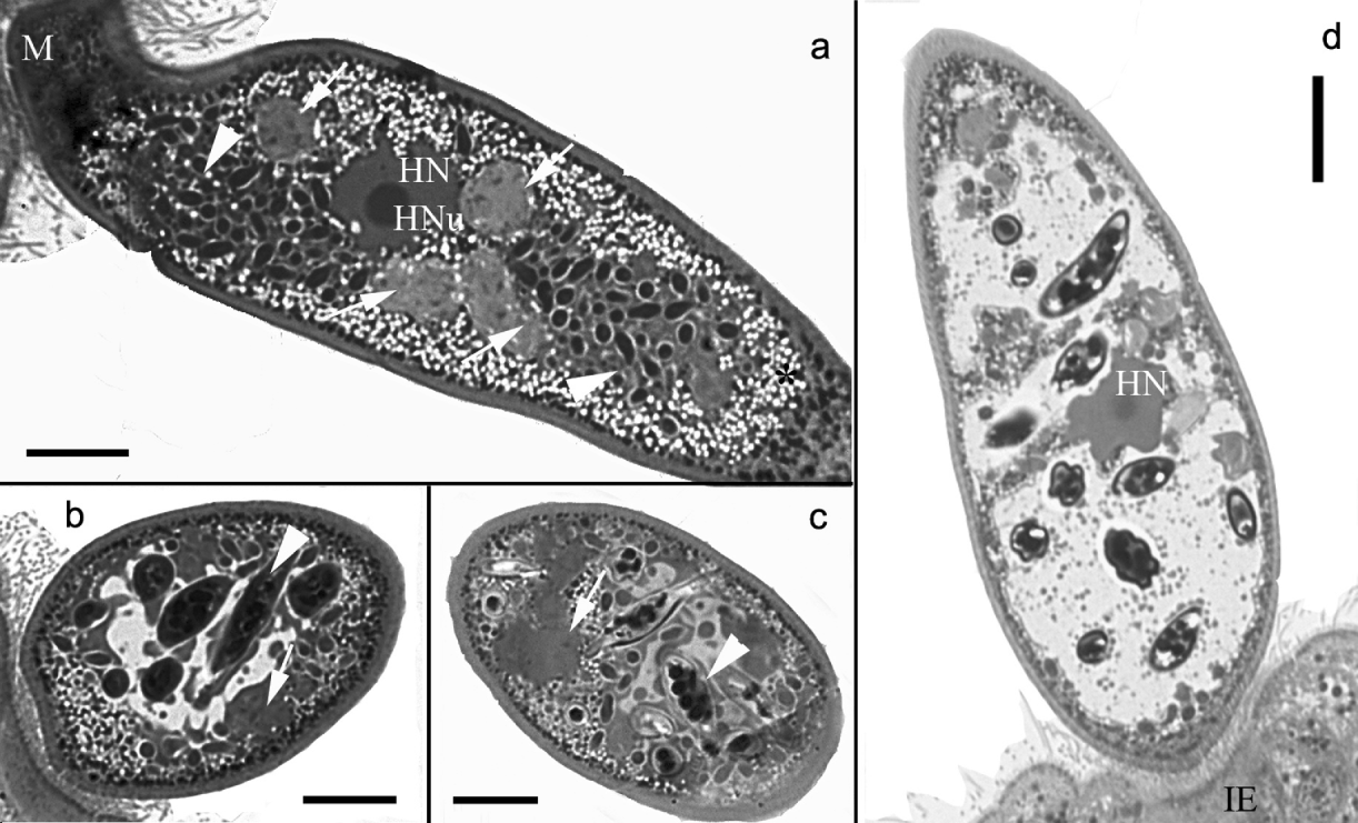
Sections through infected gregarines demonstrated plasmodia with numerous nuclei, free spores inside an amorphous mass of material, presumed pre-spore stages, and spore sacs with 2 rows of spores. Infected gregarines that did not contain spore sacs displayed relatively normal cytoplasm with amylopectin and protein granules scattered in the regions of the cell free of parasite developmental stages (Figs 1b and 2a). The host gregarine cytoplasm appeared progressively empty as the number of spore sacs increased (Figs 1c, d, 2b and d). At the end of the infection cycle the gregarine host appeared as a bag filled with metchnikovellid spore sacs (Fig. 1d). The remnants of cytoplasm were pushed to the very periphery of the cell (Fig. 2d).
Light microscopy, bright field: thick sections through gregarines stained with methylene blue. (a) Longitudinal section through the infected gregarine: plasmodia with numerous nuclei (arrows), spores and pre-spore stages of the free spore sequence (arrowheads) are displayed. Gregarine nucleus (HN) with nucleolus (HNu) is visible. Cytoplasm when free of parasites is filled with amylopectin and protein granules (asterisk); (b, c) Oblique (b) and transverse (c) sections through infected gregarines. Central region of the cytoplasm is occupied by spore sacs. Round spores inside spore sacs are arranged in 2 rows (arrowheads). Plasmodia are arrowed; (d) Gregarine at the later stage of pathogenesis. Practically no structures are left inside the cells, except for an invaginated nucleus (HN) and spore sacs. The remnants of the cytoplasm are pushed to the very periphery of the cell. IE, intestinal epithelium of the worm; M, attachment organelle of aseptate gregarines; HN, gregarine nucleus; HNu, gregarine nucleolus. Scale bar, 10 μm.
Ultrastructure
Ultrastructural study confirmed data obtained by light microscopy. The parasite life cycle included 2 types of sporogony.
Free sporogony (FS) sequence
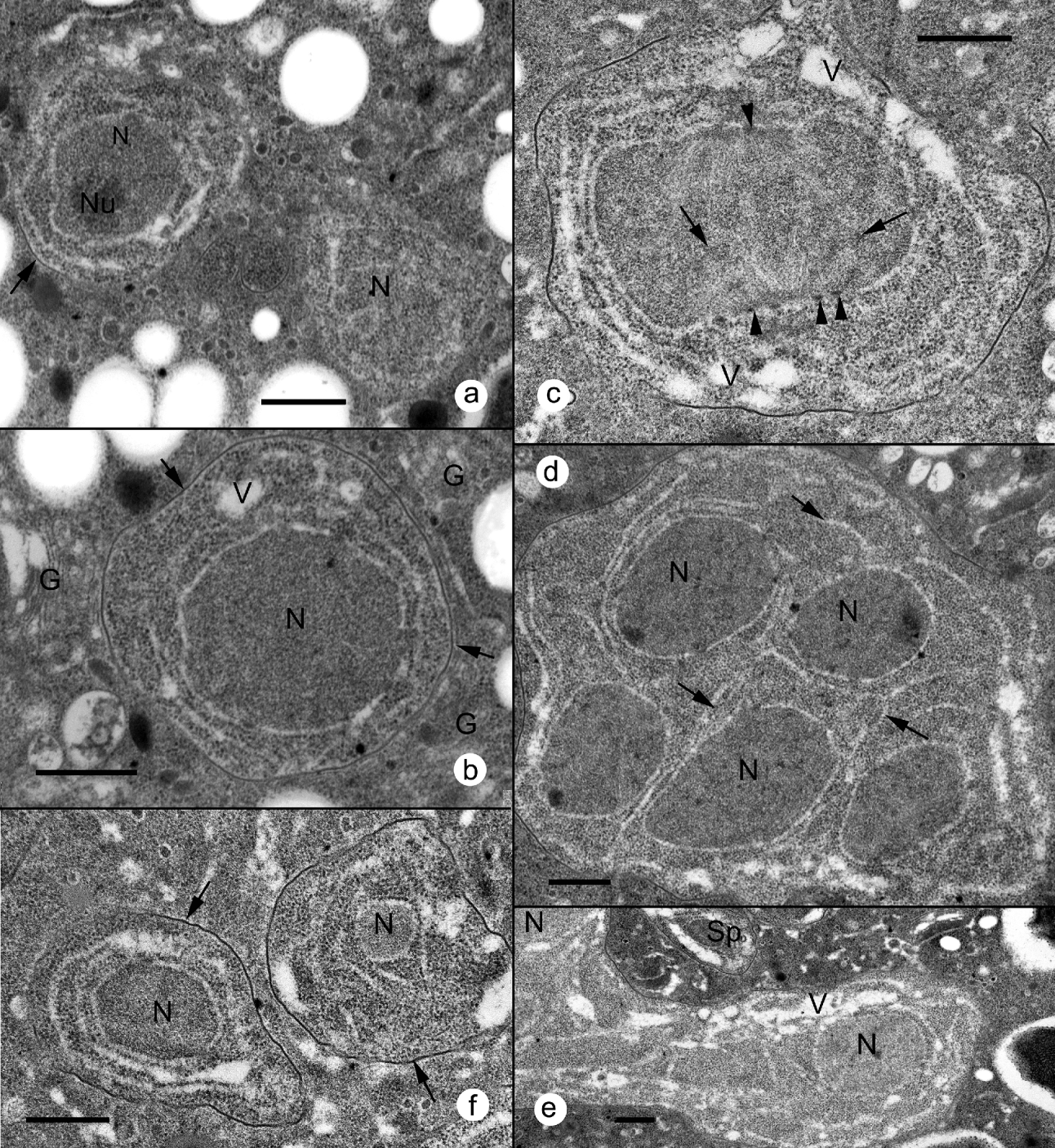
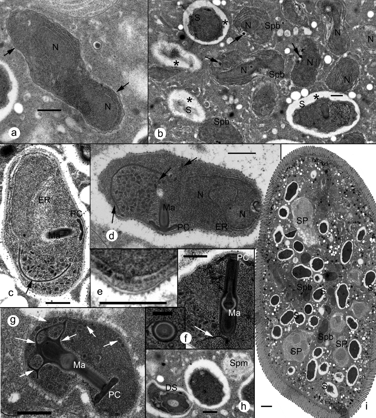
The earliest stages observed were round or oval cells, that measured 1·2–1·3 μm in diameter (Fig. 3a). We never observed nuclear divisions in cells of this type. Probably, the subsequent stage of parasite development were larger cells (1·5–1·6 μm in diameter), with vacuolated cytoplasm, and a layer of electron-dense material approximately 10 nm thick, deposited along the plasma membrane (Fig. 3b). Cells of this type (‘sporogonial mother cells’) further enlarged in volume, underwent several nuclear divisions, and formed irregularly shaped ‘sporogonial plasmodia’ measured on sections 10–20 μm across, with up to 8 rounded or oval nuclei visible in a single section (Fig. 3d shows 5 nuclei). During the further development, internal membranes segregated individual nuclei inside a plasmodium and split it into uninuclear daughter cells. Portions of cytoplasm surrounding a nucleus, often budded off the mother cell (Fig. 3e). The next stage of this sequence were sporonts (Fig. 3f) – irregularly shaped cells, 1·1–1·9 μm in diameter, covered with an electron-dense homogeneous envelope 15 nm thick. We do not know whether they multiplied further or transformed directly into sporoblasts (Fig. 4a and b). Sporoblasts displayed slightly undulating homogeneous envelopes of about 18 nm (range 15–21 nm) and electron-dense cytoplasm. Vacuoles with electron-dense bodies, probably the precursors of the ‘manubrium’ and ‘polar cap’ sensu terminology of Vivier (Reference Vivier1975) were observed in some sporoblasts (Fig. 4b).
Electron microscopy of the free sporogony: ultrastructure of proliferative stages. (a) The earliest stage observed, early sporonts surrounded by a thin inconspicuous membrane. The nucleus (N) of the left cell contains a nucleolus (Nu); (b) More advanced stage of development marked by deposition of electron-dense material on the plasma membrane (arrows). Golgi dictyosomes of the host cell (G), adjacent to the parasite interface; (c) Sporogonial mother cell, a precursor of the sporogonial plasmodium, undergoing mitosis. Spindle microtubules are attached to centriolar plaques built-in in the nuclear membrane, and with opposite ends – to centromere regions of chromosomes (arrows). The presence of numerous centriolar plaques (arrowheads) suggests multiple divisions of the nucleus; (d) Cross-section through irregular-shaped sporogonial plasmodium with 5 nuclei in the view. Numerous negatively stained membrane profiles are indicated by arrows; (e) Portion of cytoplasm with a nucleus splitting off the sporogonial plasmodium; (f) Sporonts are enclosed in electron-dense envelopes about 15 nm thick (arrow). G, host cell Golgi dictyosome; N, parasite nucleus; Nu, nucleolus of the parasite nucleus; Sp, sporont; V, vacuoles. Scale bar, 500 nm.
Electron microscopy of the free sporogony: ultrastructure of sporoblasts and spores. (a) Sporoblast with characteristic ‘undulating’ envelope (arrow). The edges of the horseshoe-shaped nucleus (N) are cross-sectioned; (b) Sporoblasts (Spb) with electron-dense cytoplasm and vacuoles containing precursors of the anchoring disk (arrows). Spores (S), unlike sporoblasts, are surrounded by electron-lucent zones (asterisks); (c, d) Spores displaying typical (for metchnikovellids) shape and sets of organelles. Note horseshoe-shaped nucleus (d) and a network of manubrial cisterns (arrows) seen occasionally as short tubules (arrows); (e) Enlarged portion of the d, displaying tripartite spore wall ornamented with electron-dense globules of about 20 nm in diameter; (f) Longitudinal section through a polar cap (PC) and manubrium (Ma), composed of layers of alternating electron density. A manubrial cistern approaches the distal thicker end of the manubrium (arrow). Insert shows transverse section through the manubrium; (g) Manubrium at its distal part splits into several branching cisterns which produce an anastomosing network of tubules (arrows); (h) Section, demonstrating an ‘empty’ discharged spore (DS), and a sporoplasm (Spm); (i) Section through a gregarine infected exclusively with the stages of free sporogony sequence: the host cytoplasm is homogeneous and looks practically normal in areas not occupied by parasites. DS, discharged spore; ER, endoplasmic reticulum; Ma, manubrium; N, parasite nucleus; PC, polar cap; S, spores, SP, sporogonial plasmodium; Spb, sporoblast; Spm, sporoplasm. Scale bar, a–e, g, h, i, 500 nm; f, 250 nm; i, 2 μm.
Spores demonstrated ultrastructure similar to that of other metchnikovellids, including the most conspicuous feature – the apical-distal axis, the line connecting the centre of the polar cap to the manubrium axis (referred to here as height), perpendicular to the long axis of the spore (Fig. 4c and d) (referred to here as width). Mature spores were surrounded by electron-lucent areas, without a delineating membrane. These ‘halos’, like in many microsporidia, probably resulted from cytoplasmic shrinkage during spore formation, an effect enhanced by chemical fixation. The spore envelope was about 20–24 nm thick and displayed 3 layers: a 6–7 nm thick plasma membrane, an electron-lucent endospore approximately twice that thick, and a thin exospore ornamented with electron-dense globules, about 20 nm in diameter each (Fig. 4d and e). On sections, the spores were roundish to oval, and slightly angular at the top of the polar cap. Spores measured in sections were 1·22–1·94 μm in height and up to 3·4 μm in width (Fig. 4c and d). The electron-dense manubrium traversed the shorter axis. Sectioned longitudinally it displayed a complex layered structure (Fig. 4f). On transverse sections the manubrium was composed of concentric circles of alternating electron density (Fig. 4f, insert). The manubrium was cylindrical and maximally 1·1 μm long, a bit thinner at the apical end (c. 190 nm) and thicker at the distal ‘bulbal’ part (up to 265 nm) (Fig. 4f). It was not connected with the polar cap (Fig. 4c, d and f). The polar cap was slightly constricted in the middle and measured about 50 nm in the centre and about 80 nm at the peripheral parts (Fig. 4d). Most spores sectioned through the polar cap–manubrium plane showed a structure referred to by previous authors as ‘a semicircular fold’ (Larsson and Køie, Reference Larsson and Køie2006) or ‘manubrial lamella’ (Ormières et al. Reference Ormières, Loubès and Maurand1981) and termed here ‘a manubrial cistern’ to highlight the shape of the organelle and its association with the manubrium (Fig. 4g). In most sections the manubrial cistern derived from the distal part of the manubrium, ran parallel to the spore wall, and was associated with a cluster of short tubular-like structures (Fig. 4f). In some sections not just one but several such cisterns stemmed from the bulbous (distal) end of the manubrium. Serial sectioning revealed that, in fact, the distal part of the manubrium split into several cisterns which branched and gave birth to an anastomosing network of tubules which, on transverse sections through this network, were seen as short tubules or vesicles (Fig. 4 g). The horseshoe shape of the nucleus (Fig. 4d) could be revealed only by examination of multiple successive sections, because on random sections nuclei were commonly irregularly shaped. The rough endoplasmic reticulum was located inside the cell compartment formed by the concave surface of the nucleus (Fig. 4c). Some spores lacked typical internal structures and contained only membrane contours (Fig. 4h). Those were probably discharged spores. In the vicinity of such ‘empty’ spores electron-lucent bodies of 1–1·5 μm in diameter, presumably sporoplasms, were occasionally observed (Fig. 4h). In 2 of 4 examined gregarines only the FS stages were observed. The cytoplasm of the gregarines in which we observed only this type of sporogony, looked practically normal in areas not occupied by the parasites (Fig. 4i).
Sac-bound sporogony
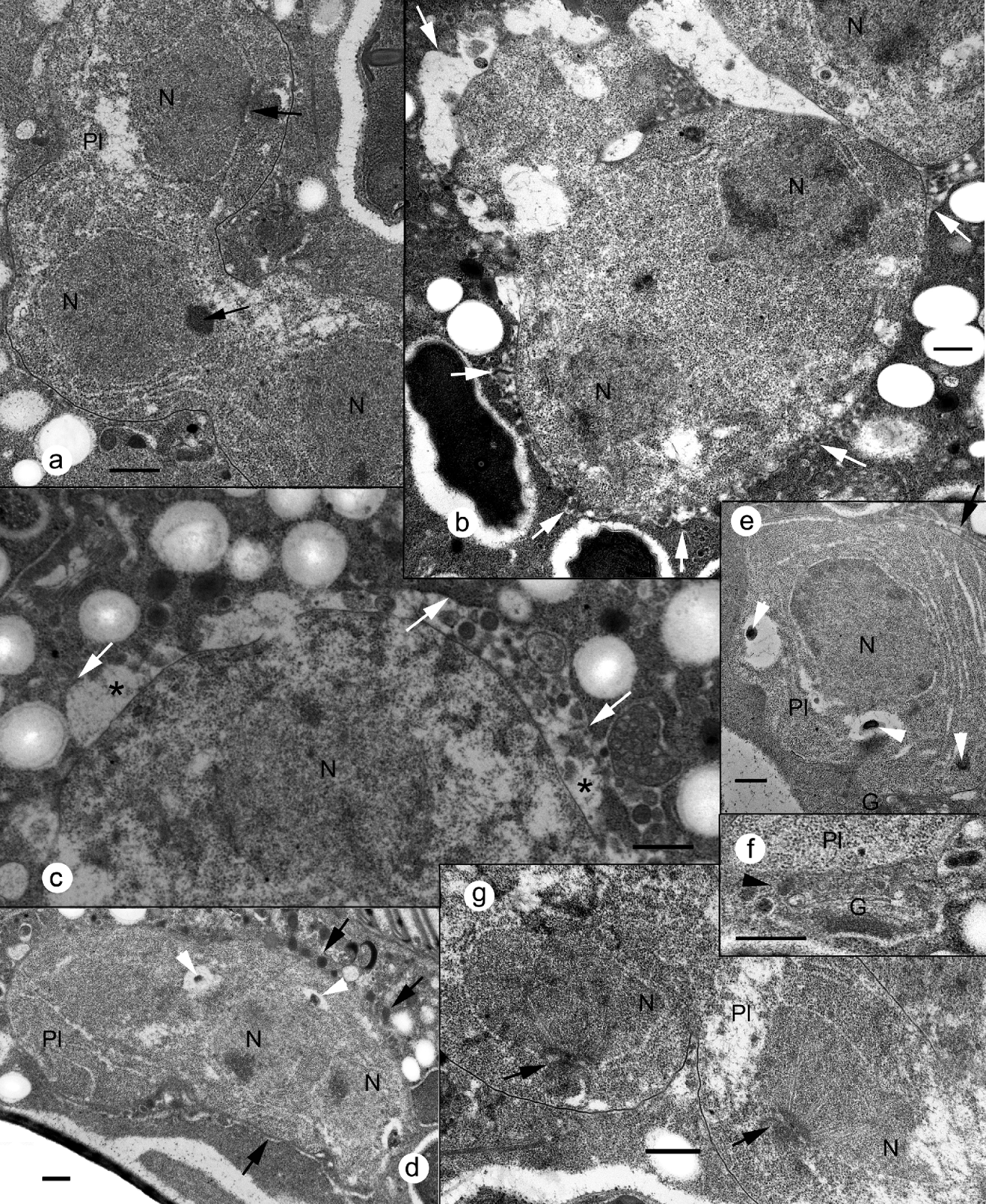
The earliest observed SBS stage were plasmodia seen on sections as chains of 2–8 large (3–6 μm in diameter) bi- or mononucleate cells interconnected by broad cytoplasmic bridges (Fig. 5a). Individual binucleate cells were occasionally observed as well (Fig. 5b). These could be either sections through a chain or a result of fragmentation of the chain into individual cells. The most conspicuous feature distinguishing these cells from the sporogonial mother cells of the FS sequence, was the structure of their interface zone: they were separated from the host cytoplasm by an electron-lucent, membrane-bound area of varied width (Fig. 5b and c). Protrusions of this zone (up to 2 μm long) penetrated the surrounding cytoplasm and formed a network around the parasite (not shown).
Electron microscopy of the sac-bound sporogony: ultrastructure of proliferative stages. (a). Sporogonial plasmodium: a chain of cells interconnected by wide cytoplasmic bridges; nuclei are undergoing divisions. Spindle microtubules and massive spindle plaques (microtubule organizing centres) are indicated by arrows; (b) A binucleate cell surrounded by an additional envelope (arrows); (c) At more advanced steps of the parasite development perivacuolar space expands (asterisks); the vacuole membrane is indicated by an arrow; perivacuolar space (asterisk) contains granules; (d, e) Longitudinal (d) and transverse (e) sections through plasmodium (Pl): vacuoles with electron-dense granules inside the cytoplasm and in vicinity of the nucleus (arrowheads); (f) Trans surface of Golgi dictyosome (G) faces the plasmodium (arrowhead); (g) Dividing nuclei: spindle microtubules radiate from microtubules organizing centre (arrows); chromosomes are attached to the distal ends of spindle microtubules. G, host cell Golgi apparatus; Pl, plasmodium. Other abbreviations are the same as in Figs 3 and 4. Scale bar, 500 nm.
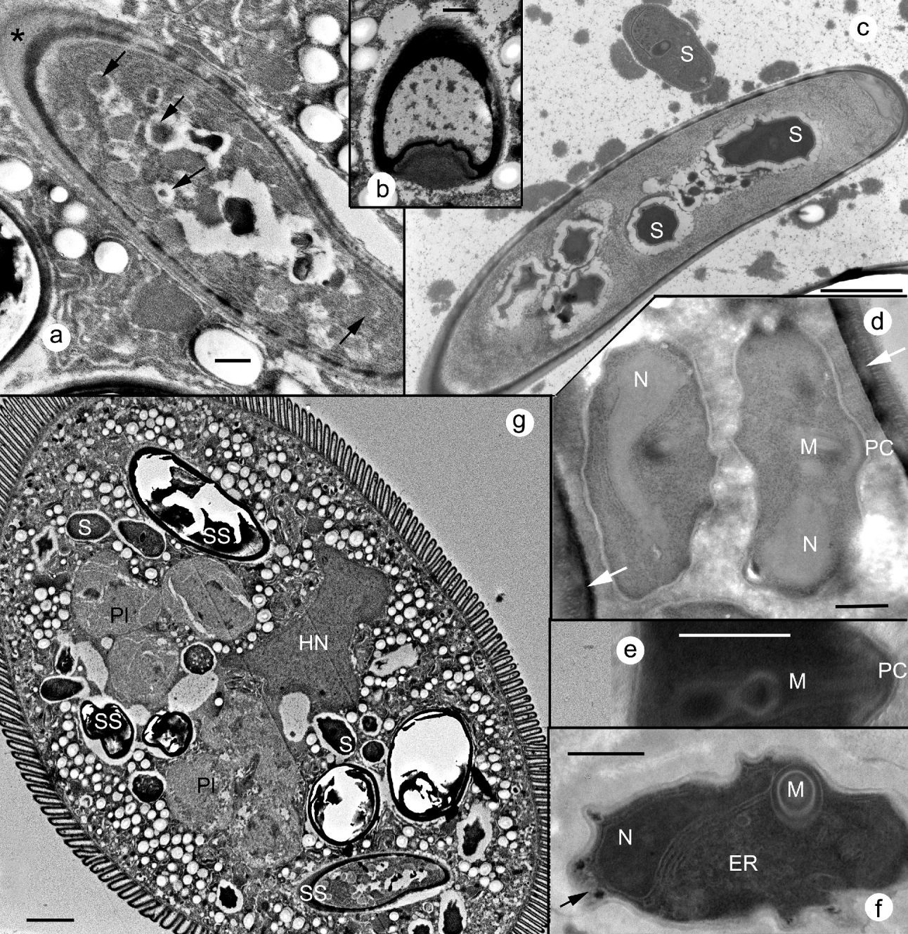
At the advanced stage of SBS, the perivacuolar space between the parasite plasmalemma and the external envelope increased; it often contained granules of various electron density (Fig. 5c and d). Vacuoles with similar electron-dense granules and/or whorls of membranes were occasionally seen budding off the perinuclear space or inside the cytoplasm of the parasite (Fig. 5d and e). Interestingly, host cell Golgi dictyosomes were constantly observed in close vicinity to plasmodia. Their trans cisternae with budding off granules faced the parasite surface (Fig. 5f). All nuclei of the plasmodium were constantly observed in the process of multiple nuclear divisions (Fig. 5a, b and g). Microtubule organizing centres with radiating microtubules (Fig. 5g) were numerous. Finally, plasmodium split into individual sporonts. The following nuclear divisions and the process of transformation of a perivacuolar space filled with electron-dense granules, into a thick rigid wall of a spore sac was not monitored. The next observed stage was a spore sac in which segregation of individual sporoblasts took place (Fig. 6a). The spore sac wall seemed nearly completed at this stage. This thick wall obstructed fixation and infiltration of spore sacs. As a result, the contents of most spore sacs were not preserved. Spore sacs on sections were about 20 μm in length and had characteristic thickenings at the ends (Fig. 6b) corresponding to the plugging structures seen on thick sections. The thickness of the sac wall which was composed of several layers of amorphous material (Fig. 6c and d), varied from 161 to 263 nm (204 nm in average). In a few relatively well-preserved spore sacs mature spores could be seen (Fig. 6c and d). Sac-bound spores were slightly smaller in size (2·54 × 1·12 μm average, range 2·41–2·65 × 1·04–1·22 μm) and had thicker envelopes (44·8 μm in average, range 32·3–64·5 μm) compared with free spores. The exospore surface was smooth, without 20 nm globule-like structures. Instead, electron-dense structures, roundish on transverse sections, were located beneath the envelope in the sites where the spore envelope formed sorts of folds (Fig. 6f). In other respects the internal structure of the spores developing inside the spore sacs looked similar to that of the free spores (compare Fig. 6d and f; Fig. 4c, d and f).
Electron microscopy of the sac-bound sporogony: spore sacs and spores. (a) Segregation of individual sporoblasts (arrows) inside a rigid wall of the spore sac (asterisk); (b) Transverse section through the thickening at the very distal end of the sac; (c) A relatively well-preserved spore sac with mature spores inside. Note a free spore outside the spore sac of similar size and structure; (d) Two mature spores inside a spore sac; arrows point to multilayered spore sac walls; (e) Longitudinal section through the manubrium and polar cap; (f) A mature spore with smooth exospore; spore envelope forms ‘folds’ with electron-dense structures inside (arrows); (g) In gregarines displaying stages of SBS, only free spores but no other stages of the free sequence could be seen. HN, host cell nucleus; HNu, host cell nucleolus; SS, spore sac. Other abbreviations are the same as in Figs 3–5. Scale bar, a–f, 500 nm; g, 2 μm.
Maturation of the spore sacs caused disintegration of the gregarine cytoplasm, preceded by migration of the host nucleus together with the remnants of the intact cytoplasm to the cell periphery. In gregarines displaying SBS stages, only free spores but no other stages of the FS sequence were seen (Fig. 6g).
DISCUSSION
The identification of microsporidia is challenging, especially when the species was described long ago, before the era of electron microscopy and sequence analysis. The shape and size of spore sacs and the number of spores within a sac do not allow differentiation of the White Sea microsporidium from the species M. incurvata Caulleri and Mesnil Reference Caullery and Mesnil1914. Metchnikovella incurvata was originally described from gregarines Polyrhabdina pygospionis (family Lecudinidae) in a polychaete Pygospio seticornis inhabiting the coasts of the English Channel. A few arguments favour the opinion that Caulleri and Mesnil misidentified the polychaete species. Without going deeply into polychaete taxonomy we assume, following Tuzet and Ormiére (Reference Tuzet and Ormières1962), that Caullery and Mesnil were dealing with Pygospio elegans, a species widely distributed along the coast of the English Channel, but not with P. seticornis, particularly given that the latter has not been recorded in the area and is missing from the list of the English Channel polychaetes (Dauvin et al., Reference Dauvin, Dewarumez and Gentil2003). Therefore, both polychaete and gregarine hosts of M. incurvata from the English Channel are either identical or closely related to those of the microsporidium from the White Sea littoral, supporting our identification.
The only discrepancy between the characters of our isolate and those mentioned in the original description of M. incurvata is the spore shape and size. The original description mentions ‘ovoid spores of about 1 μm in diameter’. Depending on the view angle, spores of our isolate, when observed inside spore sacs, could be seen as round to ovoid bodies measuring from 1·8 to 3·6 μm in diameter. We consider the discrepancy in spore size not to be decisive. After all, a linear size of 1 μm is close to the limits of resolution of the early bright-field microscopes, and in the year 1914 neither phase nor differential contrast optics were yet invented. So, we believe that measurements made in 1914 were much less accurate than nowadays. Certain size divergence might also be due to variations among geographical isolates. Considering cyst shape and size, number of spores in the cyst and host specificity, we prefer to identify our isolate as M. incurvata Caullery and Mesnil Reference Caullery and Mesnil1914.
Round or oval cells ‘delimited by plasma membrane without external reinforcement’ (Larsson, Reference Larsson2000) with 1 (Metchnikovella and Amphiamblys) or 2 (Amphyacantha) nuclei were recorded at the onset of free sporogony in all EM descriptions of metchnikovellids (Vivier and Schrével, Reference Vivier and Schrével1973; Desportes and Théodoridès, Reference Desportes and Théodoridès1979; Larsson, Reference Larsson2000; Larsson and Køie, Reference Larsson and Køie2006). Nuclear divisions were not detected at this stage. In these ‘early sporonts’ (Larsson, Reference Larsson2000) we regularly observed nucleoli (Fig. 3a), which suggested that the cells were at the interphase stage of the nuclear cycle. Similar to the descriptions given for other species (Vivier and Schrével, Reference Vivier and Schrével1973; Desportes and Théodoridès, Reference Desportes and Théodoridès1979; Larsson, Reference Larsson2000; Larsson and Køie, Reference Larsson and Køie2006), early sporonts of M. incurvata grew in size, thickened their envelopes, underwent multiple nuclear divisions and finally gave rise to multinuclear sporogonial plasmodia. Our observations support the idea that metchnikovellids lack merogony, and proliferate via 2 sporogonies, free and sac-bound, the latter following the former (Larsson, Reference Larsson2000; Larsson and Køie, Reference Larsson and Køie2006). All stages of the free sporogony developed in M. incurvata in direct contact with host cell cytoplasm, whereas in previously studied polysporoblastic metchnikovellids, the free sporogony occurs within a parasitophorous vacuole limited by the mother cell plasmalemma (Hildebrand and Vivier, Reference Hildebrand and Vivier1971; Vivier and Schrével, Reference Vivier and Schrével1973; Larsson and Køie, Reference Larsson and Køie2006). It is likely that the presence or absence of interfacial envelopes is a flexible characteristic amongst congeners: for example, the vacuole membrane persisted in M. wohlfarthi (Hildebrand and Vivier, Reference Hildebrand and Vivier1971) and M. hovassei (Vivier and Schrével, Reference Vivier and Schrével1973) until the formation of free spores, and was absent in M. incurvata. In Amphiamblys capitellides vacuoles disappeared during maturation of sporoblasts (Larsson and Køie, Reference Larsson and Køie2006), whereas they were not observed at all in Amphiamblus laubieri, in which the plasmodia were surrounded by the host ER-type membranes (Desportes and Théodoridès, Reference Desportes and Théodoridès1979). Similar to M. hovassei (Vivier and Schrével, Reference Vivier and Schrével1973), M. incurvata sporogonial plasmodia split into individual sporonts by a mechanism involving the isolation of the cytoplasm adjacent to nuclei by internal membranes, and eventual budding off the mother cell. An additional envelope with protrusions penetrating inside the host cytoplasm, built around plasmodia at the onset of bound sporogony sequence, resembled the same structure in M. wohlfarthi (Hildebrand, Reference Hildebrand1974).
The ultrastructure of M. incurvata spores was standard for metchnikovellids (Vivier and Schrével, Reference Vivier and Schrével1973; Larsson, Reference Larsson2000; Larsson and Køie, Reference Larsson and Køie2006). Because metchnikovellids possess only analogues of extrusion organelles of ‘true’ microsporidia, structurally and functionally dissimilar to the latter, we prefer to use the terminology exploited in earlier publications on metchnikovellids (reviewed by Vivier, Reference Vivier1975), such as manubrium and polar cap instead of a manubroid polar filament and polar sac (Larsson, Reference Larsson2000).
Spores contained a horseshoe-shaped nucleus, distinctive for the genus Metchnikovella (Vivier and Schrével, Reference Vivier and Schrével1973). In terms of dimensions, M. incurvata spores measured on thin sections (1·5 × 2·9 μm, free spores; 1·1 × 2·5, sac-bound spores) resembled at most A. capitellides free spores (1·7 × 2·8 μm) (Larsson and Køie, Reference Larsson and Køie2006). Metchnikovella incurvata spores were more elongated compared with M. hovassei (1·3 × 1·8 μm) (Vivier and Schrével, Reference Vivier and Schrével1973) and M. wohlfarthi (1·9 × 3·0 μm) (Hildebrand and Vivier, Reference Hildebrand and Vivier1971).
The present study shows that the posterior end of the manubrium splits into a network of branching cisterns and tubules. Sections through this network were regularly observed in metchnikovellids, and were previously described as a ‘semicircular fold in touch with a system of tubule like vesicles’ (Larsson and Køie, Reference Larsson and Køie2006) or ‘… a manubrial lamella … followed a plasma membrane …’ with … ‘circular elements observed in the concavity of this lamella’ (Desportes and Théodoridès, Reference Desportes and Théodoridès1979; Ormières et al. Reference Ormières, Loubès and Maurand1981). This network of manubrial cisterns may be a part of the late secretory compartment, the trans Golgi network, homologous both to the posterior vacuole (or posterosome, in many species containing a pool of primordial polar filament precursors), and to the containers with polar filament coils. The latter established long ago as a part of the microsporidian trans Golgi (Takvorian and Cali, Reference Takvorian and Cali1994). Recently, it has been shown that secretory transport in microsporidia occurs by progression maturation of cisterns without participation of vesicles (Beznoussenko et al. Reference Beznoussenko, Dolgikh, Seliverstova, Semenov, Tokarev, Trucco, Micaroni, Di Giandomenico, Auinger, Senderskly, Skarlato, Snigirevskaya, Komissarchik, Pavelka, De Matteis, Luini, Sokolova and Mironov2007), so, it is likely that trans Golgi cisterns with polar tube proteins inside are being transformed directly into the polar filament (Sokolova et al. Reference Sokolova, Snigirevskaya, Morzhina, Skarlato, Mironov and Komissarchik2001; Sokolova and Mironov, Reference Sokolova, Mironov, Mironov and Pavelka2008). We hypothesize that metchnikovellids demonstrate a certain evolutionary stage of transformation of Golgi cisterns into a functional polar filament. One cistern has already become a manubrium, while others still resemble the trans Golgi network. The manubrium forms a continuum with this network, so does the straight part of the polar tube of typical microsporidia with its coiled region. The polar cap, on the contrary, resides in a separate membrane container. In sporoblasts and immature spores of typical microsporidia, a vacuole with a polar disc precursor also lays separately. Larsson (Reference Larsson2000) referred to the metchnikovellidean polar cap as an ‘empty’ polar sac, with no polar disc in it. We did not find much support for such a viewpoint, and presume that the polar cap is an organelle with special organization and functions, homologous to the anchoring disc-polar sac complex of typical microsporidia. The putative function of the metchnikovellidean ‘primordial extrusion apparatus’ could be to provide a mechanical support and opening for sporoplasm exiting the spore into the surrounding cytoplasm, as well as generating membranes for a sporoplasm sac, the function accomplished by the polaroplast in typical microsporidia (Weidner et al. Reference Weidner, Byrd, Scarborough, Pleshinger and Sibley1984, Reference Weidner, Manale, Halonen and Lynn1994). Indeed, a metchnikovellidean spore displays pre-adaptations for developing the syringe-like extrusion apparatus, which is a perfect infection machinery that would allow microsporidia to switch to parasitism in multicellular organisms and to become one of the most evolutionary successful groups of eukaryotic parasites.
The regular incidence of gregarine Golgi dictiosomes near the parasite surface, never recorded for other microsporidia-host systems, might indicate a direct participation of the host secretory system in creation of interfacial envelopes, a special feature of metchnikovellid-gregarine relationships. At the same time host impact in building and maintaining the walls of parasitophorous vacuoles/sporophorous vesicles of microsporidia, as well as the parasite plasma membrane itself, always has been assumed (Weidner et al. Reference Weidner, Manale, Halonen and Lynn1994; Vavra and Larsson, Reference Vavra, Larsson, Wittner and Weiss1999).
We believe that the depletion of cell resources switches the parasite life cycle from the free to sac-bound sporogony. The sac-bound sporogony and mechanism of spore sac formation is similar in all species of metchnikovellids. It includes splitting the multinucleate plasmodium into individual cells, each producing an additional external envelope. The sac wall is formed by accumulation of material between the parasite surface and this envelope. Inside a sac, nuclear divisions generate a population of small spherical nuclei, while vacuoles and membrane whorls produce membranes to enclose each nucleus and adjacent cytoplasm. This process, termed ‘multiple division by vacuolation’ (Beard et al. Reference Beard, Butler and Becnel1990; Larsson, Reference Larsson2000), results in the production of endospores. In fact, this type of sporulation represents the so-called internal budding or endopolygeny, broadly distributed among various eukaryotic lineages of spore-forming parasitic protists. Internal budding, usually coupled with endogenous spore formation, was recorded in Haplosporida (Azevedo et al. Reference Azevedo, Casal and Montes2008), Paramyxida (Feist et al. Reference Feist, Hine, Bateman, Stentiford and Longshaw2009), Apicomplexa (Dubey, Reference Dubey, Weiss and Kim2007) and in opisthokonts Ichthyosporea (Lohr et al. Reference Lohr, Laforsch, Koerner and Wolinska2010; Nassonova et al. Reference Nassonova, Gorbunov, Naumov, Issi and Smirnov2011) and Nephridiophaga (Radek and Herth, Reference Radek and Herth1999). Given distribution among several eukaryotic lineages (not ruling out convergence), this type of proliferation may be a plesiomorphic feature, concurrent with a basal position of metchnikovellids in microsporidian phylogeny. Sac-bound sporogony has never been reported in microsporidia, other than metchnikovellids and chytridiopsids. It is likely that this beneficial adaptation for protection of infectious units in a harsh environment was lost in the process of microsporidian evolution directed towards minimization of pathogen exposure to the environment and ultimate adjustment to an intracellular lifestyle. Consequently, all known diversity of microsporidean life cycles probably derives from the free sporogony sequence of metchnikovellids.
Re-description of M. incurvata Caulleri and Mesnil, Reference Caullery and Mesnil1914
Original description (Caulleri and Mesnil, Reference Caullery and Mesnil1919)
‘Metchnikovella incurvata is characterized by fusiform cysts, wider in the middle than ends, slightly curved, measuring 18–22 × 4 μm. Cyst walls are not especially thick, but at both ends there are kinds of caps about 1·5 μm thick. There are 16 spore germs inside. They have ovoid shape and measure about 1 μm in diameter. There are spores located directly in the gregarine cytoplasm. Vegetative stages of M. incurvata were not observed …’.
Amended description. Sporogony
Two types of sporogony are known: the free sporogony precedes sac-bound sporogony. During the FS parasite cells develop in direct contact with the host cell cytoplasm; sporonts grow into multinucleate sporogonial plasmodia, which generate elongated sporoblasts further developing into free spores. In the SBS, bi- and mono-nucleate cells form chains, undergo a series of nuclear divisions, produce an external envelope, deposit electron-dense material between the plasmalemma and the envelope, and split into thick-walled spore sacs. Up to 16 endospores are produced within a spore sac.
Free spores
Live spores, oval or ovoid, measured 3·7 ± 0·4 × 1·8 ± 0·2 μm, n=12. On ultrathin sections, spores are oval, slightly angular at the top of the filament, 1·22–1·94 μm high, and up to 3·4 μm wide (sectioned), with a single horseshoe-shaped nucleus. Spore walls 20–24 nm thick, with 3 layers: plasma membrane, electron-lucent endospore, and electron-dense exospore ornamented with globules, about 20 nm in diameter. The manubrium is cylindrical, about 1 μm long, 190 nm thick at the apical and 265 nm thick at the distal ‘bulbal’ end. The polar cap is about 400 nm long, slightly constricted in the middle, 50 nm wide in the centre and about 80 nm at the periphery.
Sac-bound spores
Live spores, oval or ovoid, measured 3·6 ± 0·3 × 1·8 ± 0·1 μm, n=8. On ultrathin sections, spores oval, 1·04–1·22 μm high, up to 2·65 μm wide, with a single oval or horseshoe-shaped nucleus. Spore walls 32·3–64·5 nm, with 3 layers: plasma membrane, electron-lucent endospore, and electron-dense exospore without ornamentation. Internal structure is similar to free spores.
Spore sacs
Spore sacs elongated, slightly bent (boomerang-shaped), measured 24·2 ± 2·6 × 4·6 ± 0·3 μm (n=35); with thickenings at both ends. Spore sac walls 161–263 nm wide, composed of several layers of amorphous material.
Type host and type locality
Polyrhabdina pygospionis Caulleri and Mesnil, Reference Caullery and Mesnil1914 from Pygospio seticornis Mesnil, 1897, La Manche coast, Anse Saint-Martin, France (Caullery and Mesnil, Reference Caullery and Mesnil1919).
Host and locality used in re-description
Polyrhabdina sp. from Pygospio elegans, Chupa Inlet, Kandalaksha Gulf of the White Sea, Russia.
Material used for re-description
Slides with infected gregarines, DPX-mounted and glycerol-mounted slides are deposited with the collection of slides of the Department of Invertebrate Zoology, St. Petersburg State University (former collection of slides of the Biological Research Institution of St. Petersburg State University). Slides with semi-thin sections, grids with thin sections, and blocks, all labelled 622fa_M.incurvata, are in the collection of YS. Images of live gregarines are in the image database of the Department of Invertebrate Zoology, St. Petersburg State University under the following numbers: LM Zeiss_Metchnikovella incurvata: SNAP-172523-0001 – SNAP-173052-0016; SNAP-185719-0001 – SNAP-203243-0232; and LM Leica_Metchnikovella incurvata: leuk2011-0049, leuk2011-100015, leuk2011-100023 and 2012-1_0043.
ACKNOWLEDGEMENTS
We are grateful to Earl Weidner and William Todd, Louisiana State University, for fruitful discussions and for proofreading the manuscript, and to the personnel of the Biological Research Station of the St. Petersburg State University at the White Sea for providing facilities for our stay and sampling.
FINANCIAL SUPPORT
Supported with RFBR grant 12-04-01825 and a research grant from St. Petersburg State University (AS). The present study utilized equipment of the core facility centres ‘Culturing of microorganisms’ and ‘Development of molecular and cell technologies’ of St. Petersburg State University.