Introduction
Schistosomiasis, a parasitic disease caused by several species of schistosoma, is a significant public health problem in tropical and subtropical environments. Over 200 million people are infected with schistosomiasis, with an estimated 779 million at risk (Steinmann et al., Reference Steinmann, Keiser, Bos, Tanner and Utzinger2006; Gray et al., Reference Gray, Williams, Li and McManus2008). The three most important species affecting humans are Schistosoma haematobium, S. japonicum and S. mansoni. Recent studies have focused on the immunology, molecular biology and drug resistance of Schistosoma (Zhao et al., Reference Zhao, Lei, Liu, Zhu, Ren, Wang and Shen2008; Fitzpatrick et al., Reference Fitzpatrick, Peak, Perally, Chalmers, Barrett, Yoshino, Ivens and Hoffmann2009; Beckmann et al., Reference Beckmann, Quack, Burmeister, Buro, Long, Dissous and Grevelding2010a; Kines et al., Reference Kines, Rinaldi, Okatcha, Morales, Mann, Tort and Brindley2010; Lamb et al., Reference Lamb, Walls, Pesce, Riner, Maynard, Crow, Wynn, Schaefer and Davies2010). Several studies on female schistosomes have revealed gene expression regulation by pairing with the male of the species (Fitzpatrick & Hoffmann, Reference Fitzpatrick and Hoffmann2006; LoVerde et al., Reference LoVerde, Andrade and Oliveira2009). In addition, it has been reported that long-term pairing is necessary for females to complete and maintain their sexual maturity (Shaw & Erasmus Reference Shaw and Erasmus1981; Basch, Reference Basch1990).
The production of high-quality images is paramount for examining the morphology of schistosomes. Confocal laser scanning microscopy (CLSM) is a method widely used for imaging fixed tissue specimens (McConnell, Reference McConnell2006). Rather than sectioning tissues, it is possible to produce relatively non-invasive ‘optical sections’ using CLSM (Paddock, Reference Paddock1996). Previously, several studies used CLSM to study the morphology of S. mansoni (Neves et al., Reference Neves, Machado-Silva, Pelajo-Machado, Oliveira, Coutinho, Lenzi and Gomes2001, Reference Neves, de Lamare Biolchini, Machado-Silva, Carvalho, Branquinho, Lenzi, Hulstijn and Gomes2005; Biolchini et al., Reference Biolchini, Neves, Hulstijn, Gomes and Machado-Silva2006; Beckmann et al., Reference Beckmann, Buro, Dissous, Hirzmann and Grevelding2010b). In this study, CLSM was used to visualize the growth of S. japonicum in mice by examining reproductive developmental traits and the differences between mixed and unisexual infections.
Materials and methods
Parasites and hosts
Schistosoma japonicum cercariae (Jiangxi isolate) freshly shed from infected Oncomelania hupensis snails, the intermediate host, were provided by the Jiangxi Provincial Institute of Parasitic Diseases. Eight-week-old Balb/c mice were purchased from the Animal Centre of Anhui province, Hefei, China. The animals were kept at the animal facilities of Anhui Medical University. All procedures were carried out in accordance with the conventional guidelines for experimentation with animals.
Mixed and unisexual infections
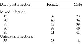
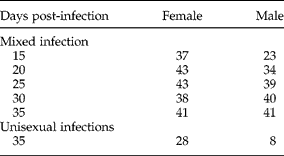
Cercariae were collected in a 50 ml beaker after exposing 50 infected O. hupensis to sunlight for 4 h (25–28°C). Mice were infected percutaneously with 40 cercariae of S. japonicum. On days 15, 20, 25, 30 and 35 post-infection (PI) all worms of S. japonicum were recovered using the perfusion technique from mice anaesthetized by intraperitoneal injection of ketamine, sexed and counted. Five to six mice were used at each time point. The total number of worms recovered at each time point is shown in table 1.
The number of male and female worms of S. japonicum recovered from mixed and unisexual infections up to 35 days post-infection.
Cercariae emitted from one infected O. hupensis were used to infect each mouse via the percutaneous route. In this infection, 30 cercariae were administered to each mouse. Animals were killed using the same procedure described above at 35 days PI. Subsequently, worms were recovered, sexed and counted mainly from the portal vein of the liver and a few from mesenteric veins.
Worm morphology
In dealing with mixed infection, paired worms were separated manually using a soft brush made from hair to enable the male and female to be analysed separately. All specimens were fixed in a fixative containing formalin (10%), alcohol (48%) and glacial acetic acid (2%), stained with hydrochloric carmine, cleared in 0.5% hydrochloric alcohol solution and preserved as whole mounts (Neves et al., Reference Neves, Pereira, de Oliveira, Gomes and Machado-Silva1998). CLSM images were obtained with a Leica TCS-SP5 Spectral Laser Scanning Confocal Microscope (Leica, Wetalar, Germany), using a 488 nm He/Ne laser. Images were obtained for the gynecophoric canal and testicular lobes in males, and for the ovary, vitelline glands, vitelline duct, oviduct and uterus in females (Neves et al., Reference Neves, Pereira, de Oliveira, Gomes and Machado-Silva1998, Reference Neves, Costa-Silva, Martinez, Branquinho, de Oliveira, Lenzi, Gomes and Machado-Silva2004). The length and width of the worm, testicular lobes and the ovary, and the area of the testicular lobes and ovary, were measured in worms from mixed and unisexual infections, using Leica Microsystems LAS AF-TCS SP5 software.
Data analysis
All parameters were measured by CLSM. The results are expressed as means and standard deviations (mean ± SD). Data from mixed infection and unisexual infections were compared by t-test using the SPSS 11.0 (SPSS, Chicago, Illinois, USA) statistics software package; a P value < 0.05 was considered significant.
Results
Mixed infection
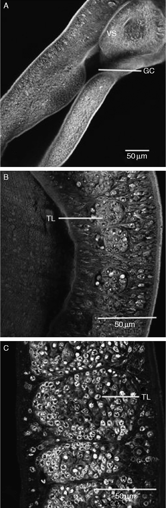
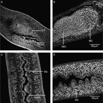
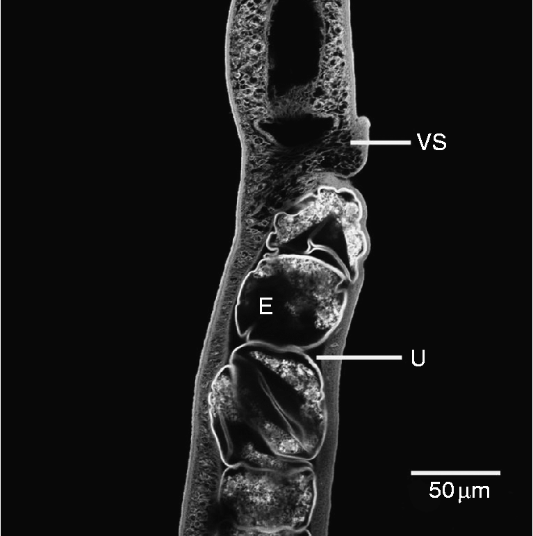
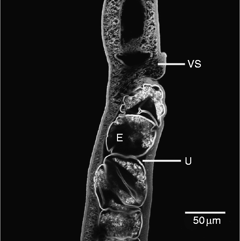
CLSM was used to visualize a total of 415 worms for this study. We were able to distinguish the sex of S. japonicum 15 days PI. At this time, male worms were easily identified because they were thicker and displayed thicker ventral suckers than females. Moreover, 33% of male worms had formed a distinct gynecophoric canal by day 15 PI (fig. 1A). Only 8% of them had testicular lobes (fig. 1B) with free areas around the few germinative cells inside. Of the females, 13% had an incipient ovary (fig. 2A) lacking cellular differentiation at day 15 PI. The ootype was covered with a single layer of cylindrical cells containing a central nucleus. Despite the presence of the ovary and ootype, the vitelline glands, vitelline duct, oviduct and uterus had not yet developed. By day 25 PI, most worms had developed reproductive systems. Of male worms, 86% had five to seven testicular lobes where more germinative cells could be observed (fig. 1C). The seminal vesicles, like saccate bags, were opened in the genital pore. In female worms, vitelline glands (fig. 2C) around the digestive canal were composed of several amorphous cells that were irregularly arranged. The ovaries with immature cells were in the middle of the worms (fig. 2B). By day 25 PI, all worms had developed complete reproductive systems. Males presented developed testicular lobes that were filled with germinative cells in varying stages of maturation (fig. 1C). In females, vitelline cell number at day 25 PI (288 ± 17/10 × 104 μm2) was increased significantly compared with that at day 20 PI (176 ± 21/10 × 104 μm2) (figs 2C and D). Ovaries (fig. 2B) were differentiated and contained cells having a central nucleus in varying stages of maturation. Smaller ovarian cells were distributed in the anterior region, while larger ones were in the posterior part of the ovary. The worms showed fully developed reproductive organs from 30 days onwards, including many eggs in the uterus (see fig. 4).
Confocal scanning laser microscope images of Schistosoma japonicum male isolated from mixed infection: (A, B) 15 days old; (C) 25 days old. GC, gynecophoric canal; VS, ventral sucker; TL, testicular lobes. Scale bars 50 μm.

Confocal scanning laser microscope images of Schistosoma japonicum female isolated from mixed infection: (A) 15 days old; (B, D) 25 days old; (C) 20 days old. O, ovary; OT, ootype; MC, mature ovary cell; IMC, immature ovary cell; VG, vitelline glands. Scale bars 50 μm.

Unisexual infection
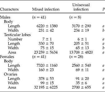
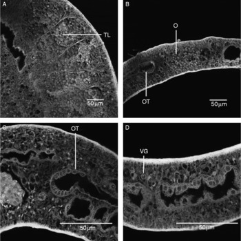
Thirty-six worms recovered at 35 days PI were used for the morphological analysis of unisexual infection. Similar to mixed infection, the males were identified as being thicker than females. However, the difference was that the length of the males was much shorter in unisexual infection (table 2). Testicular lobes (fig. 3A) were distinct in every male. The females obtained from unisexual infection had ovaries lacking cellular differentiation (fig. 3B). A single layer of cylindrical cells with a central nucleus was inside the ootype (fig. 3C). The vitelline glands (fig. 3D) were undifferentiated with a few vitellocytes.
Morphometry (mean±SD) of 35-day-old worms of S. japonicum recovered from mixed and unisexual infection analysed by confocal laser microscopy.
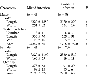
Measurements: length and width in μm; area in μm2; P values were calculated by Student's t-test; a: significant difference (P < 0.05); b: non-significant difference.
Confocal scanning laser microscope images of Schistosoma japonicum isolated from unisexual infection: (A) TL, testicular lobes; (B) O, ovary; OT, ootype. (C) OT, ootype; O, ovary. (D) VG, vitelline glands. Scale bars 50 μm.

Thirty-five days post-infection
The morphology of 36 unisexual and 82 mixed infection worms was also compared by CLSM at 35 days PI. There was a significant difference in size of the worms and main reproductive organs between the unisexual infection and mixed infection groups (table 2).
Discussion
The advantage of CLSM is its ability to acquire thin serial optical sections through a relatively thick fluorescent specimen, such as a schistosome. Every image is taken from a well-defined plane, avoiding interference by signals from unfocused areas. This significantly improves the contrast and resolution of images in comparison with conventional light microscopy (Zhang et al., Reference Zhang, Huang, Zhang, Ren, Wang, Zhang and Shen2008). Furthermore, optical sectioning eliminates artefacts that occur during physical sectioning for conventional microscopy. Moreover, CLSM enables imaging of the dynamics of organelles and proteins as well as the quantitative detection of ion mobilization in living cells. In addition, serial optical sections allow the reconstruction of three-dimensional images of specimens. In this study, we used CLSM for studying the morphological structures of S. japonicum worms recovered from mixed or unisexual infection.
In experiments with worms from mixed infections, we observed several different developmental phases of S. japonicum. We found several significant morphological changes between 15 and 25 days PI. For example, the ovaries were undifferentiated and contained immature cells at a single stage of maturation, while vitelline glands were not present by 15 days PI. As development proceeded to 25 days, however, the ovaries accumulated mature cells and vitelline glands gradually developed. These findings were similar to those of a study of S. mansoni (Biolchini et al., Reference Biolchini, Neves, Hulstijn, Gomes and Machado-Silva2006). In the present study, a fully developed reproductive system was first observed at 30 days. The uterus of S. japonicum, in which we could see many oval eggs (fig. 4), was a long and cylindrical structure and extended from the ootype to the back of the ventral sucker. The uterus of S. japonicum was filled with many more eggs than that of S. mansoni, where only one egg was observed in the uterus at any one time (Neves et al., Reference Neves, de Lamare Biolchini, Machado-Silva, Carvalho, Branquinho, Lenzi, Hulstijn and Gomes2005). Throughout our study, male worms were consistently thicker than females. This is necessary for successful reproduction because the well-muscled male carries its weaker partner to the site of egg deposition and feeding (Ribeiro-Paes & Rodrigues, Reference Ribeiro-Paes and Rodrigues1997; Kunz, Reference Kunz2001). The sole purpose of female worms is egg production, even to the extent of lacking developed muscles; hence, male worms serve as the sole mode of transportation, while the female expends most of her energy on reproduction. Fitzpatrick & Hoffmann (Reference Fitzpatrick and Hoffmann2006) provided microarray data supporting the view that the female streamlines her physiological activity towards egg production upon pairing.
The reproductive system of 35-day-old female adult Schistosoma japonicum under confocal laser scanning microscopy. VS, ventral sucker; U, uterus; E, egg. Scale bar 50 μm.
To complete sexual maturity, female worms must pair with a male (Basch & Nicolas, Reference Basch and Nicolas1989). In our experiment, both male and female worms from unisexual infections did not develop to maturity, even though the female from mixed infections was mature at 35 days PI. The sizes of testicular lobes and ovaries of worms from unisexual infections were much smaller than those from mixed infections. Vitelline glands of worms from unisexual infections did not complete maturation.
In the present study, the recovered worms were from mixed infections or unisexual infections. The morphological character of the S. japonicum worm was studied for the first time using CLSM. Our results provided a detailed comparison of different morphological traits from worms of mixed and unisexual infections throughout development.
Acknowledgements
We thank Professor Shengyun Fang and Professor Weidong Du for revising this manuscript. This investigation received financial support from the National Natural Science Foundation (no. 30771882) and the Natural Science Foundation Key Project of Anhui Education Department (no. 2005KJ045ZD).