In coronary artery congenital anomalies, the common assumption is that ectopic origin of the right coronary artery is always pre-aortic in course and implies proximal tangential origin with variable stenosis, while passing inside the aortic wall. Occasionally, this scenario − anomalous origin of the right coronary artery from the left sinus of Valsalva with intramural course (R-ACAOS-IM) − causes symptoms and even sudden cardiac death in athletes. Reference Angelini, Uribe and Monge1 The prevalence of R-ACAOS-IM is about 3 per 1000 in the general population. Reference Angelini, Cheong and Lenge De Rosen2
The present report describes for the second time in the literature an exceptional case we evaluated that was initially labelled as R-ACAOS-IM, based only on coronary catheter angiography. In a surgical South-Asian journal, Gholoobi and Poorzand Reference Gholoobi and Poorzand3 first reported a case in which the anatomy of the congenital defect seemed identical to ours but was associated with coronary artery disease. Our patient was referred to a cardiologist for further evaluation because of progressive dyspnoea on mild effort, potentially based on coronary stenosis and cardiac insufficiency; she was found to have a variant anatomy with benign physiology.
Case report
A 67-year-old African American woman with no history of structural heart disease presented with progressive dyspnoea for 5 months, without angina. She had a history of hypertension that had recently deteriorated despite regularly taking betablocker medication (metoprolol, 200 mg a day). She had a history of a multi-year diagnosis of HIV that was well-controlled on oral medications, intermittent bronchial asthma (occasionally requiring an inhaler), and mild hyperlipidaemia. Our initial physical examination showed that she had a body mass index of 40 and that her blood pressure was 160/90 mmHg.
Because of her uncontrolled, disabling dyspnoea, she had undergone a calcium score test at another institution, which showed a mildly increased calcium score, with plaque at the proximal left main trunk and left anterior descending arteries. Coronary selective angiography eventually showed a congenital coronary anomaly, initially called R-ACAOS-IM. Thus, she was sent to our Center for Coronary Artery Anomalies for further evaluation.
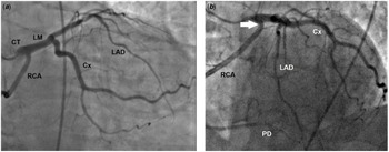
On review of the angiograms, we found that the right coronary artery was ectopically arising in a single coronary trunk located at the left sinus of Valsalva, jointly with the left coronary artery (Figs. 1a and b and 2); the right coronary artery separated from the common trunk, 5 mm from its ostium. The direction of the initial right coronary artery was straight down (caudal), and eventually turned retroaortic, directed towards the normal right coronary artery distal distribution; no suggestion of intramural aortic course or evidence of ostial stenosis was found on CT angiography (Supplemental Figure S1). Pharmacologic myocardial positron emission tomography stress testing was negative for reversible ischaemia. The angiogram did not reveal significant coronary artery acquired disease or evidence of intramural course. The patient was reassured of the findings, and her medical treatment was changed from betablockers to angiotensin II receptor blockers and chlorthalidone, which resolved the dyspnoea and controlled the hypertension.
Angiographic images of the right anterior oblique ( a ) and left anterior oblique ( b ) views of the coronary arteries (common ostium and common main trunk, two arteries) show the straight down direction of the initial right coronary artery (RCA), off the coronary common trunk (CT) located at the left sinus of Valsalva. No evidence of tangential course or ostial stenosis is apparent (white arrow in b ). Cx = circumflex; LAD = left anterior descending; LM = left main; PD = posterior descending.

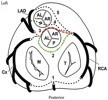
Schematic drawing of the coronary plane view in the R-ACAOS-RA anomaly in comparison with other possible anomalies of origin and course. Alternative courses are numbered: 1 = retro-cardiac; 2 = retroaortic; 3 = pre-aortic [red dotted line] or intramural; 4 = intra-septal; and 5 = pre-pulmonary. Our case is illustrated by the green dotted line, the retroaortic course of the RCA. AL = anterolateral; AR = anterior/right; Cx = circumflex; LAD = left anterior descending; M = mitral valve; P = posterior cusps/sinuses of the pulmonary (anterior) and aortic (posterior) valves; RCA = right coronary artery; T = tricuspid valve. Modified and reproduced with permission from Angelini P, Flamm SD. Newer concepts for imaging anomalous aortic origin of the coronary arteries in adults. Catheter Cardiovasc Interv 2007;69:942–954.

Discussion
This report is the first to describe in detail a case of anomalous origin of the right coronary artery from the left sinus of Valsalva with retroaortic course (R-ACAOS-RA). It is important to acknowledge this usually unmentioned anomaly in the context of the more frequent right coronary anomaly that is associated with a risk of ischaemic symptomatology from variable stenosis caused by intramural lateral compression inside the aortic media Reference Angelini, Uribe and Monge1 (Fig 2, Supplemental Figure S2). R-ACAOS-RA is the mirror image of the frequently occurring circumflex or left main coronary arteries originating from the right sinus of Valsalva ectopic origin with retroaortic course (Cx-ACAOS-RA or L-ACAOS-RA, respectively) Reference Gholoobi and Poorzand3 that has not been associated with ostial stenosis. As in the latter anomalies, congenital ostial stenosis is not associated with the retroaortic course anomalies; the proximal ectopic (retroaortic) artery is extramural (not embedded into the aortic media) and runs posteriorly, below the aortic root. In similar cases that lack tangential origin and intramural course on angiography or CT angiography, intravascular ultrasound imaging is not required. A brief mention of a probable case of R-ACAOS-RA was included in the Yamanaka large review of coronary angiography in 1990, where no ostial details were provided. Reference Yamanaka and Hobbs4 Clearly, no further testing or intervention is required for isolated R-ACAOS-RA, as for L-ACAOS-RA or Cx-ACAOS-RA, in the absence of acquired coronary artery disease. The only clinical implication of an RA coronary course is that it increases the risk of surgical complications at the time of aortic valve replacement. Reference Misawa, Saito, Oki and Fuse5 The diagnosis of R-ACAOS-RA is obvious on angiography (by selective catheter or by CT angiography). However, regarding its prevalence in the general population, we believe R-ACAOS-RA must be rare as we are reporting here the first case in the Western literature, and we did not identify a case in a large MRI-based Reference Angelini and Uribe6 or CT angiography-based series Reference Angelini, Cheong and Lenge De Rosen2,Reference Opolski, Pregowski and Kruk7 or in our extensive review of the English-language literature. Although R-ACAOS-IM ostial stenosis may be difficult to recognise by simple angiography unless it is severe (see the ostial compression in Supplemental Figure S2 for comparison), congenital ostial intramural (inside the aortic media) stenosis is generally not “possible” in the case of R-ACAOS-RA, since the right coronary artery takes off at 90° from the left main trunk, away from the aortic wall (and not with tangential intramural course to the aortic wall). It is not yet clear if the same essential anomaly can also occur with a direct independent origin of the right coronary artery from the posterior side of the left posterior sinus of Valsalva (not previously reported). Incidentally, ectopic origin of a coronary artery with stenosis can also accompany high origin of any coronary artery from the ascending aorta. Reference Angelini, Cheong and Lenge De Rosen2,Reference Angelini and Uribe6 In exceptional cases, ostial stenosis can be seen in the absence of an intramural course, like ectopic origin of the left coronary artery originating from the left ventricular outlet, which has been seen in a single case report. Reference Pirelli, Yu and Srichai8
The present new case confirms the value of having a specific nomenclature in cases of coronary artery congenital anomalies in general (Fig 2). For each different entity, we propose using a term that identifies the three essential segmental sections of each case of ACAOS: the artery involved (R for right coronary artery and L for the left one); the nature of the defect (-ACAOS- or anomalous origin of a coronary artery from an opposite sinus of the aortic valve); and the course of the proximal artery (in this case: RA for retroaortic, in others: PP for prepulmonic; IM, for intramural, pre-aortic; and RC for retrocardiac). The alternative nomenclature of “AAOCA” or even “AOLCA/AORCA” Reference Angelini, Uribe and Monge1,Reference Angelini and Uribe6,Reference Brothers, Gaynor, Jacobs, Poynter and Jacobs9 is generic and includes in a single group any one of several variants: Specific names (as indicated above) should imply their course and suggest different physiological and clinical repercussions.
Using an exclusively anatomic approach, Shirani and Roberts, Reference Shirani and Roberts10 in a general 1993 literature review of 87 cases with single coronary ostia, indicated that 20 different types of single coronary ostium are possible. Unfortunately, the quality and description of their diagrams (due to the old currently available photographic copy of the original manuscript) are poor: Our case was called IIB-3, Reference Brothers, Gaynor, Jacobs, Poynter and Jacobs9 but it seems to us that it is IIC-2. IIB-3 seems to possibly indicate “intramyocardial course.” No clinical correlations were added. In our approach, as stated, the proximal course of the ectopic artery is the critical parameter potentially associated with prognosis of each congenital coronary defect, and only the intramural course is associated with congenital compression (stenosis).
Presumably, R-ACAOS-RA has a prevalence in the general population of 1 in 1–10 million. Modern techniques used to identify this anomaly are echocardiography (frequently used but unlikely to be diagnostic in adults), CT angiography or MRI (diagnostic and more frequently used for coronary disease screening), and coronary artery catheter angiography (diagnostic but invasive and not a frequent screening method). The important message for cardiologists (paediatric or adult) is that this newly recognised entity needs to be correctly identified as benign and not requiring further testing after its diagnosis.
Supplementary material
To view supplementary material for this article, please visit https://doi.org/10.1017/S1047951121004947
Financial support
This research received no specific grant from any funding agency.
Conflict of interest
None.