Introduction
Polystomatidae of amphibians in Nigeria have, until recently, been poorly studied. According to Thurston (Reference Thurston1970) and Avery (Reference Avery1971) the first polystome reported from Nigeria was Protopolystoma xenopodis (Price, 1943). Tinsley & Jackson (Reference Tinsley and Jackson1998) reported another polystome from this region, namely Protopolystoma occidentalis from Xenopus muelleri. Recently Aisien et al. (Reference Aisien, Ajakaiye and Braimoh2003, Reference Aisien, Ayeni and Ilechie2004a, Reference Aisien, Salami, Obaro and Erakpowerib, Reference Aisien, Ogoannah and Imasuen2009) reported on other polystomes from Nigeria. Apart from Protopolystoma, other polystomes known from Nigeria include Polystoma prudhoei Saoud, 1967 from Amietophrynus regularis, P. galamensis Euzet, Bourgat & Salami-Cadoux, 1974 from Rana galamensis, Eupolystoma alluaudi (de Beauchamp, 1913) from Amietophrynus regularis and A. maculatus, Metapolystoma cachani Gallien, 1956 from Ptychadena longirostris, and unidentified Polystoma spp. from Amietophrynus sp. and Hyperolius sylvaticus. Polystoma pudhoei reported from A. regularis was a mis-identification as this parasite has now been identified as Polystoma africanum (Szidat 1932) and recently redescribed by Aisien & Du Preez (Reference Aisien and Du Preez2009).
Amphibian diversity in Nigeria has been the focus of several studies (Schiøtz, Reference Schiøtz1963, Reference Schiøtz1964, Reference Schiøtz1966, Reference Schiøtz1967, Reference Schiøtz1969; Reid et al., Reference Reid, Owens and Laney1990; Oldham, Reference Oldham2000). Opinions vary on the official number of amphibian species represented in Nigeria. Schiøtz (Reference Schiøtz1963) reported 69 species while a later study by Oldham (Reference Oldham2000) reported more than 90 species. Reid et al. (Reference Reid, Owens and Laney1990), referring to unpublished work, listed 75 species from the Calabar-Oban area alone. The most recent information by IUCN et al. (2006) put the number of species in the country at 103. In the Okomu National Park, which is a protected lowland rainforest habitat, we have recorded 40 species of amphibians, representing 10 families. Despite the rich amphibian diversity in Nigeria, very little information is available on the polystome diversity in the country. Our count to date shows that as many as 11 previously described African polystome species do occur in the country, while a number of undescribed species are still being investigated. With the high amphibian diversity present in Nigeria, we presume that there will be correspondingly rich polystome diversity.
The polystome that we describe herein was recovered from the urinary bladder of the Hyperolid frog, Phlyctimantis boulengeri Perret, 1986 collected at two seasonal lakes in the Okomu National Park, Nigeria. Apart from Nigeria, the frog occurs in Cameroon, Ivory Coast, Equatorial Guinea, Ghana, Guinea and Liberia (Schiøtz, Reference Schiøtz1999). The frog is found in subtropical or tropical moist lowland forests, intermittent freshwater marshes, and heavily degraded former forested areas where males tend to aggregate in vast numbers.
Materials and methods
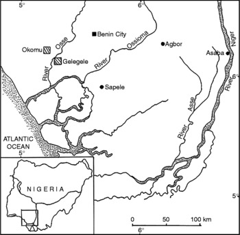
Parasites were recovered from the urinary bladder of adult P. boulengeri collected in August 2007 and May 2009 in Okomu National Park (which lies between latitudes 6°15′N and 6°25′N; longitudes 5°9′E and 5°23′E) located in Edo State of Nigeria. The park, which lies in the lowland rainforest belt of south-western Nigeria, is a protected sanctuary for flora and fauna (fig. 1). The total area of the park is 19,712 hectares. Mean annual rainfall is about 2100 mm with most of it falling between April and October. The dry season lasts from November to March. The frog specimens examined were collected from four seasonal lakes within the park which are designated as sites 1 to 4. The coordinates for these sites are as follows: site 1 (6°23′N, 5°20′E); site 2 (6°22′N, 5°21′E); site 3 (6°22′N, 5°20′E) and site 4 (6°17′N, 5°18′E). Of the four sites indicated, only site 4 retains some water in the months of the dry season.
Map of Nigeria showing the location of Okomu National Park, Edo State, Nigeria.

Frogs were anaesthetized by immersion in benzocaine solution. The parasites were recovered from the urinary bladder, fixed in 5% formol-saline under cover-slip pressure. Fixed specimens were carefully removed after 1 h and transferred to vials containing 5% formol-saline. Parasites were washed in several changes of tap water and then stained overnight in a weak solution of acetocarmine, dehydrated, cleared in xylene and permanent mounts made using Canada balsam. One specimen was fixed in 96% ethanol for molecular studies. The morphometrics of the marginal hooks were determined according to the protocols of Du Preez & Maritz (Reference Du Preez and Maritz2006).
Results
Levels of infection
Phlyctimantis boulengeri specimens were collected from four different seasonal lakes within the Okomu National Park. Of the eight specimens collected from site 1 between October 2007 and January 2008, none was infected. The three specimens caught between February and July 2008 from site 2 were also not infected. In site 3, one of the seven frogs caught between June and August 2007 was infected, giving a prevalence of 14.3% and a mean intensity of 1. At site 4, six of the 27 frogs caught in May 2009 haboured a total of nine polystomes, giving a prevalence rate of 22.2% and a mean intensity of 1.5. Of the total number of 45 frogs examined the prevalence was 15.5% and the mean intensity 1.4. Mean intensity of infection is defined as the average number of parasites/infected host.
Class: Monogenea Carus, 1863
Order: Polystomatidea Lebedev, 1988
Family: Polystomatidae Gamble, 1896
Polystoma okomuensis n. sp
Specimen studied. Nine adult specimens. Holotype LPRM15–0707–01 and two paratypes (LPRM15–0905–03 and LPRM15–0905–05) are deposited in the Parasitic Worm Collection of Professor A.B.M. Egborge Museum, University of Benin, Benin City, Nigeria; two paratype specimens (NHM 2010.1.28.1-2) in the Parasitic Worms Collection of the Natural History Museum, London; and the remaining specimens in the collection of the senior author.
Type host. Phlyctimantis boulengeri Perret, 1986 sexually mature male and female specimens (CT069002 and CT055413) are deposited in the Amphibian Collection of the Professor A.B.M. Egborge Museum, University of Benin, Benin City, Nigeria.
Type locality. Okomu National Park, Edo State, Nigeria (lies between latitudes 6°15′N, and 6°25′N; longitudes 5°9′E and 5°23′E).
Site. Urinary bladder.
Etymology. The specific name okomuensis relates to the Okomu National Park, where the host was collected.
Description. Based on adult parasites (n = 9); measurements given in micrometres. The average measurement is followed by the range in parentheses. Measurement of larval sclerites based on adult parasite specimens.
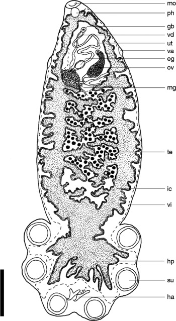
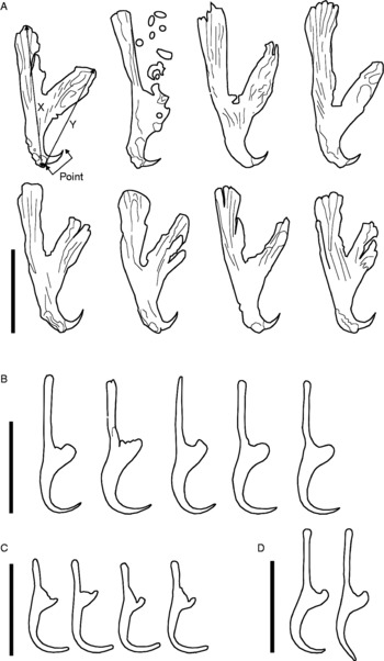
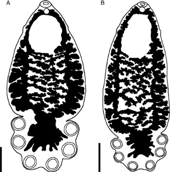
Adult. General characteristics of mature egg-producing parasite (fig. 2) typical of Polystoma. Body elongate, total length 6513 (5570–7597); greatest width 2334 (1882–3207); width at vagina 1565 (1188–1853). Haptor 1857 (1141–2153) long, 2459 (1871–2765) wide, haptor length to body length ratio 0.28 (0.20–0.31). Haptor with six suckers 403 (324–592) diameter; hamuli 369 (325–456) long, handle longer than guard and mean x/y ratio 1.43 (fig. 3); hamulus point 56 (51–67) long. Mouth subterminal, ventral. Oral sucker 472 (324–592) wide; pharynx 242 (218–277) long, 225 (204–253) wide. Intestine bifurcate with lobed lateral diverticula averaging 29.3 (20–38) in number per parasite, slender intensely branched medial diverticula numbering 17.3 (15–21) per parasite. Intestinal anastomoses infrequent, occurring in 1 (11.1%) of 9 specimens examined (fig. 4B). In two (22.2%) of the specimens two medial diverticula on one side join to form loops (fig. 4A). Caeca confluent posteriorly, extending into haptor forming haptoral caecal diverticula.
Polystoma okomuensis n. sp. Ventral view of holotype; the dashed lines indicates the outline of the vitelline system. eg, egg; gb, genital bulb; ha, hamulus; hp, haptor; ic, intestinal caecum; mg, Mehlis gland; mo, mouth; ov, ovary; ph, pharynx; su, sucker; te, testis; ut, uterus; va, vagina; vd, vas deferens; vi, vitelline distribution. Scale bar = 1 mm.

Polysoma okomuensis n. sp. (A) Hamuli from holotype and paratypes; (B) marginal hooklets 1 from adult specimens; (C) marginal hooklets 2–7; (D) marginal hooklet 8. Scale bars: (A) 200 μm; (B–D) 30 μm.

Polystoma okomuensis n. sp. ventral view showing only the pharynx, intestine and suckers. Variation in intestinal arrangement: (A) specimen with loop; (B) specimen with caecal anastomosis. Scale bar = 1 mm.

Testis follicular post-ovarian, ventral, median (fig. 2) occupying 70% of pre-haptoral region. Lots of sperm present among testis follicles. Seminal vescicle prominent and packed with sperm. Genital atrium median, ventral. Genital bulb 79 (62–97) in diameter, posterior to intestinal bifurcation, with 7–8 genital spines 29.7 (28.3–32.0) long. Ovary very prominent, sinistral, submedian, 775 (675–1040) long, 267 (169–402) wide and packed with oocytes. Ootype well developed. Genito-intestinal canal present on same side as ovary, joining intestinal caecum posterior to ovary. Uterus confined to area anterior to ovary, holding one egg in six polystomes and three polystomes with respectively two, three and seven eggs. Eggs operculate. Egg capsule length 249 (256–297), width 167 (145–192). No intrauterine development of eggs observed. Vitellaria distributed throughout most of the body (fig. 2).
Larval sclerites. Marginal hooklet 1 43 (39.8–44.7) long; marginal hooklets 2–7 28 (21–33) long, marginal hooklet 8 38.5 (37–40) long.
Discussion
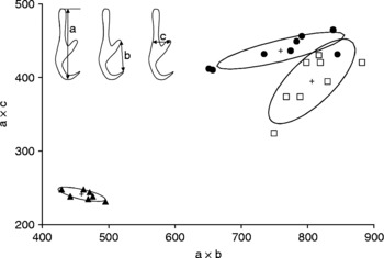
In Africa, the genus Polystoma is at the moment represented by 33 described and several undescribed species. Of these, six species have been reported from Nigeria while information on undescribed species is emerging. Polystoma okomuensis n. sp. is the first species to be described from the country. The separation of polystomes based on morphological characters is complicated because of the limited interspecific morphological variation and large intraspecific variation. Murith (Reference Murith1981) has, however, shown that marginal hooklets of polystomes display little intraspecific variation and assist species separation. A plot of the products of the total length (a in fig. 5) and the width at the level of the guard (c in fig. 5) versus the product of the total length versus the length of a tangent between the tip of the blade to the guard (b in fig. 5) of marginal hooklet 1 (Du Preez & Maritz, Reference Du Preez and Maritz2006) showed an overlap of P. okomuensis n. sp. with P. dorsalis Maeder, Euzet & Combes, Reference Maeder, Euzet and Combes1970 (fig. 5). Polystoma perreti, which infests Hylarana albolabris (Hallowell, 1856) in the same environment, occupied a distinct position separated from P. okomuensis and P. dorsalis in the plot.
Scatter diagram of a × b plotted against a × c for Polystoma okomuensis n. sp. (●), P. dorsalis (□) and P. perreti (▲). Measurements for P. dorsalis and P. perreti were obtained from Murith (Reference Murith1981).

Despite the overlap of P. okomuensis n. sp. and P. dorsalis, a number of features differ between the two parasites. With a mean total body length of 6513 (5570–7597) P. okomuenisis n. sp. is larger than P. dorsalis described by Maeder et al. (Reference Maeder, Euzet and Combes1970) from Liberia (4.0–5.5) and much larger than those specimens recovered from the Ivory Coast (1.7–2.7) by Maeder (Reference Maeder1973). In P. okomuensis n. sp. the medial intestinal diverticula are highly branched (figs 2 and ) while those of P. dorsalis have fewer branches with a higher frequency of forming pre-haptoral anastomoses. The presence of a loop formed by two adjacent medial diverticula (fig. 4A) in P. okomuensis n. sp., which is absent in P. dorsalis, is another distinguishing difference between the two parasites. The shape of the hamuli, which is an important systematic character for separating polystome species further differentiates P. okomuensis n. sp. from P. dorsalis. Whereas the handle and guard of the hamulus in P. okomuensis n. sp. are well separated, those of P. dorsalis are, in most cases, poorly divided and, in some others, lacking a division between handle and guard.
Another factor that must be taken into consideration in separating P. okomuensis n. sp. from P. dorsalis is the host specificity of the two parasites. Polystoma okomuensis parasitizes P. boulengeri while P. dorsalis is parasitic in Afrixalus dorsalis (Peters, 1875). Host specificity is a well-documented phenomenon for the genus Polystoma (see Combes, Reference Combes1966, Reference Combes1968; Tinsley, Reference Tinsley1973, Reference Tinsley1974; Euzet et al., Reference Euzet, Combes and Knoepffler1974a, Reference Euzet, Bourgat and Salami-Cadouxb; Bourgat & Salami-Cadoux, Reference Bourgat and Salami-Cadoux1976; Combes & Channing, Reference Combes and Channing1979; Murith, Reference Murith1981, Reference Murith1982; Kok & Van Wyk, Reference Kok and Van Wyk1986; Kok & Du Preez, Reference Kok and Du Preez1987; Du Preez & Kok, Reference Du Preez and Kok1992, Reference Du Preez and Kok1993, Reference Du Preez and Kok1997). Invoking strict host-specificity along with some key morphological characteristics of African polystomes, Aisien & Du Preez (Reference Aisien and Du Preez2009) recently concluded that P. africanum, which was thought to be a multi-host parasite, was exclusively parasitic in A. regularis and that polystomes retrieved from Ptychadena mascareniensis (Duméril & Bibron, 1841) hitherto described as P. africanum were most likely P. pricei Vercammen-Grandjean, 1960 and those from Amietia angolensis (Bocage, 1866) most likely represented another species. In the same vein, we conclude that P. okomuensis n. sp. is a different species from P. dorsalis and should be admitted as a separate species parasitic in P. boulengeri. We hypothesize that Nigerian polystomes will display the same degree of host-specificity displayed by other anuran polystomes; however, experimental infection and cross-infection experiments are needed to confirm this.
Unfortunately, the dimension of the testis in P. dorsalis was not discussed by Maeder et al. (Reference Maeder, Euzet and Combes1970) nor by Maeder (Reference Maeder1973). The testis of P. okomuensis n. sp. is rather large, occupying about 70% of the pre-haptoral space (fig. 2). The only other African polystome known with such a large testis is P. testimagna (Du Preez & Kok, Reference Du Preez and Kok1993) described from Stongylopus fasciatus (Smith, 1849). Whereas the testis follicles are restricted to the area between the intestinal caeca in P. okomuensis n. sp., the testis follicles in P. testimagna extend beyond the intestinal caeca, reaching as far as the body wall and occupying three-quarters of the pre-haptoral body. The huge testis, vast amount of sperm among testis follicles, prominent ovary and relatively few eggs in the uterus reflect on the mode of reproduction. These features are typical of a polystome that can produce a large number of eggs per day over a long period. This fits in with the reproductive strategy of the host, which has an extended breeding season.
Polystoma okomuensis n. sp. is further distinguished from other African polystomes by the length of its marginal hook 1. A length of 43 μm in this regard separates it from P. africanum Szidat, 1932 (38), P. sodwanensis Du Preez & Kok, Reference Du Preez and Kok1992 (38), P. togoensis Bourgat, 1977 (40), P. baeri Maeder et al., Reference Maeder, Euzet and Combes1970 (41), P. mangenoti Gallien, 1956 (42). While marginal hooklet length does overlap with that of P. prudhoei, the hosts belong to different families.
The mean length of marginal hooklets 2–7 in P. okomuensis n. sp. is of the same size as those reported for Polystoma claudecombesi Du Preez & Kok, 1995 (28 μm) but they are longer than those of P. testimagna (24), P. sodwanensis (23), P. dawiekoki Du Preez, Vaucher & Mariaux, 2002 (20) and P. africanum (19). Irrespective of the size similarity to those of P. claudecombesi and their disparity with those of the other listed African polystomes, a peculiar feature of marginal hooklets 2–7 in P. okomuensis n. sp. that separates them from those of other African polystomes is the length and shape of hooklet tips. While the tips of these hooklets are enlongated and rounded in P. okomuensis n. sp. (fig. 3C), in other African polystomes, the tips of these marginals are short and pointed.
Although the general body features and morphometrics remain the fundamental basis of polystome species identification and description, there is a need for a comprehensive molecular study resolving taxonomic complexes and relationships among African polystomes.
Acknowledgements
We thank the National Park Service of Nigeria for permission to undertake this study at the Okomu National Park. We also thank Matthias Enaberue, Okechukwu Egonu, Elisha Enabulele and Festus Arijode for assistance during the field collections. We deeply appreciate the logistic and financial support of Mr Y.M. Kolo, Comptroller, Okomu National Park, Edo State. We are indebted to the South African National Research Foundation for financial support.