Introduction
Radiotherapy is often a local therapy in which the main objective is to deliver the maximum recommended dose to the tumour while preserving the surrounding healthy organs.Reference Strbac and Jokic1 Usually the radiotherapy dose received by the patient is given fractionally. Hence, the reproducibility of daily therapy sessions is important.Reference Boyer, Antonuk and Fenster2 The possible actions that can cause error in the treatment include patient movement, non-compliance of the delivered point of the prescribed dose to tumour centre, opening treatment fields incorrectly by the technician, the incorrect positioning of the multi-leaf collimators in conformal treatmentsReference Castadot, Lee, Parraga, Geets, Macq and Grégoire3 and changes in tumour volume in the last sessions compared with the first sessions.Reference Goitein4
Each step of the treatment processes has several opportunities for set-up error sources; however, treatment must be delivered with the highest accuracy. Uncertainty in each step can affect the next steps and the total of these errors could affect the treatment results.Reference Podgorsak5 There are some errors that can occur in the treatment processes including initial errors (such as wrong dose prescription, target contouring, fixation and machine technical errors), treatment field errors (such as fields overlapping, incorrect field size, incompatibility between anterior–posterior (AP) and posterior–anterior (PA) fields and incompatibility between lateral fields), angle errors (such as wrong gantry angle, collimator angle and table angle), beam modification errors (such as wedge errors, bolus errors and shield errors).Reference Asnaashari, Gholami and Khosravi6 For these and also other reasons, an effective way to reduce set-up errors is by using portal imaging. Portal imaging is commonly used to check the position and verification of the patient positioning relative to the isocentre by using bony landmarks just before radiation therapy.Reference Herman, Kruse and Hagness7
In the current study, set-up error in patient treatment was defined as the difference between the intended and actual position of the treatment fields delivered to the patient. The reference or patient position is imaged and recorded and is known as a reference image. This can be either a digitally reconstructed radiograph (DRR) or a simulator image; and the bony structures, body contour and radio-opaque markers used to verify the position of the treatment fields can be observed on the reference image. Set-up errors are evaluated in all directions separately, using Cartesian coordinates, and are divided into two main groups: (1) systematic or intra-fraction errors that are the same in deviation, which are repeated in each fraction in the same direction in all of the treatment fractions.Reference Strbac and Jokic1 As these uncertainties can be related to mechanical inaccuracies in medical devices, such as an incorrect setting of laser lights, a problem in the collimator system and changes in machine efficiency. (2) Random or inter-fraction errors include those that can occur day to day and can vary for each patient; as these errors can relate to incorrect patient position, block shields and the beam(s).Reference Strbac and Jokic1
Gross tumour volume (GTV), clinical target volume (CTV) and planning target volume (PTV) are defined as the main types of volume described in radiotherapy planning. The GTV is defined as the extent of the gross tumour that can be seen and imaged and this volume can be shown on computed tomography (CT) images. The CTV includes the GTV and the area of sub-clinical disease and it contains the GTV plus a margin,Reference Burnet, Thomas, Burton and Jefferies8 and this volume can be observed in molecular images such as positron emission tomography; treating this volume, the CTV, can prevent the recurrence of disease. The PTV is the third volume applied that considers the uncertainties in treatment planning and this margin has a geometric concept designed to ensure that the radiotherapy dose is actually delivered to the CTV. It is important to get the best PTV margin because of the potential to underdose the tumour and overdose the nearby critical organs.Reference Kang, Lovelock, Yorke, Kriminiski, Lee and Amols9
In this study, the main aim was to evaluate systematic and random errors in pelvic radiation therapy using an electronic portal imaging device (EPID) and propose the optimum CTV to PTV margin in pelvic cancer patients. Furthermore, the performances of the linear accelerator (linac), EPID and MOSAIQ software were assessed to verify the validity of these margins. By this software, images can record electronically that which provides clinical and administrative oncology management solution. This software can communicate between Linac and treatment planning system (TPS) automatically.
Materials and Methods
Patient selection and definition of the target volume
The present study was retrospectively carried out on randomly selected 22 patients with pelvic cancer treated using three-dimensional conformal radiotherapy (3DCRT) at Imam Reza Radiation Oncology Center (Mashhad, Iran). There was no particular change in the routine treatment steps of the patients, so no ethical approval was sought. All patients were scanned using a CT scanner (16 slices; Neusoft Medical System Co., Shenyang, China) with 5 mm slice thickness in supine position and using three radio-opaque labels under laser beams guidance in CT planning step. It is notable that these markers were tattooed on the patient’s body or patient’s thermoplastic just to be stable until the last session. Then the CT images were imported into the Isogray (Dosisoft, Cachan, France) TPS and the DRRs were computed. These DRRs were considered to be the reference images. The target values and the surrounding organs were contoured by the oncologist physician; the CTV to PTV margins of 10 mm were added to the defined CTV.
All the patients were irradiated by 6, 10 and 15 MV photon beams from an Elekta Compact linear accelerator (Elekta AB, Stockholm, Sweden). This machine was equipped with amorphous silicon EPID that was mounted on it at the same isocentre with a detector size of 40 × 40 cm2 and multi-leaf collimators having 40 leaves on each side. The prescription dose to PTV was 70 Gy with 2 Gy per fraction.
Treatment process
There was no particular change in routine treatment steps of the patients, except that the port image was taken on certain days of treatment. Before starting the treatment, the patients were positioned with their own immobilisation device. The, they were set-up with the treatment room lasers using the tattoo markers as a guide. By adjusting the gantry angles at 0° (anterior–posterior [AP]) and 90° (lateral [LAT]), the orthogonal portal images were obtained using 6 MV photon beams and a typical exposure time of 3 monitor units (MU) per field at a dose rate of 400 MU/minutes.
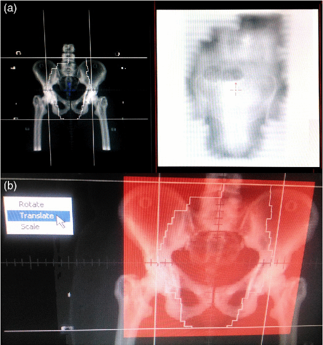
For the first three fractions, the portal images were obtained as pre-treatment images per patient. The portal images were compared with DRRs as the reference images. Afterward if the displacements were acceptable (correction standard was set at >5 mm), the next image would be taken every week. Displacements between the DRRs and the images obtained by EPID in each anterior and lateral projections were estimated along three major axes by matching rigid bony landmarks. The reference landmarks used in electronic portal images were the coccyx bone for lateral image and pubic symphysis for AP projection (Figure 1).
(a) MOSAIQ offline review, anterior–posterior (AP) images (portal and digital reconstructed radiograph [DRR]), bony landmarks using megavoltage X-rays and electronic portal imaging device (EPID). The portal image obtained immediately before the radiotherapy fraction using the EPID. (b) Fused images and calculated deviation.

Total port images taken from 26 pelvic cancer patients consisted of 204 images of which 182 images were acceptable. Four patients (22 images) were removed from this study because they continued their treatment until the middle of treatment sessions and decided not to complete their treatment. For the analysis process, posterior, inferior and left-sided shifts are implied as negative shifts and anterior, superior and right-sided shifts as positive. Rotational errors are not evaluated in the current study.
Statistical analysis
Random and systematic errors combine in µ deviation. So µ is defined as a patient set-up deviation recorded for all 22 patients for the three directions separately. σ represents random errors that occurred day to day in each set-up position and Σ is the systematic errors defined as average set-up deviation per patient. To obtain these errors, the total number of patients P and total images used in this study N are needed. In the following equation, m p is the mean deviation of n p images which is defined as systematic set-up deviation for a patient P Reference Strbac and Jokic1:
 $${m_p} = {1 \over {{n_p}}}\sum\nolimits_{i = 1}^{{n_p}} {{\mu _{{{\left( {PI - DRR} \right)}_i}}}} $$
(1)
Random set-up deviation for a patient P in a given direction is obtained by Equation (2)Reference Strbac and Jokic1:
 $${\sigma _{{\rm{rand}},p}} = \sqrt {{1 \over {{n_p}}}\sum\nolimits_{i = 1}^{{n_p}} {{{\left( {{\mu _{{{\left( {PI - DRR} \right)}_i}}} - {m_p}} \right)}^2}} } $$
(2)
Overall, the mean systematic errors in a given direction for all the patients P are as followsReference Strbac and Jokic1:
 $${m_{{\rm{overall}}}} = {1 \over N}\sum\nolimits_{p = 1}^p {{n_p}} {m_p}$$
(3)
The random set-up errors of the σ r and, p distribution for all the patients P in a given direction can be obtained from Equation (4)Reference Strbac and Jokic1:
 $${\sigma _{{\rm{set}} - {\rm{up}}}} = {1 \over P}\sum\nolimits_{p = 1}^p {{\sigma _{{\rm{rand}},p}}} $$
(4)
And the final equation is the systematic set-up errors for all the patients P in a given directionReference Strbac and Jokic1:
 $$\sum\nolimits_{{\rm{set}} - {\rm{up}}} = {1 \over P}\sum\nolimits_{p = 1}^p {{m_p}} $$
(5)
CTV to PTV margin
To achieve the CTV-PTV margins, there are numerous mathematic formulas given by the International Commission on Radiation Units and measurements (ICRU) report No. 62 (sqrtΣ 2+σ 2),Reference Wambersie and Landgerg10 Stroom and Heijmen (2Σ+0·7σ)Reference Stroom and Heijmen11 and Van Herk et al. (2·5Σ+0·7σ)Reference van Herk, Remeijer, Rasch and Lebesque12. Σ systematic and σ random are the symbols to show the standard deviations of the systematic and random population errors, respectively. According to the ICRU 62 formula, the systematic and random uncertainties have the same contribution to the dose distribution; hence, to product the CTV–PTV margins, they should be added in quadrature. It should be noted that random errors cause blurring in dose distribution, while the systematic errors shift the cumulative dose distribution.Reference Gupta, Chopra and Kadam13 Stroom and HeijmenReference Stroom and Heijmen11 and Van Herk et al.Reference van Herk, Remeijer, Rasch and Lebesque12 suggested the formula incorporating these differential effects by using probability matrices and dose population histograms. The formula of Stroom and Heijmen (2Σ+0σ) guarantees that 99% of the CTV receives ≥ 95% of the prescribed dose. Van Herk et al. reported that by the margin recipe (2·5Σ +0·7σ) it can ensure that a minimum cumulative dose received in the CTV will be at least 95% of the prescribed dose in 90% of patient population.Reference van Herk, Remeijer, Rasch and Lebesque12 In another study, Van Herk introduced the random errors as the motion in organs and systematic errors as the set-up uncertainties. He mentioned that an increase in the margin by three to four times is required to cover the systematic errors compared to random errors; so by using the correct CT scan procedures, multimodality imaging and electronic portal imaging as image-guided tools, these margins could be reduced.Reference Van Herk14
Validation of linac and EPID
A phantom study was performed to investigate the uncertainty in the devices (linac and EPID). In this part of the research, a Rando phantom (Phantom Laboratory, Salem, NY, USA) was used, and the phantom CT images were taken in supine position. Then the phantom was placed on the room couch in the position that its CT images had been taken and it was set on the coordinate’s origin of the patient (source to axis distance (SAD) = 100) and its port image was taken in the AP field. Afterward the linac gantry was rotated 90° and another port image was taken under the same conditions to produce a lateral phantom image. These two port images of the phantom were registered with DRRs and displacement of the two images (reference and port) was investigated. In this part of the study, the movements of the patient are omitted, so it is like to consider a patient without moving. It is expected that performing this work can show parts of systematic errors such as the deviations of EPID and what is displayed in MOSAIQ software.
Validation of MOSAIQ software
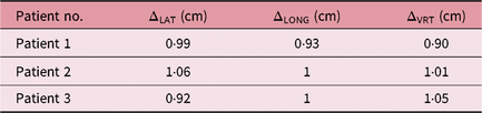
Six reference images from three patients were used to validate MOSAIQ software. The DRR images of patient in AP position were shifted as much as 1 cm in two orthogonal directions X and Y. This process was also performed for LAT position in which the Z direction can be displayed. The two obtained images (reference DRR and changed DRR) were sent to the MOSAIQ software and registered with MOSAIQ, and displacements in three directions were determined. These registrations have a problem, that is, for MOSAIQ software, the association of two DRR images was not defined. To solve this problem, the shifted image was first saved in the name of “EPI Image” in the MOSAIQ software so that it is recognised as a port image and then it was registered. This procedure was done for three patients to reduce the probability of errors.
Results
All the results of this study are related to the Imam Reza Therapeutic Center and are presented to compare with the results obtained from other centres. The number of initial days of portal imaging measurement depends on the magnitude of the random set-up error. To obtain a 95% confidence level in prediction, an experiential formula n = min {9, 4 + 2(σ − 1)} was used, where σ predicts random error and n is the number of daily portal image needed. For any σ ≥ 1 mm, we should take portal imaging for 4–9 days to achieve a confident prediction.Reference Yan, Ziaja and Jaffray15 In this study, all the displacements in the 182 portal images were measured, including those at 96 AP and 86 LAT positions.
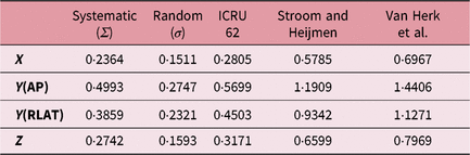
Equations (1–5) were used to calculate the systematic set-up deviation for a patient P, the random set-up deviation for a patient P, the overall mean systematic errors, the random set-up errors for all the patients and the systematic set-up errors for all the patients, respectively. These data are presented in Table 1. There were systematic errors during the whole course of treatment. The population systematic errors (Σ) in lateral, longitudinal and vertical axes were 0·2364, 0·4993 and 0·2742 cm, respectively. The random errors (σ) happened in set-up of day-to-day patient. The population random errors (σ) in the corresponding axes were 0·1511, 0·2747 and 0·1593 cm, respectively.
The brief results of the population systematic Σ and random σ errors in all patients with pelvic cancer were based on the portal images in the caudocranial longitudinal and left–right lateral direction measured by anterior–posterior (AP) field and dorsoventral and caudocranial field measured by lateral (LAT) field

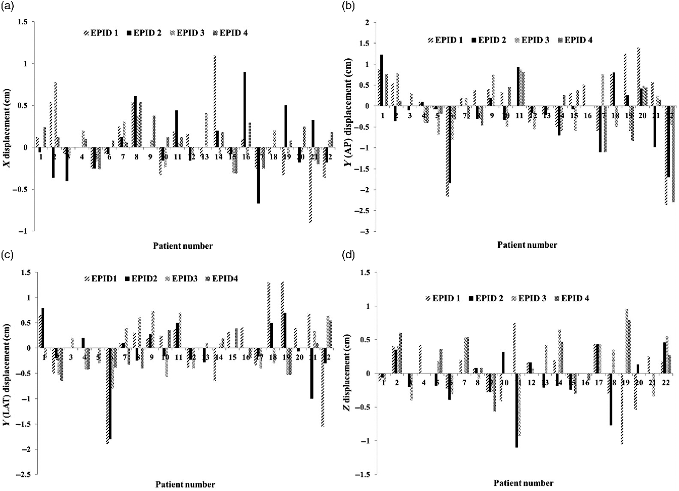
The distribution of the pelvic set-up deviations at mediolateral, superoinferior (AP and LAT) and AP directions was demonstrated in Figure 2. As seen from this figure, the largest amount of movement for each patient is related to the port images of the first sessions (brown and black column).
The distribution of the pelvic set-up deviations at (a) mediolateral, (b) superoinferior from the anterior–posterior field, (c) superoinferior from the lateral field and (d) anteroposterior directions.

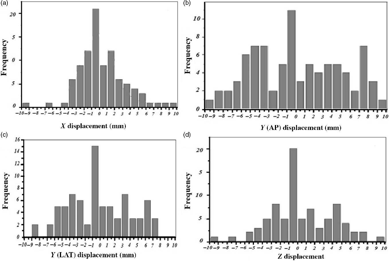
Figure 3 shows the total deviations in three directions of caudocranial longitudinal, left–right lateral direction from the AP field and dorsoventral vertical direction from the LAT field. As shown from this figure, the redundancy of displacements in X and Z directions is around −1 to 1 mm in the form of Gaussian curve.
Distribution of total deviations at (a) left–right lateral direction, (b) caudocranial longitudinal direction from the anterior–posterior field and (c) dorsoventral vertical direction from lateral field.

The findings (Table 2) demonstrate that the obtained CTV-PTV margins based on the ICRU 62 recommendation (Wambersie and Landgerg, 1999) at the lateral, longitudinal and vertical directions were 0·2805, 0·5699, and 0·3171 cm, respectively. Using the formula of Stroom and Heijmen (2002), the corresponding values were 0·5785, 1·1909, and 0·6599 cm; and using the formula presented by van Herk et al. (2000), the values were 0·6967, 1·4406 and 0·7969 cm.
Population systematic Σ, random σ errors, and CTV to PTV margins (cm)

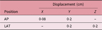
Furthermore, the results related to the performance accuracy of linac and EPID and MOSAIQ are presented in Tables 3 and 4, respectively.
The uncertainties in linear accelerator and EPID

The uncertainties in MOSIAQ software

Discussion
In the current study, the systematic and the random set-up errors in the patients treated with pelvic 3DCRT were investigated by EPID. Furthermore, the CTV-PTV margin was obtained in these patients. By considering this margin, the target volume will be covered by radiation. Moreover, the performance accuracy of linac, EPID and MOSAIQ software were evaluated.
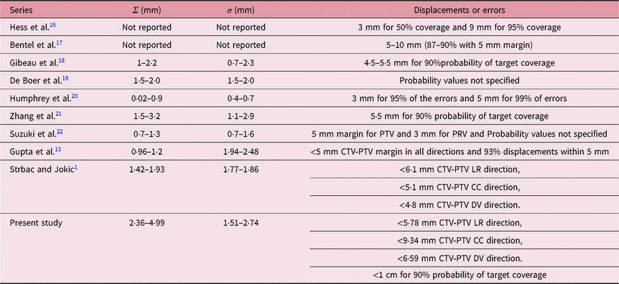
In our institution, the action level for translational direction for the pelvic cases is 5 mm. The findings demonstrated that 88, 55, and 81% of set-up deviation in lateral, longitudinal and vertical axes was less than 5 mm. Table 5 represents a comparison between the results obtained in present study with similar works.Reference Hess, Kortmann, Jany, Hamberger and Bamberg16, Reference Bentel, Marks, Hendren and Brizel17, Reference De Boer, de Koste, Creutzberg, Visser, Levendag and Heijmen19–Reference Zhang, Garden and Lo21 As seen in this table, there is a good agreement between our data and other similar studies. As a recommendation, it should be noted that using appropriate immobilisation methods, improving laser alignment and table and gantry stability is necessary to reduce errors and achieve more reliable results. In a study by Ippolito et al.,Reference Ippolito, Mertens, Haustermans, Gambacorta, Pasini and Valentini23 the set-up accuracy in radio therapeutic treatment was assessed, and they mentioned that it depends on the treatment site, the device of immobilisation and the institution. Furthermore, they reported the importance of systematic errors ranges from 1·1 to 4·7 mm for pelvis cases, 1·6 to 4·6 mm for head and neck cases and 1·0 to 4·7 mm for breast cases. Also, the positioning errors in pelvic cancer treatment were due to the filled bladder and rectum.
Population systematic (Σ) and random errors (σ) in some other relative studies with the recommendation of target volume coverageReference Gupta, Chopra and Kadam13

The findings (Table 2) show that the most differences in positioning errors belonged to the Y axis. Khosa et al.Reference Khosa, Nangia, Chufal, Ghosh, Kaul and Sharma24 stated that if the reference images were based on an implanted marker or bone marker, the types of markers have a role in the displacement of the Y axis. Osei et al.Reference Osei, Jiang, Barnett, Fleming and Panjwani25 mentioned that the implanted marker was the most significant variation in the Z axis, followed by the Y axis. In present study, the bone markers were used for registration and it can be concluded that if the bone markers are used as reference points, displacement is most significant in the Y axis, followed by the Z axis.
According to the ICRU 62Reference Wambersie and Landgerg10, Stroom and HeijmenReference Stroom and Heijmen11 and Van Herk et al.Reference van Herk, Remeijer, Rasch and Lebesque12 formulas, the margins in all axes were equal to 4·04, 8·40 and 10·01 mm, respectively, as these margins should be considered for the pelvic cancer patients for full coverage of the target. It is notable that these values were obtained by averaging the margins in three directions. Furthermore, the set-up margins were <6, <12 and <15 mm at all three directions, according to ICRU 62,Reference Wambersie and Landgerg10 Stroom and HeijmenReference Stroom and Heijmen11 and Van Herk et al.Reference van Herk, Remeijer, Rasch and Lebesque12 formulas, respectively. Hence, by 15-mm extension of CTV to achieve PTV, it can be ensured that 90% of the pelvic cancer patients will receive ≥95% of the prescribed dose. By 12-mm extension in CTV to PTV margin, it can be ensured that 99% of the clinical target area receives ≥95% of the prescribed dose. An adequate correction strategy is needed to reduce the margins. Furthermore, it is suggested that before considering the margin size, all errors that can potentially affect the margins should be considered. However, random errors have several uncertainty sources. The decrease in the PTV margins can reduce the normal tissue complication probability.Reference Gilbeau, Octave-Prignot, Loncol, Renard, Scalliet and Grégoire18
As previously mentioned, a phantom study was implemented to validate the performance accuracy of linac and EPID. The results (Table 3) revealed a 0·8-mm shift in X direction and 2 mm shift in Y and Z directions; as these values were well confirmed within the acceptable range. The results of validation the MOSAIQE software (Table 4) showed that the average displacements of X, Y and Z axes were 0·99, 0·93, and 0·98 cm for 1 cm shift, respectively; as the differences were 0·1, 0·7 and 0·2 mm, respectively, and these values were within the acceptable range. Therefore, these results demonstrate that the measurements of the MOSAIQ software are acceptable and within the tolerance uncertainties.
There were several limitations in the current study. First, rotational errors were not considered. Second, the portal images were just taken in two projections (AP and LAT). The next one is about organ motion that could not be displayed by the portal images. Therefore, these kinds of errors were not accounted for in calculating the PTV margins. Suzuki et al. stated that the effects of organ motion in random and systematic set-up errors ranged from 0·3 to 0·6 mm and 0·2 to 0·8 mm, respectively.Reference Suzuki, Nishimura and Nakamatsu22
Conclusion
In this retrospective study, the range of random and systematic errors in inter-fraction set-ups of pelvic radiation therapy was investigated. The findings demonstrated that the set-up accuracy of patients treated with 3DCRT pelvic radiotherapy is somewhat good in comparison with the errors reported in other studies. Furthermore, it was found that extension of about 6·9–14·4 mm in CTV margin can ensure that 90% of the pelvic cancer patients will receive ≥ 95% of the prescribed dose in the CTV area. The measurements to validate the performance accuracy of linac, EPID and MOSAIQ software were acceptable and within the tolerance uncertainties.
Finally, EPID is suggested as a reliable device for the correction of geometrical inter-fraction errors in radiotherapy departments where the common treatment is 3DCRT. Moreover, it is proposed that to overcome systematic and random errors, the portal images must be taken every week.
Acknowledgements
The authors would like to extend their highest gratitude to radiation oncology department of Imam Reza Hospital for allowing us to use their systems and their sincere co-operation.
Financial support
This research was financially supported by Mashhad University of Medical Sciences (Mashhad, Iran).
Conflicts of interest
None.