Introduction
Advanced maternal age is an established prognostic factor associated with decreased fertility and reduced assisted reproduction and infertility treatment outcomes (Tan et al., Reference Tan, Betts, Mason, Edwards, Campbell, Royston and Jacobs1992; Minaretzis et al., Reference Minaretzis, Harris, Alper, Mortola, Berger and Power1998; Engmann et al., Reference Engmann, Maconochie, Bekir, Jacobs and Tan1999; Wang et al., Reference Wang, Chembers and Sullivan2004; Lintsen et al., Reference Lintsen, Eijkemans, Hunault, Bouwmans, Hakkaart, Habbema and Braat2007; Malizia et al., Reference Malizia, Hacker and Penzias2009; Van Loendersloot et al., Reference Van Loendersloot, Van Wely, Limpens, Bossuyt, Repping and Van Der Veen2010; Liu et al., Reference Liu and Case2011; Harris et al., Reference Harris, Fitzgerald, Paul, Macaldowie, Lee and Chambers2016; McLernon et al., Reference McLernon, Maheshwari, Lee and Bhattacharya2016b). Many studies demonstrated a negative relation between age, especially above 35 years, and embryo quality, pregnancy rates and live birth rates (Tan et al., Reference Tan, Betts, Mason, Edwards, Campbell, Royston and Jacobs1992; Engmann et al., Reference Engmann, Maconochie, Bekir, Jacobs and Tan1999; De Brucker et al., Reference De Brucker, Haentjens, Evenepoel, Devroey, Collins and Tournaye2009; Nazemian et al., Reference Nazemian, Esfandiari, Javed and Casper2011; American College of Obstetricians and Gynecologists Committee on Gynecologic Practice and Practice Committee, 2014). This process has been related to the physiologic decline in both oocyte quantity and quality attributed to ageing (Navot et al., Reference Navot, Bergh, Williams, Garrisi, Guzman, Sandler and Grunfeld1991; Te Velde and Pearson, Reference Te Velde and Pearson2002; Bartmann et al., Reference Bartmann, Romao, da Silveira Ramos and Ferriani2004; Wilding et al., Reference Wilding, Di Matteo and Dale2005; Tatone et al., Reference Tatone, Amicarelli, Carbone, Monteleone, Caserta, Marci, Artini, Piomboni and Focarelli2008), including the increase in aneuploidy and chromosomal anomalies (Piette et al., Reference Piette, De Mouzon, Bachelot and Spira1990; Munné et al., Reference Munné, Chen, Colls, Garrisi, Zheng, Cekleniak and Cohen2007; Franasiak et al., Reference Franasiak, Forman, Hong, Werner, Upham, Treff and Scott2014; Etebary et al., Reference Etebary, Yari, Golestan Jahromi and Khalili2015). While female age-related fertility decline is in consensus (Te Velde and Pearson, Reference Te Velde and Pearson2002; American College of Obstetricians and Gynecologists Committee on Gynecologic Practice and Practice Committee, 2014), a controversy exists regarding the age effect in young women, aged 18–25 years, undergoing assisted reproduction treatments. Several studies have shown that clinical pregnancy and live birth rates in women under 25 years are lower than expected and significantly decreased compared to 25–35 years age group (Templeton et al., Reference Templeton, Morris and Parslow1996; Lintsen et al., Reference Lintsen, Eijkemans, Hunault, Bouwmans, Hakkaart, Habbema and Braat2007; Humm et al., Reference Humm, Dodge, Wu, Penzias, Malizia, Sakkas and Hacker2015). Surprisingly, the risk of aneuploidy and genetic anomalies by woman’s age was also shown to be increased in very young and older women compared to the mid-range of 25–35 years, following a bimodal pattern (Franasiak et al., Reference Franasiak, Forman, Hong, Werner, Upham, Treff and Scott2014; Grøndahl et al., Reference Grøndahl, Christiansen, Kesmodel, Agerholm, Lemmen, Lundstrøm, Bogstad, Raaschou-Jensen and Ladelund2017). These findings were emphasized in the young 22–23 years age group, in which the chances for aneuploid embryos were nearly as high as those of women aged 42 years (Franasiak et al., Reference Franasiak, Forman, Hong, Werner, Upham, Treff and Scott2014). Similarly, young women were also shown to have higher occurrence of zygotes with three pronuclei (PN) (Nazemian et al., Reference Nazemian, Esfandiari, Javed and Casper2011), an observation that might suggest impaired completion of the second meiosis (Grøndahl et al., Reference Grøndahl, Christiansen, Kesmodel, Agerholm, Lemmen, Lundstrøm, Bogstad, Raaschou-Jensen and Ladelund2017).
Nevertheless, previous studies that investigated in vitro fertilization (IVF) outcomes of women up to 25 years of age demonstrated improved outcomes with regard to the number of oocytes retrieved, fertilization rates and pregnancy and live-birth rates, compared to the reference age group of 26–35 years, along with similar aneuploidy rates (Piette et al., Reference Piette, De Mouzon, Bachelot and Spira1990; Dicker et al., Reference Dicker, Goldman, Ashkenazi, Feldberg, Shelef and Levy1991; Tan et al., Reference Tan, Betts, Mason, Edwards, Campbell, Royston and Jacobs1992; Engmann et al., Reference Engmann, Maconochie, Bekir, Jacobs and Tan1999; Hoyos et al., Reference Hoyos, Cheng, Brennan, Hubert, Wang, Buyalos, Quinn and Shamonki2020).
Additional factors, such as infertility aetiology, duration of infertility, parity and ovarian response to stimulation, were also shown to be associated with pregnancy and live birth rates in the general infertile population (Lintsen et al., Reference Lintsen, Eijkemans, Hunault, Bouwmans, Hakkaart, Habbema and Braat2007; Wang et al., Reference Wang, Healy, Black and Sullivan2008; Bhattacharya et al., Reference Bhattacharya, Maheshwari and Mollison2013; McLernon et al., Reference McLernon, Maheshwari, Lee and Bhattacharya2016a; McLernon et al., Reference McLernon, Steyerberg, Te Velde, Lee and Bhattacharya2016b). However, only scarce data exists in the literature regarding their effect within the group of young women. A possible relevant factor in this regard is the infertility aetiology factor, as the combination of a female infertility aetiology with the woman’s young age might modify reproductive outcomes compared to young women without infertility-related factors.
The aim of this study was to investigate the young IVF patients’ population and evaluate the effect of age and infertility aetiology on reproductive outcomes within this group.
Methods
Study population
This retrospective cohort study was conducted at the IVF unit of a university-affiliated tertiary medical centre between January 2003 and November 2020. The study included women between the ages of 17 and 25 years who underwent ovarian stimulation with a consequent fresh embryo transfer. Cycles for whom there were missing data regarding the embryo transfer cycles and outcomes were not included in the study. We included patients with female factor, such as ovulation disorders, polycystic ovary syndrome, endometriosis with and without mechanical factors and unexplained infertility. Cases of combined aetiologies (male and female factor or several female infertility factors), fertility preservation and preimplantation genetic testing (PGT) were excluded in order to allow evaluation of the effect of a sole female or male factor on outcomes.
Data collection
General and gynecological medical history was collected systematically from the electronic medical records of the IVF unit. Collected data included male and female age at cycle initiation, age at menarche, infertility aetiology, duration of infertility, parity, gravidity, previous miscarriages and number of previous IVF cycles, day 3 follicle-stimulation hormone (FSH) and maternal body-mass index (BMI). Cycle-related data included endometrial thickness, estradiol (E2) levels and number of 14 mm or more follicles at the day of ovulation trigger administration, number of oocytes retrieved, number of mature oocytes, fertilization rate (the number of fertilized oocytes divided by the total number of mature (MII) oocytes retrieved) and technique (intracytoplasmic sperm injection (ICSI) vs. standard IVF), percent of mature oocytes, fertilization rate, embryo quality, day of embryo transfer and number of transferred embryos. Rates of 1PN and 3PN embryos were also recorded.
IVF protocol and ET technique
Controlled ovarian stimulation was initiated by gonadotropin-releasing hormone (GnRH), long or short protocols, or a GnRH antagonist protocol. Oocyte retrieval was performed using transvaginal ultrasound guidance under general anaesthesia. Embryos were cultured in Global medium (Life Global, Guilford, Connecticut, US), Sage Cleavage/Blast medium or Sage One-Step medium (‘SAGE 1-step’) (SAGE, Al-Rad Medical, Nes Ziona, Israel). Fertilization technique was by either IVF or ICSI (with the use of ejaculated or surgically extracted testicular sperm). In our centre, ICSI was performed for all couples with male factor infertility or any known sperm abnormalities. ICSI was also performed in selected non-male factor cases – such as low oocyte yield, unexplained infertility or prior IVF cycle failure.
Fresh embryo transfer was performed on day 2, day 3 or at blastocyst stage according to physician’s evaluation and patient and laboratory convenience. The SART grading (Racowsky et al., Reference Racowsky, Vernon, Mayer, Ball, Behr, Pomeroy, Wininger, Gibbons, Conaghan and Stern2010) was used for embryos morphologic evaluation with ‘A’, ‘B’ and ‘C’ grades determined by cell number, symmetry and fragmentation. Embryo transfer was performed using either the SIVF catheter (K-Jets-7019-SIVF; Cook IVF, Eight Miles Plains, Queensland, Australia) or the Edwards-Wallace catheter (Classic Embryo Replacement Catheter; Smiths Medical, Hythe, Kent, UK) according to the physician’s preference.
Outcomes
The primary outcomes of this study were clinical pregnancy and live birth. Clinical pregnancy was defined by an ultrasound measurement at 6 weeks of pregnancy or later that demonstrated evidence of a fetal heartbeat. Secondary outcomes were maturation and fertilization rates and rates of 1PN and 3PN embryos. Outcomes were compared according to two separate analyses. The first analysis was according to female partner’s age. Age as a continuous parameter was assessed to explore possible correlations with the ART cycle outcomes using Pearson’s correlation coefficient (R). The age factor was also evaluated as a categorical parameter – by dividing the study population into two age groups using a woman’s age cut-off of 23 years – the 17–22 years group and 23–25 years group, according to previous studies that showed alteration in aneuploidy rates between patients aged 22, 23 and 24 years or more (Franasiak et al., Reference Franasiak, Forman, Hong, Werner, Upham, Treff and Scott2014).
The second analysis was according to allocation to two main infertility factors – male-only vs. female-only infertility factors. We further performed two logistic regression analyses to predict clinical pregnancy and live birth.
The study was approved by the Investigation Review Board of Hebrew University – Hadassah Medical Center (IRB number 0714-20-HMO).
Statistical analysis
A priori power analysis was performed to estimate the sample size needed to detect a clinically meaningful difference of approximately 15 percentage points in the primary outcome (clinical pregnancy or live birth). Using a two-sided α of 0.05 and 80% power, this required roughly 150–200 patients per group based on published data in comparable cohorts. Testing the association between two categorical variables was carried out using either the chi-square test or the Fisher’s exact test. Quantitative variables were compared using the Student’s t-test for the two independent groups or the Mann–Whitney U test for not normally distributed samples. To test the association between two continuous variables (age and non-categorical cycle outcome) we calculated Pearson’s correlation coefficient (R).
Univariate analysis was performed to identify factors associated with clinical pregnancy and live birth. A logistic regression model was used to simultaneously assess the effect of several variables on the dichotomous dependent variable.
Variables found significantly associated with the dependent variable in the univariate analysis were entered into the multivariate logistic regression model. In this model, the significance of each independent variable was reported, and the adjusted odds ratio and 95% confidence interval were calculated. A p-value of <0.05 was considered statistically significant for all comparisons.
Results
Study population
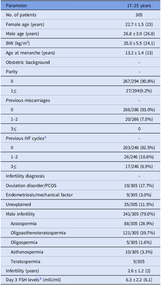
Between 2003 and 2020, 414 women aged 17–25 have undergone a controlled ovarian stimulation cycle at our IVF unit. One hundred and nine cases of a combined (female and male) factor, unexplained infertility, PGT and fertility preservation were excluded from the final analysis. The basic characteristics and reproductive background of the entire study population are presented in Table 1.
Basic characteristics of the entire study population – in vitro fertilization patients 17–25 years of age

Data presented as mean ± SD (Median) or n (%). p-Value < 0.05 was considered statistically significant.
Note: BMI, body mass index; FSH, follicular stimulating hormone; IVF, in vitro fertilization; PCOS, polycystic ovary syndrome.
a Data was available for 246/305 patients only.
b Baseline FSH levels measured in hormone panel test at day three of menstrual period (prior to initiation of hormonal therapy).
In the entire study population, male factor (only) infertility was the most prevalent diagnosis (241/305 (79.1%)), with oligo-astheno-teratozoospermia (OAT) being the most common subtype of male factor infertility (121/241 (50.2%)).
Age and cycle outcomes
Assessment of the correlation between age and cycle outcomes (Figure 1A–D) showed no correlation between age and the following parameters: number of retrieved oocytes, number of mature and fertilized oocytes and the maturation and fertilization rates (correlation coefficients (R) of 0.01, 0.03, −0.01, 0.02 and −0.05, respectively; NS for all).
Correlation between age and cycle outcomes: (A) number of retrieved oocytes, (B) number of mature oocytes, (C) percent of mature oocytes and (D) fertilization rate.

An additional evaluation of the age factor was done using allocation of patients to two age groups – 17–22 years (n = 117) and 23–25 years (n = 188). Comparison of basic characteristics between two women’s age groups demonstrated that in the younger group the male partner’s mean age as well as the rates of previous miscarriage were lower, the duration of infertility was shorter and rates of previous IVF cycles and day 3 FSH levels were also decreased (Suppl. Table 1). The younger group had higher prevalence of male factor aetiology compared to the 23–25 years group (95.6% vs. 85.5%, p = 0.04). Evaluation of cycle outcomes between the age groups (Suppl. Table 2) showed similar outcomes with the exception of one parameter – the rates of 3PN embryos. The 17–22 years group had significantly higher rates of 3PN embryos compared to the 23–25 years group (3.7% (28/759) vs. 0.7% (15/1193), p = 0.001).
Comparison according to infertility etiology – male vs. female factor
We further analyzed our entire cohort according to infertility diagnosis – male factor (MF) only (n = 241) vs. female factor (FF) only (n = 64).
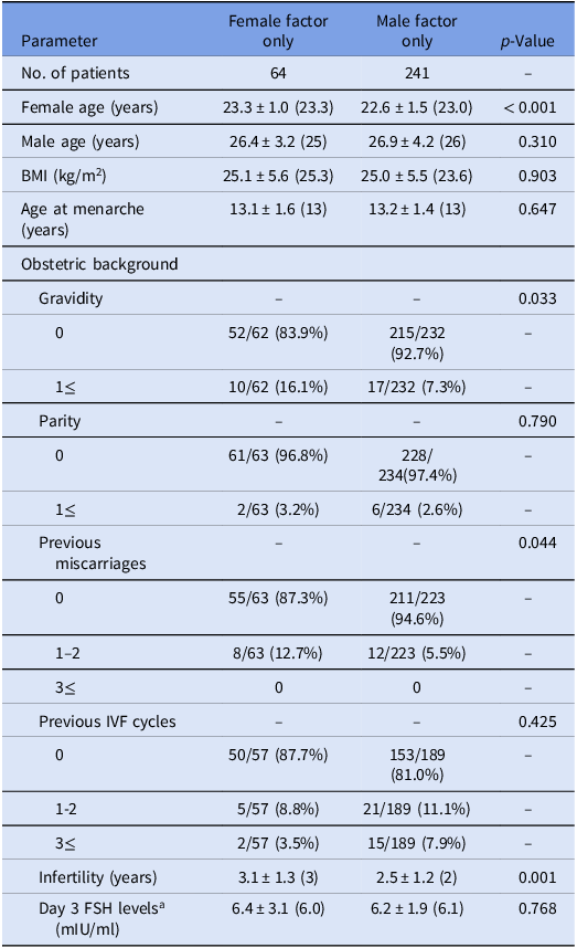
Table 2 presents the comparison of basic characteristics between the two infertility diagnoses groups – the FF only and MF only. Patients in the MF group were slightly younger (22.6 ± 1.5 vs. 23.3 ± 1.0, respectively; p < 0.001), with higher rates of women without previous pregnancies (92.7% vs. 83.9%, respectively; p = 0.033) or miscarriages (94.6% vs. 87.3%, respectively; p = 0.044) and a shorter duration of infertility (2.5 ± 1.2 vs. 3.1 ± 1.3, respectively; p = 0.001).
Basic characteristics of the study population according to infertility aetiology – female vs. male factor only

Data presented as mean ± SD (Median) or n/N (%). p-Value < 0.05 was considered statistically significant.
Note: BMI, body mass index; FSH, follicular stimulating hormone; IVF, in vitro fertilization.
a Baseline FSH levels measured in hormone panel test at day three of menstrual period (prior to initiation of hormonal therapy).
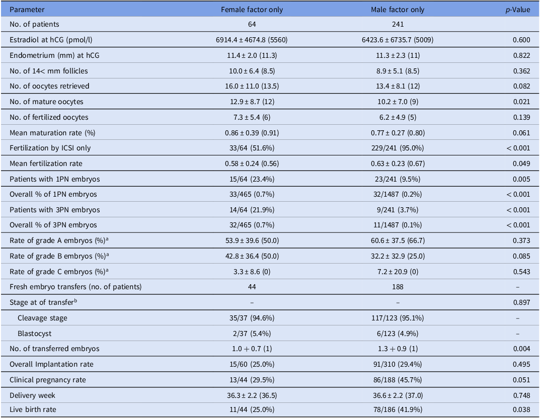
Comparison of cycle outcomes (Table 3) demonstrated preferable results in the MF group vs. the FF group, with higher mean fertilization rates (0.63 ± 0.23 vs. 0.58 ± 0.24, respectively; p = 0.049) and lower percent of 1PN and 3PN embryos (0.2% vs. 0.7% and 0.1% vs. 0.7%, respectively; p < 0.001 for both). The exception was the mean number of mature oocytes, which was higher in the FF group compared to the MF group (12.9 ± 8.7 vs.10.2 ± 7.0, p = 0.021).
Embryo transfer cycle outcomes according to infertility aetiology – female vs. male factor only

Data presented as Mean ± SD (median) or n/N (%). p-Value < 0.05 was considered statistically significant.
ICSI, Intracytoplasmic sperm injection; hCG, human chorionic gonadotropin; PN, pronuclei.
a Embryo quality was determined according to the SART grading.
b Data regarding stage at transfer was available for 37/44 patients in the female factor group and for 123/188 patients in the male factor group.
The mean number of transferred embryos was slightly higher in the MF group compared to the FF group (1.3 ± 0.9 vs. 1.0 ± 0.7, respectively; p = 0.004).
While implantation rate and gestational week at delivery were similar, The MF group presented significantly higher live birth rate compared to the FF group (41.9% vs. 25.0%, p = 0.038) and nearly significant improved clinical pregnancy rate (45.7% vs. 29.5%, p = 0.051) (Table 3).
Univariate logistic regression analyses were performed for the parameters of clinical pregnancy and live birth. The logistic regression models for these outcomes included the following covariates: male vs. female factor infertility, maternal age, number of oocytes retrieved, proportion of mature oocytes, fertilization rate (for the clinical pregnancy model only) and number of embryos transferred. The regression analysis for clinical pregnancy did not identify any parameter significantly associated with pregnancy, although male factor infertility nearly reached statistical significance (OR = 2.01 (95% CI 0.99–4.08), p = 0.051). The univariate logistic regression for the dependent parameter of live birth showed that the only factor associated with live birth was MF infertility aetiology (OR = 2.17 (95% CI 1.03–4.55), p = 0.038) and therefore a multivariate logistic regression was not performed.
An additional subgroup analysis was performed, comparing the FF group with MF cases in which fertilization was done using ejaculated sperm only (n = 153), excluding cases of azoospermia and fertilization done with testicular sperm. Results were similar in this comparison, with younger age, higher nulliparity rate and shorter infertility duration in the MF group. Lower percent of 1PN and 3PN embryos were also noted in the MF ejaculated sperm group compared to the FF group. A trend of higher clinical pregnancy and live birth rates in the MF group was additionally shown, though statistical significance was not reached. (Data available upon request).
Discussion
This study aimed to investigate the population of young women undergoing IVF treatments with regard to age and infertility aetiology. The unique feature of this group – the IVF outcomes of young female partner’s age – was evaluated as both a continuous and categorical parameter. This analysis did not demonstrate a correlation between age and oocyte number, oocyte maturation, fertilization rate, implantation rate and clinical pregnancy and live birth rates. Allocation to age groups did not reveal a significant difference in these outcomes as well, except for the percentage of 3PN embryos which was higher in the younger group. These results suggest that within the young age group of 25 years or younger, the age effect on IVF outcomes is less substantial, though it is possibly related, in some cases, to abnormal fertilization reflected by the rate of 3PN embryo, as demonstrated previously (Franasiak et al., Reference Franasiak, Forman, Hong, Werner, Upham, Treff and Scott2014).
We then evaluated the impact of the infertility aetiology in this population by comparing couples with female infertility to those with male factor infertility only.
Interestingly, this analysis revealed several significant differences in cycle outcomes and demonstrated poorer outcomes for young females that additionally had a female infertility factor. This was reflected in lower fertilization rates, higher percent of 1PN and 3PN embryos and lower live birth rate. This was also emphasized in a logistic regression analysis which found that the only parameter associated with live birth is male factor infertility.
This study focuses on the possible additive effect of the infertility factor in the young IVF female patients’ population. We have shown that the age factor within this unique population is not associated with altered results. However, the combination of a female’s young age along with a female infertility factor is a major element, associated with poorer IVF outcomes, compared to young females without infertility, but rather a male factor infertility solely.
Previous studies that evaluated the age factor were contradictory regarding its association with reproductive outcomes in IVF patients. While some studies demonstrated higher live birth rate in very young patients compared to older patients (Piette et al., Reference Piette, De Mouzon, Bachelot and Spira1990; Dicker et al., Reference Dicker, Goldman, Ashkenazi, Feldberg, Shelef and Levy1991; Tan et al., Reference Tan, Betts, Mason, Edwards, Campbell, Royston and Jacobs1992; Engmann et al., Reference Engmann, Maconochie, Bekir, Jacobs and Tan1999; De Brucker et al., Reference De Brucker, Haentjens, Evenepoel, Devroey, Collins and Tournaye2009), others showed poorer IVF treatment outcomes in the young population (Templeton et al., Reference Templeton, Morris and Parslow1996; Lintsen et al., Reference Lintsen, Eijkemans, Hunault, Bouwmans, Hakkaart, Habbema and Braat2007; Humm et al., Reference Humm, Dodge, Wu, Penzias, Malizia, Sakkas and Hacker2015; Grøndahl et al., Reference Grøndahl, Christiansen, Kesmodel, Agerholm, Lemmen, Lundstrøm, Bogstad, Raaschou-Jensen and Ladelund2017), as well as with young egg donors (Humphries et al., Reference Humphries, Dodge, Kennedy, Humm, Hacker and Sakkas2019).
A possible explanation to this discrepancy might be related to other confounding factors at the young age group. One of the factors shown to affect outcomes when comparing results according to age is the infertility aetiology (Tan et al., Reference Tan, Betts, Mason, Edwards, Campbell, Royston and Jacobs1992; Alsalili et al., Reference Alsalili, Yuzpe, Tummon, Parker, Martin, Daniel, Rebel and Nisker1995; Bancsi et al., Reference Bancsi, Huijs, den Ouden, Broekmans, Looman, Blankenstein and te Velde2000; Lintsen et al., Reference Lintsen, Eijkemans, Hunault, Bouwmans, Hakkaart, Habbema and Braat2007; Ottosen et al., Reference Ottosen, Kesmodel, Hindkjær and Ingerslev2007; Garrido et al., Reference Garrido, Bellver, Remohí, Simón and Pellicer2011; McLernon et al., Reference McLernon, Maheshwari, Lee and Bhattacharya2016 Reference McLernon, Maheshwari, Lee and Bhattacharyaa).
In the current study, male factor aetiology was the most common type of infertility (79%). This high prevalence of MF couples in our young population was significantly higher than shown in most studies evaluating the effect of age in the entire range of female’s reproductive years. This finding, representing the unique group of young IVF patients, may explain why the age factor was not shown to be a significant factor in our population, as opposed to previous studies that had approximately half of couples with MF infertility. The comparison of two groups based on infertility factor which showed significant differences between the aetiology groups may support this suggested assumption.
The current study showed that even though the female factor group had a higher number of retrieved and mature oocytes, the male factor group presented higher fertilization rate and lower 1PN and 3PN rates. This may be related to poorer oocyte maturation and quality and decreased optimal fertilization potential in young women with a female infertility compared to those without infertility other than MF.
Moreover, comparison of the two age group revealed higher percentage of MF cases in the younger (17–22 years) group and despite that, this group had higher 3PN embryos. It is possible that this data caused an underestimation of a more substantial difference in 3PN rate between the age groups.
Normal fertilization is established by the appearance of 2PN. Two main anomalies reported in the process of fertilization are monopronuclear (1PN) and tripronuclei (3PN) fertilized oocytes (Staessen and Van Steirteghem, Reference Staessen and Van Steirteghem1997). According to a meta-analysis that evaluated several small studies, which performed cytogenetic analyses of abnormally fertilized oocytes, 28–57% of 1PN embryos completed their division to normal diploid embryos, while up to 86% of 3PN embryos eventually developed to polyploid or mosaicism embryos (Feenan and Herbert, Reference Feenan and Herbert2006). Therefore, higher rate of 3PN embryos may indirectly reflect higher aneuploidy potential.
Young women were previously shown to exhibit increased rates of aneuploidy (Franasiak et al., Reference Franasiak, Forman, Hong, Werner, Upham, Treff and Scott2014) and embryos with abnormal process of pronuclei appearance. Some investigators suggested that these findings derive from the process of the follicular atresia during infancy and childhood, a process which gradually recedes towards the beginning of the third decade of life. Accordingly, young IVF patients have a relatively high portion of oocytes in different stages of apoptosis and though fertilized, they may exhibit lower-quality embryos with higher rates of aneuploidy and abnormal meiosis process reflected in higher percentage of 1PN and 3PN embryos.
Our finding of significantly higher rates of 3PN embryos in the younger, 17–22 years group compared to the older group (23–25 years) is in agreement with those studies. This difference might have been more emphasized if the percentage of female infertility aetiology (shown to be associated with higher rate of 3PN embryos in our population) was not lower in the 17–22 years group compared to the 23–25 years group.
In our study, the female factor population exhibited significantly higher rates of both 1PN and 3PN embryos and a higher percentage of patients in this group had at least one 1PN or 3PN embryo, compared to the male factor infertility group. These results may support a hypothesis that very young patients with an underlying ovulation disorder or other female infertility are at higher risk for the development of aneuploid embryos, perhaps due to a possible disturbance in the hypothalamic-pituitary-ovarian axis maturation in these patients. These observations are of value to the present literature in this field, and further evaluation of young patients with female factor infertility is needed with regard to aneuploidy in order to establish the abovementioned association.
The differences in fertilization rates and number of embryos transferred between the MF and FF groups likely reflect a combination of biological and clinical factors rather than inherent differences in embryo quality, which was similar between groups. Higher fertilization rates in the MF group resulted in more embryos available for transfer, and in cases of severe male factor infertility requiring surgical sperm retrieval, local policy permits transferring two embryos even in the first cycle. This combination of factors – higher fertilization, more embryos and two-embryo transfers – may contribute to the observed trend towards higher clinical pregnancy rates and the significantly higher live birth rate in the MF group.
Moreover, in our cohort, the most common diagnosis within the FF group was PCOS, a condition in which oocyte developmental competence may be reduced even when oocytes appear mature. Previous studies (Patel et al., Reference Patel and Carr2008; Qiao et al., Reference Qiao and Feng2011) have shown that women with PCOS often exhibit compromised oocyte quality, including abnormalities in cytoplasmic maturation, meiotic spindle structure and metabolic function that can diminish fertilization potential. These intrinsic impairments provide a possible explanation for the lower fertilization rates observed in the FF group. Thus, subtle deficits in oocyte competence associated with PCOS may have contributed to the reduced fertilization rate in this group.
A possible clinical implication is that young women with female factor infertility particularly those with ovulatory disorders such as PCOS – may benefit from early consideration of adjunctive strategies to optimize oocyte quality before IVF (e.g., metabolic stabilization, lifestyle changes and weight optimization when relevant).
A possible explanation for the observed differences between the youngest age group (17–22 years) and the older cohort (23–25 years) relates to both the type of infertility and the clinical treatment pathways. Severe or combined male factor infertility is typically identified quickly through routine semen analysis, and such couples – particularly when IUI is unlikely to succeed – often proceed directly to IVF/ICSI. In contrast, suspected female factor infertility, even with normal ovarian reserve, usually involves several cycles of ovulation induction (with aromatase inhibitors or clomiphene citrate) and, when appropriate, gonadotropin with IUI before IVF is considered. These differing diagnostic and treatment trajectories may contribute to the shorter infertility duration, fewer prior treatments and miscarriages, lower day-3 FSH and younger female age observed in the male factor and younger cohorts. While these patterns primarily reflect standard clinical practice, they may also indicate a subtle gender bias, whereby male factor cases are more rapidly preferred for IVF referral, potentially leading to earlier treatment. This highlights how clinical pathways may unintentionally influence apparent age and infertility-related differences in observational studies. Due to small subgroup sizes, these interpretations cannot be fully confirmed but provide a reasonable clinical context for the findings. Besides its retrospective nature and the long period of the study, this study has several other limitations. The female factor group was heterogenous, combining different female factor aetiologies, without isolation of a single type of female infertility for analysis. This heterogeneity may represent a limitation of the study. However, due to the small sample size, subgroup analyses were not feasible. Future larger studies should explore these differences to better understand their impact.
Despite the planned a priori sample-size calculation, the final cohort did not reach the target numbers, particularly in the female factor group (n = 64), resulting in reduced statistical power. Consequently, the study may have been underpowered to detect more modest differences, which likely explains the absence of statistical significance for several parameters. This limitation should be considered when interpreting the findings and highlights the need for validation in larger, prospective cohorts. The higher percentage of MF cases (shown to result in improved outcomes and lesser 1PN and 3PN) in the younger 17–22 years group could have confound possible differences between the 17–22 years and 23–25 years and this should be considered when assessing the generalizability of this comparison’s results. Also, our analysis did not account for the difficulty of embryo transfer, as this parameter is not captured consistently in our records and is noted only in infrequent, markedly difficult procedures. Finally, the division of the cohort into 17–22 vs. 23–25 years was based on a mid-range cutoff to explore potential differences within this early-adult population. While female age was also included as a continuous variable in our regression models, allowing gradual age-related effects to be accounted for, we note that this grouping may not fully reflect underlying differences across the age range.
Despite these potential caveats, our study has several strengths of which are the relatively large group of young women evaluated and the analysis of IVF cycle outcomes according to both age and a single infertility aetiology.
In conclusion, we found that in young women undergoing IVF cycles, the age factor is not associated with the cycle’s outcomes, except for higher rates of 3PN embryos. However, couples with a young female partner (under 25 years) who has female infertility have poorer outcomes compared to couples with male factor infertility. This reflects in lower fertilization rates, higher rates of 1PN and 3PN embryos and lower live birth rates in the young female factor infertility group.
Supplementary Material
The supplementary material for this article can be found at https://doi.org/10.1017/S0967199426100306.
Acknowledgements
The authors thank all the dedicated embryologists of the Fertility and IVF Unit at Hadassah Medical Organization.
Competing interests
The authors declare that they have no competing interests.
Funding information
None.
Ethical standards
The authors assert that all procedures contributing to this work comply with the ethical standards of the relevant national and institutional committees on human experimentation and with the Helsinki Declaration of 1975, as revised in 2008. This study was approved by the Human Research Ethics Committees (IRB reference 0714-20-HMO). No consent form was required for this anonymous retrospective cohort study.