INTRODUCTION
Within the framework of an environmental monitoring study conducted in 2007 in a tuna farm off the south-east coast of Spain, many benthic samples were collected in order to assess the disturbance caused by this facility on the bottom communities. The key factor of the study was that these communities were settled on a rhodolith bed, a habitat well known for being extremely rich in species but also very fragile to human activities (Bellan-Santini et al., Reference Bellan-Santini, Lacaze and Poizat1994). Besides characterizing several abiotic features of the affected bottoms, the polychaetous annelids were selected as a target group since their sensibility to disturbance in the environment can cause changes in their populations that reflect accurately the existence of any alteration (Giménez-Casalduero et al., Reference Giménez-Casalduero, Rodríguez-Ruiz, Vivas and Ramos-Esplá2001). Over 6000 specimens were identified to family level, several of which resulted to belong to Fauveliopsidae, a family never before recorded on the Iberian Mediterranean coasts. More detailed examination of these specimens revealed them to belong to Fauveliopsis glabra (Hartman, Reference Hartman, Hartman and Barnard1960).
The family Fauveliopsidae comprises about 20 species of small-sized polychaetous annelids belonging to the genera Fauveliopsis McIntosh, Reference McIntosh1922 and Lauberiopsis Petersen, Reference Petersen2000, of which the latter includes only four species (Petersen, Reference Petersen2000). They are mainly found on very deep sedimentary bottoms down to 6000 m (Levenstein, Reference Levenstein1970) although a few species can be found above 100 m depths (Katzmann & Laubier, Reference Katzmann and Laubier1974). The species of Fauveliopsis are almost completely sedentary, dwelling within abandoned mollusc shells or tests of large-sized foraminiferans (Blake & Petersen, Reference Blake, Petersen, Blake and Petersen2000).
All the fauveliopsids possess a retractile head, which is very difficult to examine, and a cylindrical or club-shaped body. The body wall can be smooth or rough, bearing more or less numerous transverse ridges and papillae of variable size (even minute, the so-called micropapillae). Some species also have genital papillae, whose number (one or two) and segment of occurrence are important taxonomic characters (Petersen, Reference Petersen2000); when only a single papilla exists, it is usually located in the right side of the corresponding segment. In all the known species, every segment bears biramous parapodia, although lobes may be reduced or even lacking, so chaetae are inserted in the body wall. A large, spherical interramal papilla is always present, which can be sessile or stalked. The chaetae can show only two forms: more or less straight capillaries and curved spines with acute tips, also known as acicular chaetae (Petersen, Reference Petersen2000).
Subsequently to the family description, the most important contributions to the systematics and the knowledge of the biology and anatomy of the fauveliopsids have been those by Levenstein (Reference Levenstein1970), Katzmann & Laubier (Reference Katzmann and Laubier1974), Riser (Reference Riser1987), Blake & Petersen (Reference Blake, Petersen, Blake and Petersen2000), Petersen (Reference Petersen2000) and Zhadan & Atroshchenko (Reference Zhadan and Atroshchenko2010).
MATERIALS AND METHODS
Sampling stations were selected under the tuna cages and in their vicinity and marked in all the cases by GPS, in order to precisely locate the coordinates. The samples were collected by means of a 400 cm2 wide Van Veen grab, replicating the procedure three times in each station. In order to assess the seasonality of the impacts two sampling campaigns were performed. The first set of samples was collected in July 2007 and the second one in November of the same year, when the very same stations were located and studied by means of their GPS coordinates. In all samples, the extracted sediment was immediately sieved through a 0.5 mm gauge mesh and fixed in 5% formalin. Subsequently, the retained material was examined in the laboratory under a stereomicroscope to sort the polychaetous annelids into families. Specimens of Fauveliopsis glabra were examined using a stereomicroscope and a compound microscope with differential contrast optics (Nomarski) equipped with a drawing tube. Two specimens were critical point dried and gold coated for scanning electon microscopy (SEM) study following standard procedure and the remainder were preserved in 70% ethanol and deposited in the Museo Nacional de Ciencias Naturales de Madrid (MNCNM).
RESULTS
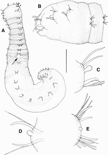
Fauveliopsis glabra (Hartman, Reference Hartman, Hartman and Barnard1960). (A) Entire animal, dorso-lateral view showing position of genital papilla (arrow); (B) anterior part of body, lateral view; (C) parapodium of chaetiger 1 showing duplication of neurochaetae, anterior view; (D) parapodium of chaetiger 10, posterior view; (E) parapodium of chaetiger 21, posterior view. Scale bars: (A) 0.7 mm; (B) 0.3 mm; (C, D, E) 0.1 mm.

Additional material of Fauveliopsis glabra (Hartman, Reference Hartman, Hartman and Barnard1960). (A) Anterior end, dorsal view showing position of genital papilla (arrow), first scanning electron microscopy (SEM) specimen; (B) detail of prostomium and first four chaetigers, ventro-lateral view, second SEM specimen; (C) chaetigers 6 and 7, lateral view, first SEM specimen; (D) chaetiger 10, dorsal view, showing detail of genital papilla (arrow), same specimen; (E) body wall of the posterior region of the body showing position of micropapillae (arrows), second specimen; (F) detail of a micropapilla (arrow) of the posterior region of the body, same specimen. Scale bars: (A) 0.2 mm; (B, C) 0.1 mm; (D, E) 50 µm; (F) 10 µm.

MATERIAL EXAMINED
Isla del Fraile, Murcia, Spain (37°24′41″N–1°32′3″W); rhodolith bed under tuna farming cages; 40 m depth; 10 July 2007; seven specimens plus two additional specimens prepared for SEM. Same station; 7 November 2007; one specimen.
DESCRIPTION
Body club-shaped, anterior part narrower, posterior distinctly swollen (Figure 1A); straight or coiled depending on housing shell (scaphopod or gastropod); yellowish after fixation. Largest specimen is 3.8 mm long for 28 chaetigers; body width 0.34 mm in middle of its narrower part and 0.70 in middle of its swollen part. Prostomium usually invaginated causing partial contraction of chaetiger 1 and making the anterior end look rounded and truncate (Figures 1A, B & 2B). Intersegmental furrows well defined from chaetigers 1 to 8; absent in segments of the posterior, swollen part of body (Figures 1A & 2A). Body surface more or less rough in the anterior narrower part of the body, due to the presence of numerous fine transverse ridges (Figures 1A & 2A); relatively smooth in the swollen part, with transverse ridges only present over its ventral surface (Figure 1A); last 4–6 chaetigers again with transverse ridges extending all around the body. Body wall is quite thick in the anterior part; very thin in the swollen part, allowing the observation of the gut (Figure 1A); ventral scutum indistinct. Parapodial lobes well developed and biramous, clearly projecting only in the transitional area between anterior and posterior part of the body (Figure 2C). Pygidium bent ventrally and covering the anus. Body divided into three regions based on chaetal shape and distribution. Anterior region formed of the first two or three chaetigers; parapodial lobes bearing only spines; notopodia of chaetiger 1 each with two similarly sized spines, lower spine slightly falcate, upper one straight (Figures 1B & 2B); corresponding neuropodia each with two spines clearly thicker than their notopodial counterparts, upper one very large and strongly falcate, lower one thinner and not so curved (Figures 1B & 2B); noto- and neuropodia of chaetiger 2 and sometimes of chaetiger 3 with spines resembling those of chaetiger 1; in several specimens the number of chaetae appears duplicated on some parapodia (Figure 1C). Middle region extending from chaetigers 3–4 to 19–23, thus including most of the swollen part of the body; besides occasional doubling, each parapodial ramus bearing a slightly falcate spine and a thinner, straight capillary chaeta (Figures 1D & 2C); difference in thickness between spine and capillary chaeta becoming less more posteriorly; spines at first clearly thinner and straighter than those of anterior region, then becoming thicker and more strongly curved. Posterior region extending from chaetiger 20–24 to pygidium and characterized by higher number of chaetae on neuropodia and occasionally also on notopodia; each neuropodial ramus with 2–4 spines similar in size to those of the middle region and a more or less equal number of capillary chaetae; chaetae of notopodial rami resembling those in the posterior chaetigers of the median region but more numerous (Figure 1E). Interramal papilla stalked; relatively large in chaetigers of anterior region (Figures 1B & 2B); smaller and more centrally located in the following chaetigers (Figure 2C). In some specimens, pigmented segmental organs are immediately behind the median line of parapodial lobe in chaetigers 9 to 20; organs more conspicuous after methyl green staining. Genital papilla usually retracted; when everted, present in right side only, as a single oval process near the rear end of chaetiger 10 (Figures 1A & 2A, D). Micropapillae (Figure 2E, F) piriform, scattered over ridged surfaces of the body, being more abundant on the flanks; more conspicuous after methyl green staining.
REMARKS
Although separate from the known geographical distribution and inhabiting a totally different environment, i.e. a rhodolith bed, the specimens described herein correspond quite well with former descriptions of F. glabra, especially with the most recent and detailed one (Blake & Petersen, Reference Blake, Petersen, Blake and Petersen2000). Some of the Mediterranean specimens bear paired intersegmentary organs located just behind the median line of the parapodial lobe and appearing from chaetiger 9 onwards. Although the function of these structures remains unclear, they might correspond with the multicellular, epidermal glands recorded by Riser (Reference Riser1987) for Lauberiopsis arenicola (Riser, Reference Riser1987).
The species is found from deep shelf and slope (Hartman, (Reference Hartman, Hartman and Barnard1960) to bathyal and abyssal zones (Blake & Petersen, Reference Blake, Petersen, Blake and Petersen2000), with most of the records collected from depths ranging from 135 to 2500 m in the Pacific Ocean and down to 5000 m in the western Atlantic. It usually inhabits soft bottoms, especially those of a silty composition, where it uses abandoned scaphopod or gastropod shells as refuge. Blake & Petersen (Reference Blake, Petersen, Blake and Petersen2000) examined the gut of one (the largest) specimen and found it filled with ‘fine silt and a few coarser particles including foraminiferans’, so they presumed the species to be a non-selective sedimentivore feeder with a rather sedentary way of life.
The studied material herein was collected from a quite different environment. Albeit being also shell-dwellers and using scaphopod or gastropod shells (both very abundant in the sediment where the worms were collected), these Mediterranean specimens were collected from a rhodolith bed at 40 m depth. Apart from the distinctly shallower collecting depth compared with those mentioned in most of the literature, the sediment structure of this kind of bottom is very different to that of the previously recorded silty ones. Instead of a more or less compact and uniform structure, rhodolith beds are composed of layers of biogenic nodules formed by the growing of calcareous red algae and placed over a layer of coarse to very coarse calcareous debris (Foster, Reference Foster2001). The rhodoliths are 1–15 cm wide and are branched to a degree which depends on hydrodynamic conditions (more irregular under slow current forces), thus creating a net of interstitial spaces that allows a fast interstitial water movement (Luque & Templado, Reference Luque and Templado2004). These conditions seemingly do not favour the settlement of fine particles available to be ingested by sedimentivores; however, the sampling was conducted close to a tuna farm so the presence of otherwise unusual fine particles of organic matter cannot be discarded.
The original description of the species (Hartman, Reference Hartman, Hartman and Barnard1960) was based on material collected from the Pacific coast of the United States (Northern California), but subsequently the species has been recorded from the Galápagos Islands and from several localities in the eastern Pacific, ranging from the type locality to Peru (Salazar-Vallejo & Londoño-Mesa, Reference Salazar-Vallejo and Londoño-Mesa2004). It has also been recorded from deep waters in the north-western Atlantic, near Bermuda and off the east coast of the United States (Hartman, Reference Hartman1965; Hartman & Fauchald, Reference Hartman and Fauchald1971). This is the first record of the species for the Mediterranean Sea.
ACKNOWLEDGEMENTS
Part of the present work was financially supported by the Spanish Ministerio de Ciencia y Tecnología through the research project ‘Fauna Ibérica VIII, Anélidos Poliquetos III, Scolecida, Amphinomida y Phyllodocida, CGL2004–04680–C10–02/BOS’. The author also wishes to express his gratitude to the Marine Ecology Division of Laboratorios Munuera S.L., especially to Alejandro Polo and Alberto Echeita, who provided the specimens described herein, as well as useful data on the environmental parameters of the samples.